Types of protein-ligand interactions and how to read an interaction diagram
Why one structure can give several different contact lists

Matic Broz, PhD19 min
What are protein-ligand interactions?
Protein-ligand interactions are the noncovalent contacts between a bound molecule and the amino acids around it. The main types are hydrophobic contacts, hydrogen bonds, salt bridges, pi stacking, cation-π interactions and halogen bonds, with water bridges and metal coordination added where the structure has waters or metal ions. A 2D interaction diagram draws the contacts that a program finds in a 3D structure by testing distances and angles.
The test is where the trouble starts. The crystal structure 1EVE holds donepezil, the Alzheimer's drug, in acetylcholinesterase from the electric ray Torpedo californica.[7] We ran that one file through PLIP and ProLIF with their default settings. PLIP lists 10 contacts with seven residues. ProLIF lists seven contacts with six residues, or eight if the drug's amine is entered as charged, and the two lists share five residues.
Neither program is wrong. Each applies its own distance and angle limits, its own ring definitions and its own assumptions about hydrogens, waters and charge, and a line in a diagram records only that a contact passed those rules. It does not record what the contact is worth. Literature values for a single hydrogen bond run from 1.5 to 4.7 kcal/mol, which is a 13-fold to 2,800-fold change in Kd at 298 K, and a buried methyl group adds about 0.7 kcal/mol, or about 3-fold.[2][3]
The sections below give the geometry and typical value of each interaction type, then look at pi stacking in detail, read a diagram line by line and return to 1EVE to see where the programs part ways.
The main types of protein-ligand interactions
Ferreira de Freitas and Schapira counted contacts in 11,016 crystal structures of small molecules bound to proteins at 2.5 Å resolution or better, covering 750,873 ligand-protein atom pairs within 4 Å.[2] Table 1 lists the types they found, most frequent first, with what the literature says each is worth. The counts show what crystallographers see, which favors ligands that chemists made lipophilic during optimization. They are not a ranking of importance.
| Interaction | Contacts | Typical geometry | What it is worth |
|---|---|---|---|
| Hydrophobic contact | 66,772 | Carbon to carbon, halogen or sulfur within 4 Å | About 0.7 kcal/mol per buried methyl, roughly 3-fold in Kd; ranges from a loss to a hundredfold gain |
| Hydrogen bond (N-H···O, O-H···O, N-H···N) | 28,577 | Median donor-acceptor distance about 3.0 Å, angle at the hydrogen mostly 130 to 180° | 1.5 to 4.7 kcal/mol, or 13-fold to 2,800-fold; can fall to zero once desolvation is paid |
| Pi stacking | 17,241 | Ring pairs face to face (8,537) or edge to face (8,704) | Benzene dimer: 2.5 kcal/mol calculated, 1.6 to 2.4 measured, for an isolated pair |
| Weak hydrogen bond (C-H···O) | 13,600 | Median distance 3.4 Å, heavy atoms rarely closer than 3.2 Å | About half an N-H···O bond by calculation |
| Salt bridge | 7,276 | Positive nitrogen to negative oxygen; arginine is the cation in 83.6% | Small in solution, decisive when buried |
| Amide-π stacking | 4,967 | Amide plane 3.2 to 3.7 Å from the ring plane; 88.5% involve a backbone amide | Comparable to nearby hydrogen bonds in model peptides |
| Cation-π | 2,577 | Positive nitrogen over an aromatic ring | Ammonium desolvation can outweigh the gain |
| Halogen bond | 351 | C-X···O, median C-X···Y angle 156°; Cl, Br and I only | Grows from Cl to Br to I |
Hydrophobic contacts
Hydrophobic contacts are the most common by a wide margin: 66,772 contacts, about 190 times the number of halogen bonds. Most pair an aromatic carbon on the ligand with an aliphatic side chain, and leucine, valine, isoleucine and alanine supply most of the protein side.[2]
Each contact is small. The hydrophobic effect is about 30 cal/(mol·Å²) of buried nonpolar surface, which works out to 0.7 kcal/mol for a methyl group.[3] The average hides a wide spread. Adding a methyl group raises potency tenfold in 8% of cases and a hundredfold in 0.5%, and losses are as common as gains.[2]
The balance between contact types shifts with ligand size. Hydrogen bonds reach 62% of the hydrophobic count in fragments of 20 or fewer heavy atoms, against 31% in drug-like molecules of 30 to 50. Among the 1,500 most efficient binders they fall to 34% of it, against 59% among the 1,500 least efficient.[2] Polar contacts give a small ligand most of its efficiency, and hydrophobic contacts are what optimization adds.
Hydrogen bonds and their weak cousins
Hydrogen bonds are second in frequency, with N-H···O the commonest. The surveyed literature puts one at 1.5 to 4.7 kcal/mol, but the contribution can be very modest or even negative when the new bond does not repay the cost of stripping water from the partners.[2] Bissantz and colleagues cite a penalty of 4.3 to 5.3 kcal/mol for a hydroxyl group that binds in a hydrophobic pocket, from desolvation.[3]
A weaker class gets less attention. C-H···O contacts, at a median 3.4 Å, are the fourth most frequent type in the census, more than salt bridges and cation-π interactions combined.[2] PLIP's eight interaction classes have no category for them, so a PLIP diagram never draws one.[1]
Salt bridges, cation-π interactions and halogen bonds
Salt bridges add little in water, because the energy gained does not repay desolvating the two charges, but a buried one can decide potency. In an EGFR inhibitor, replacing the nitrogen of the dimethylamino tail that pairs with Asp831 by a carbon cut potency more than 800-fold.[2]
Cation-π interactions put a positive nitrogen over an aromatic ring. In more than 90% of them the nitrogen belongs to the protein and the ring to the ligand, and arginine takes part three times as often as lysine.[2] Bissantz and colleagues point out that the desolvation cost of an ammonium group can far outweigh the gain from a direct NH···π contact.[3]
Halogen bonds are rare, 351 in the census (222 chlorine, 91 bromine, 38 iodine), and fluorine does not form them. The halogen points at an oxygen, most often a backbone carbonyl, along a nearly straight line. In a series of PDE2a inhibitors, potency rose in the order H and F, then Cl, Br and I.[2]
Water bridges and metals
The census counts 4,888 hydrogen bonds between protein and ligand that run through a water molecule.[2] Donepezil is a case in point: Kryger and colleagues found that it reaches the catalytic triad and the oxyanion hole of its enzyme only through solvent molecules.[7] A program can draw a water bridge only if the structure keeps its crystal waters, and docked poses and predicted structures have none.
Metal coordination needs a metal ion in the file and, in PLIP, a ligating atom within 3.0 Å of it.[11]
Pi stacking in protein-ligand complexes
Pi stacking is the attraction between two aromatic rings that lie over or against each other. In a protein-ligand complex it means a ring of the ligand next to the ring of phenylalanine, tyrosine, tryptophan or histidine. Phenylalanine supplies almost half of all pi stacking contacts in the census, tyrosine 36.8%, tryptophan 8.7% and histidine 5.1%.[2]
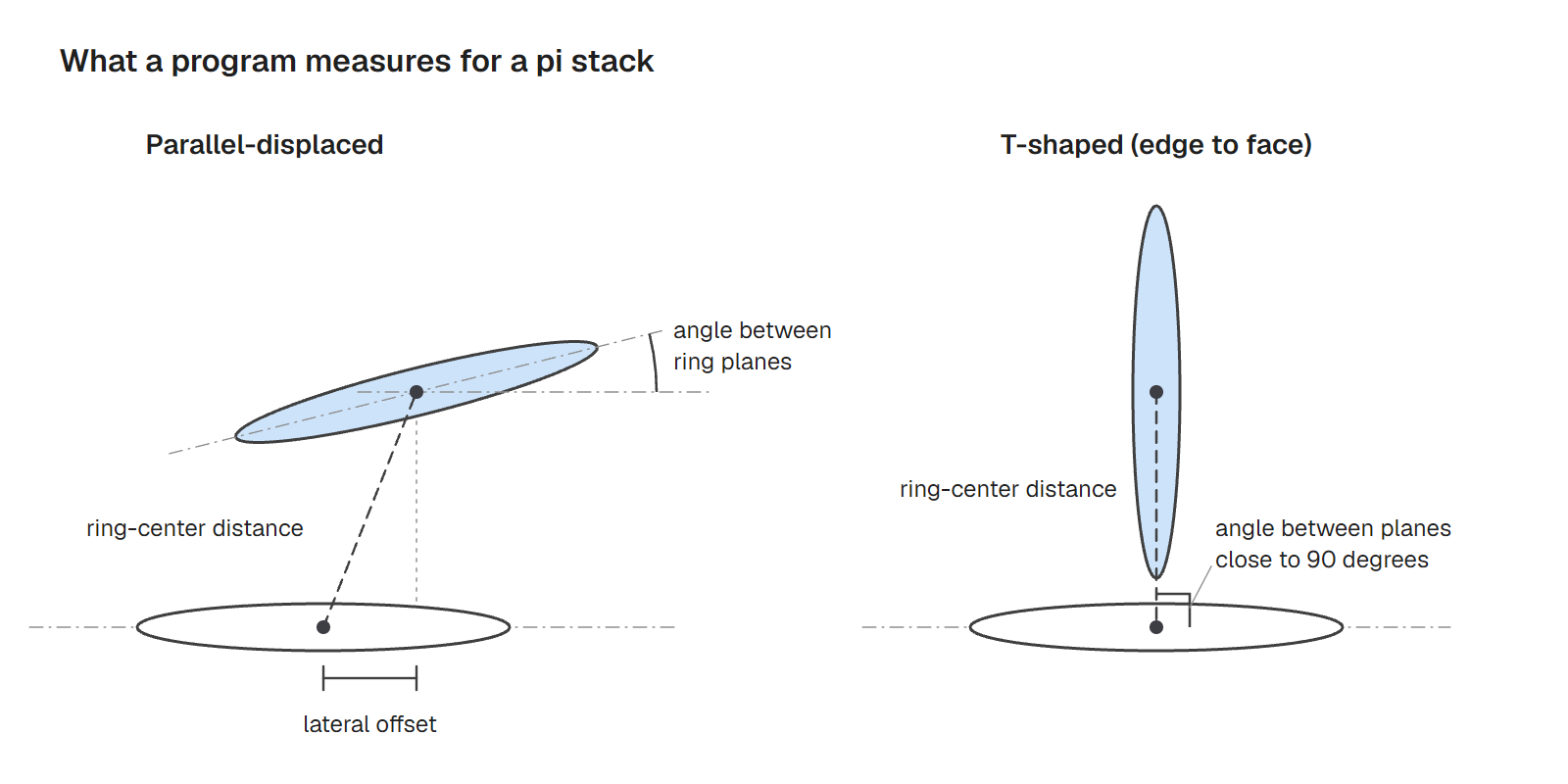
Two arrangements dominate, and the PDB shows no preference between them: 8,537 contacts are face to face and 8,704 edge to face.[2] Calculations on the benzene dimer agree. The parallel-displaced and T-shaped forms have the same energy, about 2.5 kcal/mol, against 1.6 to 2.4 kcal/mol measured, and the fully eclipsed sandwich is less stable.[2][3]
Those are values for an isolated pair of benzenes. In a protein the ring pair also sheds water, which is one reason aromatic contacts are treated as a special case of the hydrophobic ones.[2] The gain can still be large. In a series of soluble epoxide hydrolase inhibitors, a phenyl ring that stacks on His524 goes with an IC50 of 7 nM, while the analog without it, 700 nM, is 100-fold weaker.[2]
Martinez and Iverson reviewed the experimental and computational evidence and argued that the terms "pi-stacking" and "pi-pi interaction" mislead, because direct electrostatic interactions between polarized atoms and solvation effects can dominate how aromatic rings associate.[4] The term stays in use, and a program still needs a rule for it. Each program writes one in terms of three measurements, set out in Table 2.
| Test | PLIP | ProLIF |
|---|---|---|
| Distance between ring centers | Under 5.5 Å | 5.5 Å face to face, 6.5 Å edge to face |
| Angle between ring planes | Within 30° of parallel, or 60 to 90° for T-shaped | 0 to 35° face to face, 50 to 90° edge to face |
| Lateral offset | Under 2.0 Å | Ring normal within 33° (face to face) or 30° (edge to face) of the line between centers, plus an intersection test for edge to face |
| Which rings count | Aromatic rings flat to within 5° | Rings matching a SMARTS pattern for six- or five-membered aromatic rings |
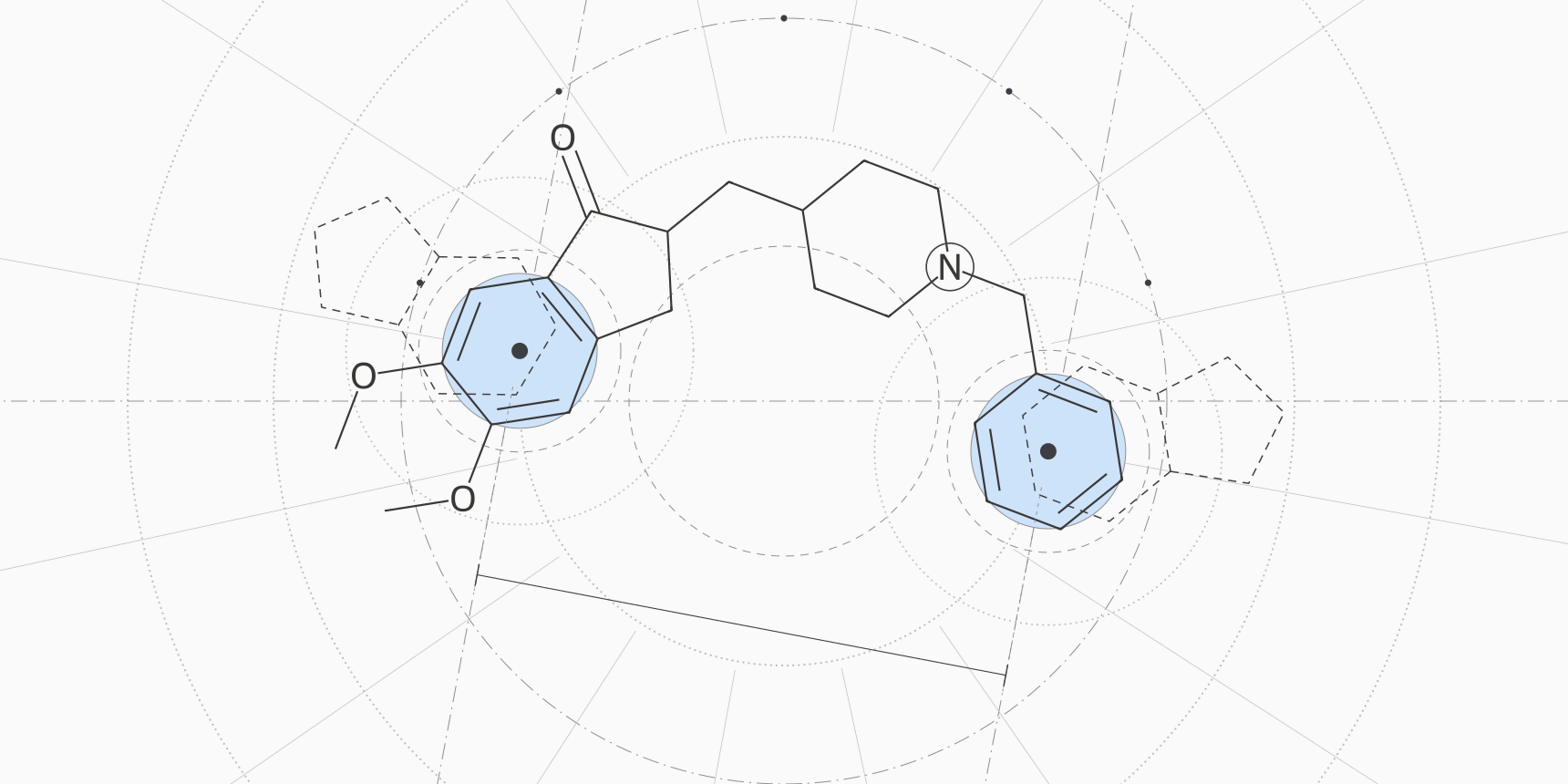
Donepezil in 1EVE has two stacks, one at each end of the drug. Kryger and colleagues describe it as extending from the anionic subsite at the bottom of the active-site gorge to the peripheral anionic site at the top, linked by aromatic stacking.[7]
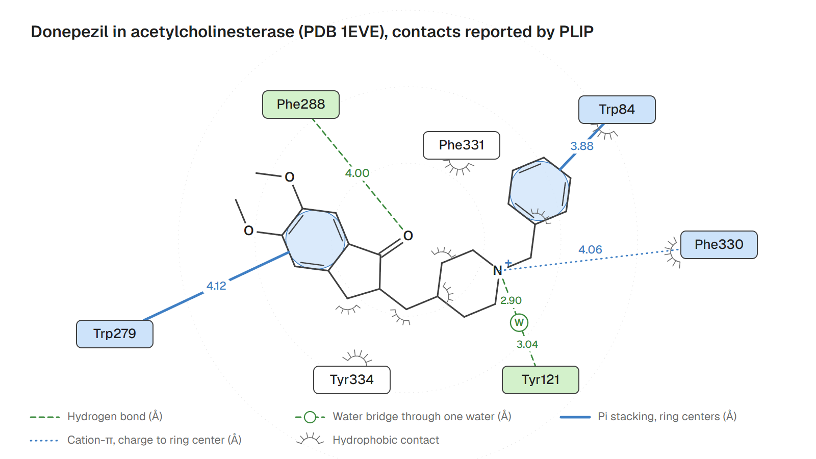
In the PLIP report the benzyl ring lies over Trp84, with ring centers 3.88 Å apart, planes 15° apart and an offset of 0.59 Å. The dimethoxy-indanone ring lies against Trp279, 4.12 Å apart, 13° and an offset of 1.82 Å, which passes PLIP's 2.0 Å limit by 0.18 Å.
ProLIF's defaults find only the first stack, and measure it to the wrong ring: 4.37 Å for Trp84, with Trp279 missing.
The cause is the ring pattern. The six-ring pattern requires an atom whose smallest ring has six members, and the two fusion atoms of indole belong to the five-membered ring, so tryptophan's benzene ring never matches. We checked: the pattern matches nothing in either tryptophan, so only their five-membered rings are tested. Widened to any aromatic six-membered ring, ProLIF reports both stacks at 3.88 Å and 4.12 Å, the same distances PLIP gives.
How to read a 2D interaction diagram
A 2D interaction diagram redraws the ligand flat in the middle of the page and arranges the contacting residues around it. In the LIGPLOT convention, ligand bonds are drawn thick and the protein residues that hydrogen-bond to it thinner. Hydrogen bonds are dashed lines, and hydrophobic contacts are spoked arcs on the residue, with matching spokes on the ligand atoms they touch.[9]
LigPlot+, its successor, draws the same two kinds of contact between the ligand and the main-chain or side-chain parts of the protein,[5] and PoseView draws every molecule at atomic level as a structure diagram.[6] By default LIGPLOT takes its hydrogen bonds and nonbonded contacts from a separate program, HBPLUS, so the cutoffs are not its own.[10] Colors and symbols differ between programs and can be changed.
Figure 2 is our diagram for 1EVE, drawn from the PLIP report.
Read a diagram in this order:
- Find which ligand it shows and which model. A crystal structure can hold several ligands, symmetry copies and additives.
- Find which program and settings drew it. The cutoffs decide what can appear at all.
- Separate the polar lines from the arcs. Dashed lines and ring bars are specific and testable; arcs mostly count how much of the ligand is buried.
- Read the numbers on the lines and the residues they end on, and not just the count of lines.
- Ask what could not be drawn: waters, hydrogens, alternative side-chain positions, charges and contacts for which the program has no class.
Table 3 turns that into a lookup for each element.
| Element | What it records | What it leaves out |
|---|---|---|
| Dashed line with a distance | A donor and acceptor that passed the program's distance and angle test | The angle, the energy, and whether the hydrogen was placed or observed |
| Spoked arc or residue label with no line | At least one nonpolar pair within the cutoff | How many atoms touch, and the fact that larger ligands collect more of them |
| Bar or line to a ring | Two rings that passed the distance, angle and offset tests | Whether the ring is face on or edge on, unless the program prints the type |
| Water in the middle of a line | One crystal water bonded to both partners | Chains of two or more waters, and every water that was not modeled |
| Charge symbol on a group | The program's assumption about that group | The real protonation state in that pocket |
| Residue with nothing attached | A residue near the ligand | Nothing; absence of a line is not absence of an interaction |
One crystal structure, five contact lists
Table 4 puts the 1EVE runs side by side, at the 2.5 Å resolution of the deposited structure.[13] ProLIF's main output is not a picture but an interaction fingerprint, a table of residues by interaction type, which suits comparing many poses.[8]
We ran PLIP 3.0.1 on the crystal file as downloaded, then without its crystal waters, then without its added hydrogens. We ran ProLIF 2.2.0 with the nine default interaction classes of the ProteinIQ tool, waters removed and implicit hydrogens, with donepezil's piperidine nitrogen first neutral and then protonated.
| Run | Hydrophobic | H-bond | Water bridge | Pi stack | Cation-π | Contacts | Residues |
|---|---|---|---|---|---|---|---|
| PLIP, default | 5 | 1 | 1 | 2 | 1 | 10 | 7 |
| PLIP, no waters | 5 | 1 | 0 | 2 | 1 | 9 | 6 |
| PLIP, no added H | 5 | 0 | 0 | 2 | 1 | 8 | 5 |
| ProLIF, neutral amine | 6 | 0 | 0 | 1 | 0 | 7 | 6 |
| ProLIF, charged amine | 6 | 0 | 0 | 1 | 1 | 8 | 6 |
The aromatic core agrees once the rules are aligned. With the amine protonated and the tryptophan ring pattern widened, ProLIF reproduces both of PLIP's stacks and its cation-π contact with Phe330, at 4.06 Å in both programs. The remaining differences come from the lines that sit near a cutoff or depend on something the file does not hold.
The hydrogen bond exists only in PLIP. It runs from the backbone N-H of Phe288 to the carbonyl oxygen of the indanone, with a donor-acceptor distance of 4.00 Å and an angle of 139° at the hydrogen. That passes PLIP's 4.1 Å limit and fails ProLIF's 3.5 Å. It also needs a hydrogen to exist: The polar hydrogens PLIP adds put it on the list, and with hydrogen addition off it disappears.
The water bridge to Tyr121 needs the crystal waters. Remove them and it goes, as it would in a docked pose of the same drug. ProLIF's extra residue, Tyr70, makes a hydrophobic contact at 4.26 Å, which is outside PLIP's 4.0 Å limit but inside its own 4.5 Å.
PLIP also counts the piperidine nitrogen twice. It is a positive charge over Phe330 in the cation-π row and a lone-pair acceptor behind a water in the bridge to Tyr121. The rules run separately, and nothing in the report says a protonated amine cannot do both. ProLIF takes the charge from the file, so the same ligand gives the cation-π contact only when it is entered with a plus sign.
The script and the per-contact output for every run are in the guide's data folder. None of the lists says what any contact is worth. The Phe288 row at 4.00 Å and the stack on Trp84 are one line each. Kryger and colleagues describe the drug as spanning the gorge through aromatic stacking, which the diagram does show, but only the stacking residues and the geometry behind them tell you so.[7]
Checks before you trust an interaction diagram
Table 5 collects the checks that settled the 1EVE comparison, each with a quick way to run it.
| What could mislead | Why | Quick check |
|---|---|---|
| A line near a cutoff | Programs differ by 0.6 Å or more in the limit for the same bond | Sort by distance and look at rows within 0.5 Å of the limit; run a second program |
| Contact count | Hydrophobic rows grow with ligand size | Divide by heavy atoms, and compare only ligands of similar size |
| Charged groups | Some programs assign charge by group and some read it from the file | Check the protonation state of the ligand and of His, Asp and Glu near it |
| Missing hydrogens | A hydrogen bond needs a donor hydrogen to exist | Repeat the run with hydrogens added and without |
| Missing waters | Water bridges appear only where waters were modeled | Treat crystal and docked structures with the same protocol |
| Aromatic ring perception | A ring pattern can skip a ring, as it did for indole | List ring pairs with centers 3.5 to 4.5 Å apart and compare them with the stacks the program reported |
| Contacts as evidence for affinity | A line records geometry and no energy | Use a score or a free-energy method for energy and the contact list for explanation |
A contact list explains a pose and does not rank it. A docking score carries an energy estimate, MM/GBSA and free energy perturbation carry better ones, and the contact list says which residues and which kinds of contact the number depends on. Before reading contacts at all, check that the pose is physically valid and, for docking, how far it sits from the reference by RMSD.
Protein-ligand interaction tools on ProteinIQ
Start by generating or cleaning the complex, check it, then analyze the contacts. Poses from docking come as a receptor file and a separate ligand file, and PLIP needs them merged into one PDB with the pose as HETATM records.
| Task | Tool |
|---|---|
| Dock a ligand into a receptor and keep several poses | AutoDock Vina, GNINA, DiffDock |
| Repair missing atoms and residues in a PDB file | PDBFixer |
| Estimate protonation states of protein residues | PROPKA |
| Protonate a ligand at a chosen pH | Dimorphite-DL |
| Merge a docked pose into a receptor PDB | SDF to PDB |
| Check that a pose is physically plausible | PoseBusters |
| List the interactions of one complex with distances and angles | PLIP |
| Compare interaction fingerprints across many poses | ProLIF |
| Browse a deposited structure as a linked summary report | PDBsum |
Frequently asked questions
What are the main types of protein-ligand interactions?
The main types are hydrophobic contacts, hydrogen bonds, pi stacking, weak C-H···O hydrogen bonds, salt bridges, amide-π stacking, cation-π interactions and halogen bonds. In 11,016 PDB complexes the first three account for 66,772, 28,577 and 17,241 contacts.[2] Water-mediated bonds and metal coordination come on top of these where the structure has waters or metals.
What do the dashed lines and arcs in a 2D interaction diagram mean?
In the LIGPLOT convention, dashed lines are hydrogen bonds and spoked arcs on a residue are hydrophobic contacts.[9] Other programs add symbols for pi stacking, charges and water bridges, and their colors differ. A line means the contact passed the program's distance and angle test, not that it contributes a given amount of energy.
Is pi stacking the same as a pi-pi interaction?
In everyday use, yes, and both terms describe an aromatic ring lying over or against another. Martinez and Iverson argue that both terms mislead, because direct electrostatic interactions between polarized atoms and solvation effects can dominate aromatic association.[4] In structures, face-to-face and edge-to-face geometries are equally common: 8,537 and 8,704 contacts.[2]
Which interaction contributes most to binding?
No single type does. Hydrophobic contacts are the most frequent and each adds about 0.7 kcal/mol for a methyl group, while a hydrogen bond can contribute 1.5 to 4.7 kcal/mol or nothing, depending on desolvation.[2][3] Fragments lean on polar contacts and optimized ligands on hydrophobic ones.[2]
Why do two programs show different interactions for the same structure?
They apply different distance and angle limits, different ring and charge rules, and different treatments of hydrogens and waters. In our test on 1EVE, PLIP listed 10 contacts with seven residues and ProLIF seven with six, and the two shared five residues. The differences traced to a 4.1 Å against 3.5 Å hydrogen bond limit, a ring pattern that skips tryptophan's benzene ring and a ligand charge that one program assigns and the other reads.