Docking score, binding energy and binding affinity: what the numbers mean
Three terms that share a unit and mean different things

Matic Broz, PhD17 min
Docking score vs binding energy vs affinity
Binding affinity is how tightly a ligand binds a target, measured as a dissociation constant (Kd). Binding free energy (ΔG) is that same fact as an energy, ΔG = RT ln Kd. A docking score is a software estimate of ΔG for one pose. "Binding energy" is looser: it can mean the free energy, a force-field interaction energy or, in chemistry and physics, a bond or nuclear binding energy. Only affinity and ΔG are measured.
A shared unit hides the gap. AutoDock Vina prints −12.51 kcal/mol for imatinib on c-Abl, while its measured apparent Kd of 106 nM is −9.52 kcal/mol.[11] MM/PBSA stretches it further: for three charged biotin analogues in avidin it gives −35 to −53 kcal/mol, while the tightest of seven measured avidin ligands reaches −20.4.[6] The numbers look alike and are not.
Which number is which
Table 1 lists the quantities that get called affinity or energy. Only the first two are the same thing in different units, and only the first is measured directly.
| Quantity | What it is | Unit | Where it comes from |
|---|---|---|---|
| Binding affinity (Kd, Ki) | How tightly a ligand binds one target at equilibrium; lower Kd means tighter | Molar concentration, or pKd | Calorimetry, surface plasmon resonance, binding assays |
| Binding free energy (ΔG) | The affinity as an energy, ΔG = RT ln Kd; more negative means tighter | kcal/mol or kJ/mol | Converted from Kd, or read from calorimetry |
| Binding energy, interaction energy | Force-field energy of the complex minus its parts, with or without solvation | kcal/mol | Minimization, MM/PBSA, MM/GBSA |
| Docking score | Value of a scoring function for one pose | kcal/mol for some programs, arbitrary units for others | Docking programs |
| Predicted affinity | A model's estimate of pK or log IC50 for a predicted complex | pK or log units | Machine-learning scoring such as GNINA and Boltz-2 |
Binding affinity and the dissociation constant
Affinity is a statement about equilibrium. For a protein P and ligand L that form a complex PL, the dissociation constant is Kd = [P][L]/[PL], the free ligand concentration at which half the protein is occupied.[16] A smaller Kd means tighter binding, so a 1 nM ligand is a thousand times stronger than a 1 µM one. Kd also belongs to the complex, not to the experiment, unlike an IC50, which shifts with substrate and enzyme concentration.
Affinity is often quoted as a negative logarithm, pKd = −log10 Kd in molar units, so that bigger numbers mean tighter binding. A Kd of 10 nM is a pKd of 8, and pK is the scale that machine-learning tools such as GNINA predict on.
Affinity is also conditional. A Kd belongs to one protein construct, one buffer and one temperature, and it describes one ligand binding one site. Two labs measuring the same complex under different conditions commonly differ by a few-fold, a spread the error section below returns to.[4]
Converting ΔG to Kd and back
Kd and ΔG are the same information on different scales. The standard binding free energy is
where R is the gas constant, T the absolute temperature and c° the standard concentration of 1 M.[1] At 25 °C, RT is 0.592 kcal/mol (2.48 kJ/mol), so every tenfold change in Kd moves ΔG by 1.364 kcal/mol (5.71 kJ/mol).
Table 2 shows the conversion at 25 °C. The scale is steep: a docking score that is off by 2.4 kcal/mol is off by a factor of about 57 in Kd.
| ΔG (kcal/mol) | ΔG (kJ/mol) | Kd |
|---|---|---|
| −4 | −16.7 | 1.2 mM |
| −6 | −25.1 | 40 µM |
| −8 | −33.5 | 1.4 µM |
| −10 | −41.8 | 47 nM |
| −12 | −50.2 | 1.6 nM |
| −14 | −58.6 | 55 pM |
| −16 | −66.9 | 1.9 pM |
| −18 | −75.3 | 64 fM |
Imatinib shows the conversion in practice. Its apparent Kd of 106 nM gives ΔG = 0.5925 × ln(106 × 10−9) = −9.52 kcal/mol.[11] Run it backwards and the Vina score of −12.51 for the same compound would mean a Kd of 0.68 nM, about 150 times tighter than the measurement. That gap is why a docking score should never be converted and reported as an affinity.
Most wrong conversions come from units and inputs. Putting kJ/mol into a formula built for kcal/mol is a factor of 4.184, and the temperature matters little, since 37 °C changes RT by only 4%. The bigger trap is the input: an IC50 is not a Kd, and the IC50 to Ki calculator applies the assay correction first. Femtomolar values like the last row of Table 2 are usually kinetic or displacement estimates with large uncertainty.
What binding energy means in each field
"Binding energy" has three common meanings, and which one applies depends on who is writing.
In drug design and simulation
In papers about docking and molecular dynamics, binding energy usually means an energy difference between the bound complex and the separated partners, in kcal/mol. When the authors have included solvation and entropy it is a binding free energy, and when they have not it is an interaction energy. Most MM/PBSA and MM/GBSA studies leave the entropy term out, because it is costly and noisy, so their values are not free energies.[6]
The difference is large. In the avidin calculations above, the MM/PBSA values of −35 to −53 kcal/mol for charged biotin analogues lie far past the −20.4 kcal/mol that the best of seven measured avidin ligands reaches.[6] Such numbers can rank related ligands on one target, but they are not affinities. Much of the excess is the entropy term that MM/PBSA and MM/GBSA studies usually leave out.
In chemistry
For a chemist, binding energy is the energy needed to break something apart, usually written as a bond energy or bond dissociation energy. Splitting the C-C bond of ethane takes about 90 kcal/mol, and splitting H2 takes about 104 kcal/mol.[9] Both are enthalpies of breaking a covalent bond in the gas phase, a few electronvolts per bond.
A drug sticking to a protein is a far smaller event. A ΔG of −10 kcal/mol is a ninth of the C-C bond energy, and it is only the net of many weak contacts (hydrogen bonds, van der Waals contacts and buried hydrophobic surface) after paying to strip water from both partners.
In physics
A physicist means nuclear binding energy: the energy released when separate protons and neutrons form a nucleus, equal to the mass defect times c². Helium-4 is bound by about 28.3 MeV in total, and the curve of binding energy per nucleon peaks near iron-56 at 8.79 MeV and nickel-62 at 8.7945 MeV, the highest of any known nuclide.[7] The same word also describes the energy that holds an electron in an atom: 13.6 eV for hydrogen.[8]
Table 3 puts the scales side by side, using 1 eV per particle = 96.485 kJ/mol. A helium nucleus binds about 65 million times more energy than a drug does per complex, which is why binding energy in nuclear physics and in drug design never meet in the same equation.
| What is bound | Energy per particle | Energy per mole |
|---|---|---|
| Drug in a protein pocket (ΔG) | 0.43 eV | 41.8 kJ/mol |
| C-C bond in ethane | 3.9 eV | 377 kJ/mol |
| H-H bond in H2 | 4.5 eV | 436 kJ/mol |
| Electron in a hydrogen atom | 13.6 eV | 1,312 kJ/mol |
| Helium-4 nucleus | 28.3 MeV | 2.7 billion kJ/mol |
Why a binding energy is not a binding free energy
Affinity responds to the free energy, which is the energy change minus a temperature-weighted entropy term, ΔG = ΔH − TΔS. Binding between a protein and a ligand gives off heat and, at the same time, costs the molecules freedom of motion. The two terms are large and nearly cancel, so the small difference between them is what sets Kd.
Streptavidin and biotin show it at the extreme. Calorimetry on wild-type streptavidin at 300 K gives an enthalpy of −29.4 kcal/mol, an entropy cost of +11.4 kcal/mol and a net ΔG of −18.0 kcal/mol, which corresponds to a Kd of about 8 × 10⁻¹⁴ M by our calculation. Kinetic measurements in the same study put the Kd of biotin at 6.5 × 10⁻¹⁴ M.[5]
| Term | Value (kcal/mol) | What it represents |
|---|---|---|
| ΔH | −29.4 | Heat released: contacts formed minus contacts broken |
| −TΔS | +11.4 | Cost of freezing motion and ordering the complex |
| ΔG | −18.0 | Net free energy; sets Kd |
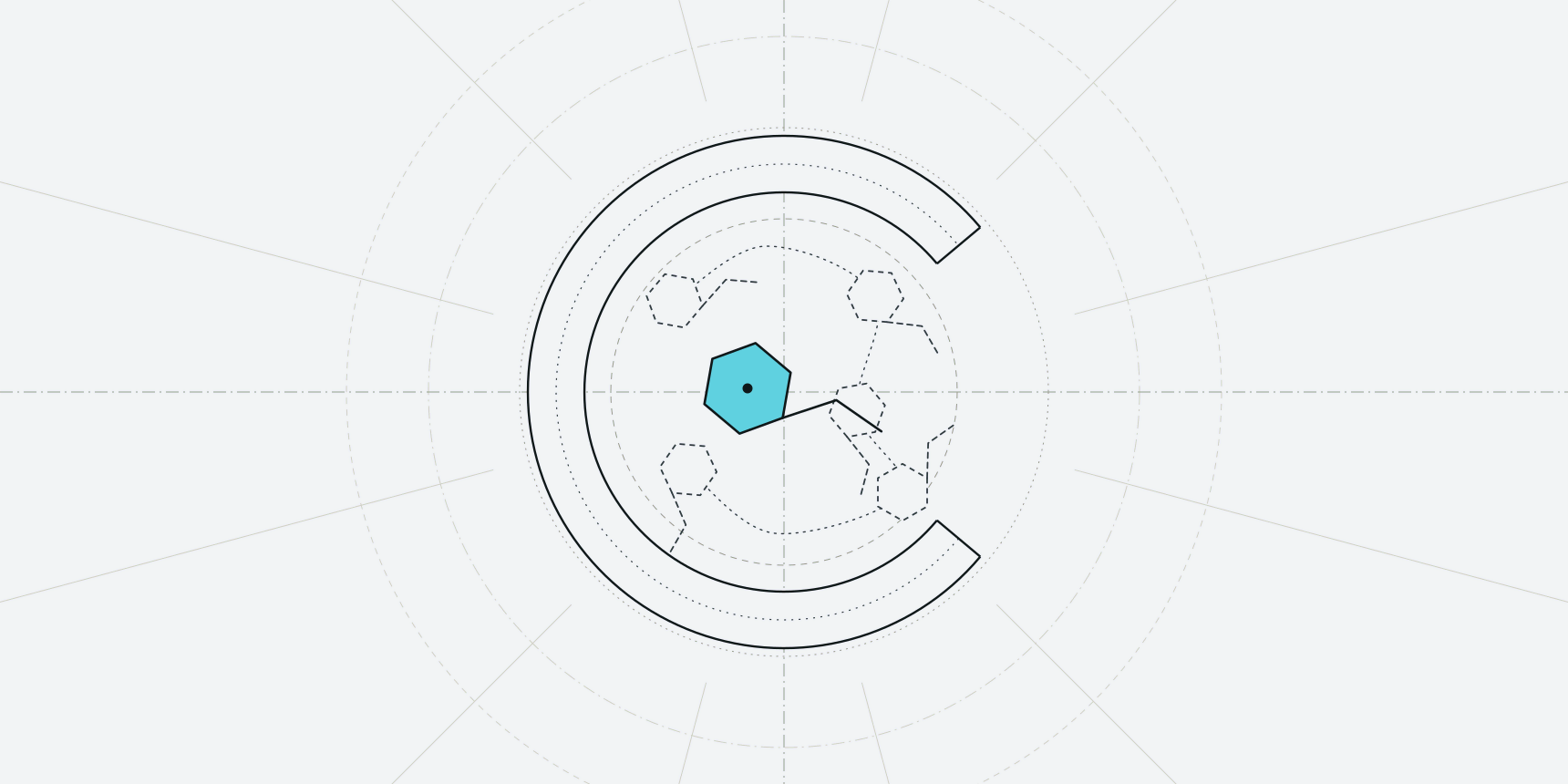
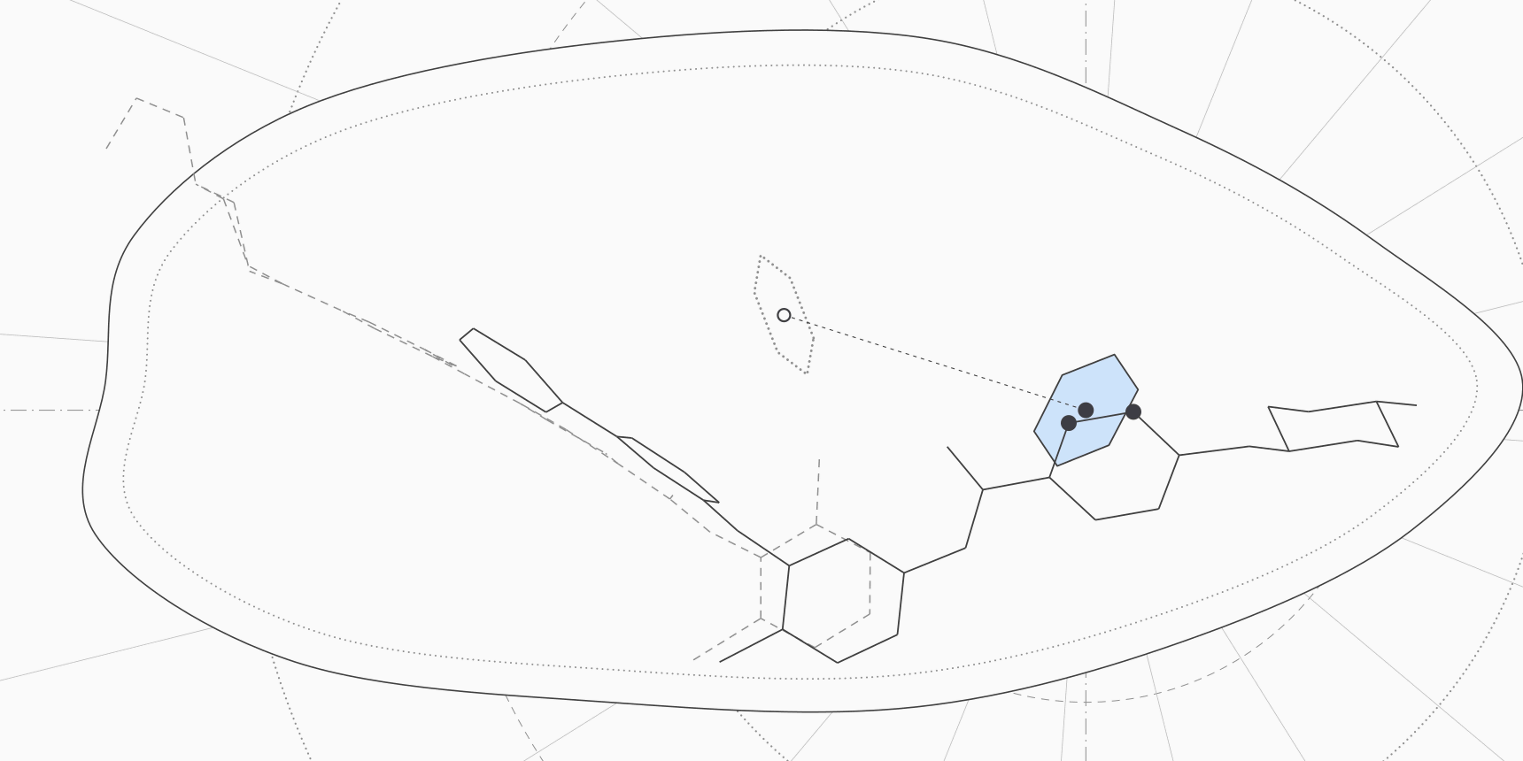
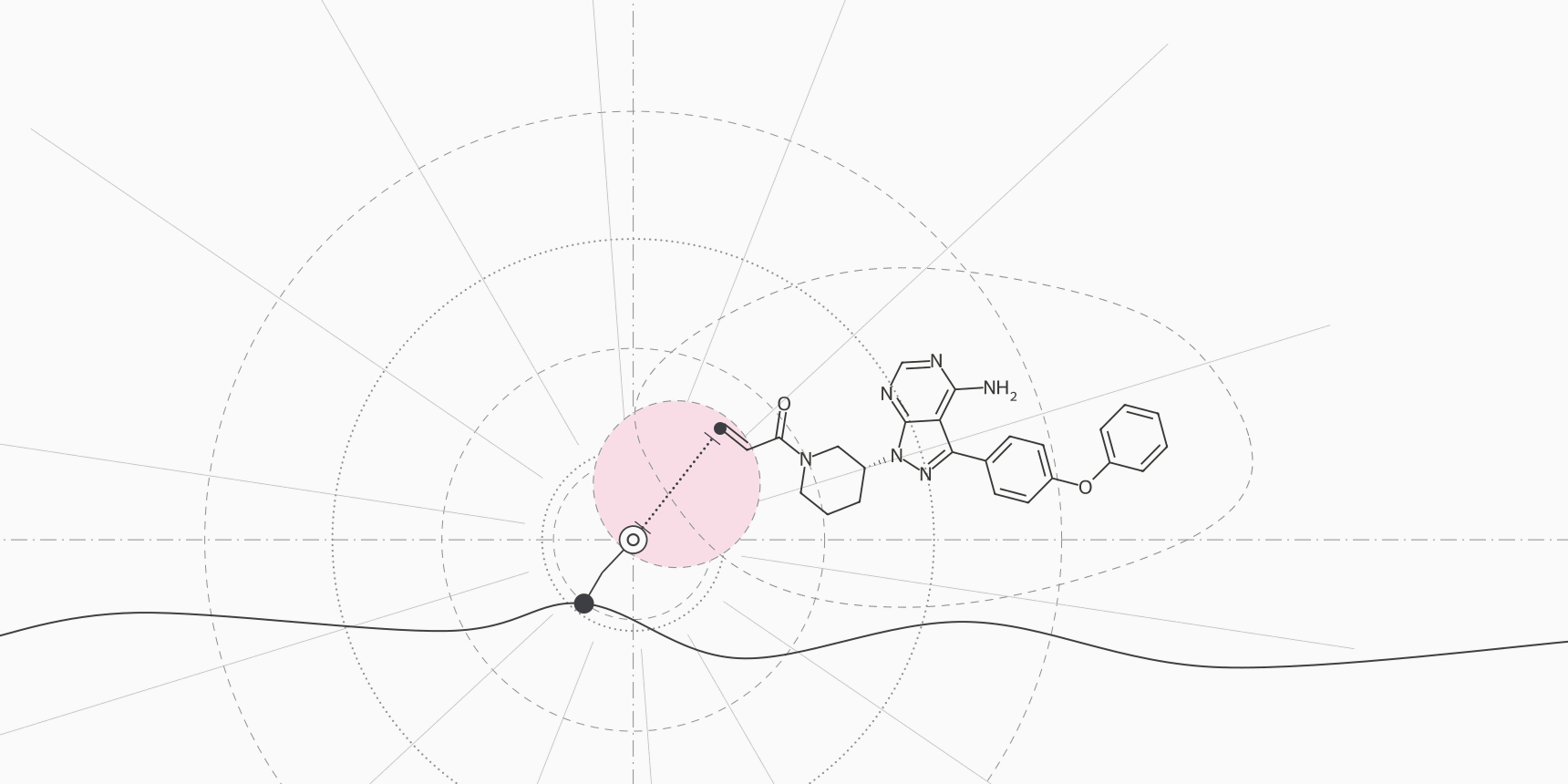
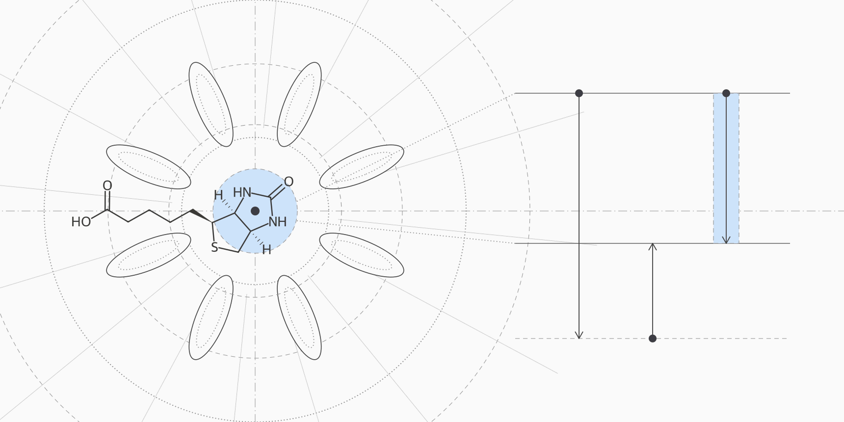
A calculation that captured only the enthalpy would overshoot the free energy by more than half. That is the pattern in MM/PBSA output, and it is why interaction energies run well past measured ΔG values. The drawing at the top of this page shows the three steps to scale.
What a docking score is
A docking program searches for ligand poses in a receptor and ranks them with a scoring function. Scoring functions are fast approximations, summing simple terms for contacts, hydrogen bonds, clashes and sometimes flexibility. They look at one static structure, so they leave out most of what separates ΔH from ΔG: all but a rotatable-bond penalty for entropy, the cost of moving water and the energy of relaxing the protein around the ligand.
Some programs fit their function to measured affinities so the output lands on a kcal/mol scale. AutoDock Vina does this, and the Vina manual calls its score a predicted binding affinity.[12] Others do not. GOLD's fitness functions, including ChemPLP, are dimensionless, with higher meaning better.[13] The unit on the page tells you little about whether the number is an energy.
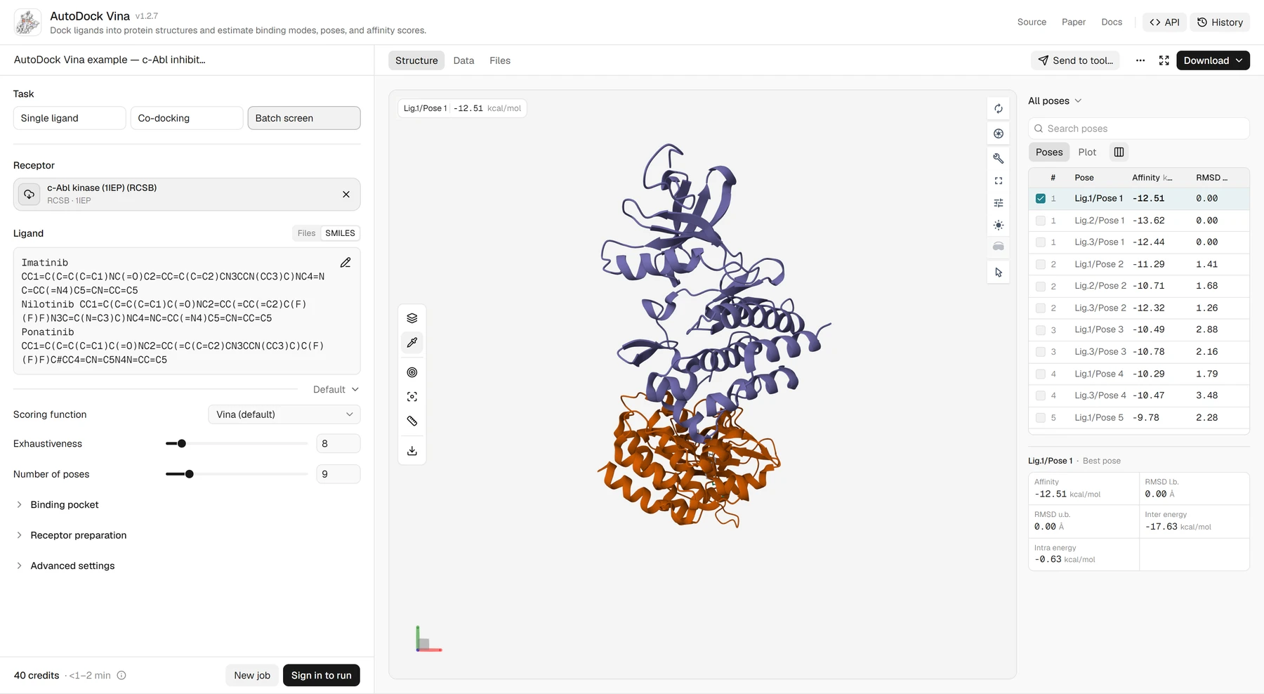
In Figure 1 the Affinity column is the score, and the energy terms underneath it show what the score is made of. Other tools report on other scales, and some count better in the opposite direction (Table 5).
| Tool | Value reported | Unit | Better is |
|---|---|---|---|
| AutoDock Vina | Vina score per pose | kcal/mol | Lower |
| GOLD (ChemPLP) | Fitness score | Dimensionless | Higher |
| GNINA | CNNaffinity, a predicted pK | pK (−log10 of molar Kd) | Higher |
| Boltz-2 | affinity_pred_value, log10 of an IC50 in µM | Log units | Lower |
| PRODIGY | ΔG and Kd for protein-protein complexes from interface contacts | kcal/mol and M | Lower ΔG, lower Kd |
| gmx_MMPBSA | End-point binding energy from simulation snapshots | kcal/mol | Lower |
Converting a predicted pK is simple arithmetic: a CNNaffinity of 7 is a nominal Kd of 100 nM and a ΔG of −9.5 kcal/mol. The arithmetic adds no accuracy, though. A CNNaffinity typically misses by about 1.4 pK units, and Boltz-2 predicts a log IC50, which needs an assay correction before it means anything like a Kd.
How close a docking score gets to the measured affinity
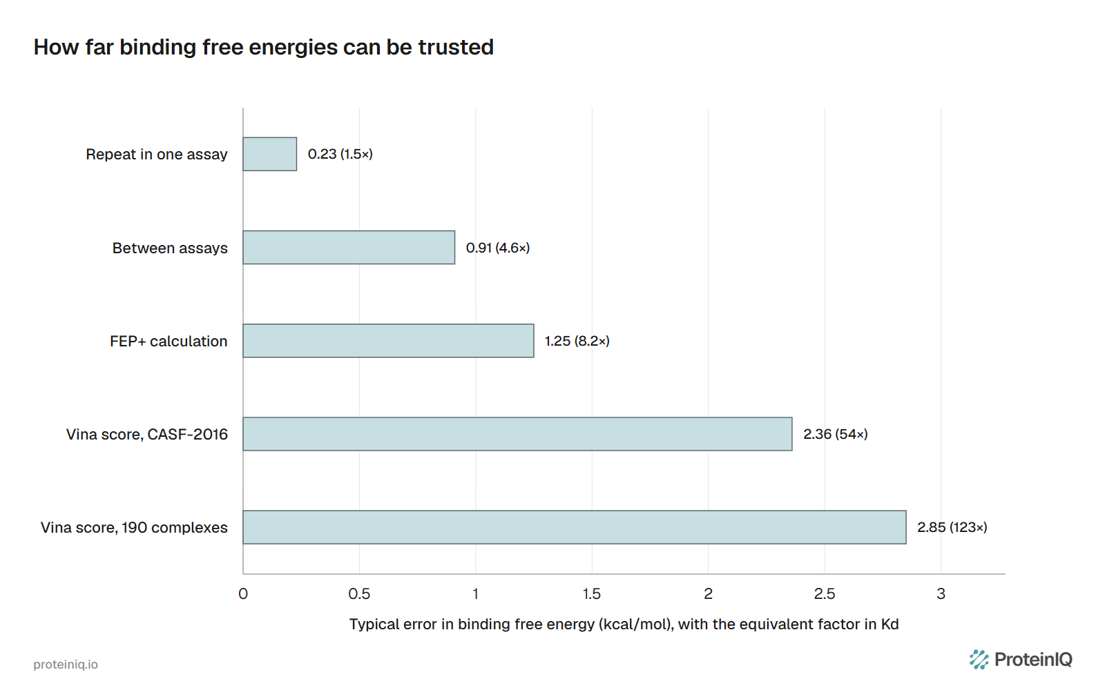
Binding free energies have a noise floor even when measured. Ross and colleagues compared compounds that were tested in more than one assay and found a pairwise RMSE of 0.91 kcal/mol between assays of the same molecules, against 0.23 kcal/mol for repeats within one assay.[4] In the same study, FEP+ free energy perturbation calculations on 1,237 ligands reached a pairwise RMSE of 1.25 kcal/mol, not far above the disagreement between two assays.[4]
Docking is in a different league. Trott and Olson reported a standard error of 2.85 kcal/mol between Vina scores and experimental free energies for 190 complexes.[2] On the CASF-2016 benchmark of 285 complexes, Vina correlated with measured affinity at R = 0.604, with a standard deviation of 1.73 pK units, equal to about 2.4 kcal/mol.[3] A 2.4 kcal/mol error is a factor of 54 to 57 in Kd.
The consequence for any conversion is that a Vina score of −10, or 47 nM, is consistent with a Kd from about 1 nM to 2.5 µM. Docking is good at telling a plausible binder from an implausible one and at proposing poses. It is a poor instrument for ordering potent binders: for the three c-Abl inhibitors in Figure 1, Vina's ranking is the reverse of the measured one. Rigorous methods such as free energy perturbation cost far more compute and still reach only about 1 kcal/mol.
What is a good docking score
No score is good in itself. The same −8 kcal/mol is strong for a fragment and weak for a drug-sized molecule, because every extra heavy atom in contact adds favorable terms. A common way to remove that size effect is ligand efficiency, the free energy divided by the number of heavy atoms. About 0.3 kcal/mol per heavy atom is the floor often proposed for drug candidates.[10]
| Heavy atoms | ΔG at 0.3 per atom | Equivalent Kd |
|---|---|---|
| 15 | −4.5 kcal/mol | 500 µM |
| 25 | −7.5 kcal/mol | 3.2 µM |
| 35 | −10.5 kcal/mol | 20 nM |
| 40 | −12.0 kcal/mol | 1.6 nM |
Read the table as a way to judge a docking score against the size of the ligand. A 25-atom compound scoring −7.5 is as efficient as a 40-atom compound scoring −12, so the second is not the better lead. Experimentally, the strongest binders gain about 1.5 kcal/mol per heavy atom up to roughly 15 atoms and then level off.[17] Docking scores keep rising with size, which is why large ligands score better whether or not they bind better.
For a threshold you can defend, use your own target. Redock the co-crystallized ligand and use its score as the bar, dock known actives against decoys and choose the cutoff that separates them, or take the top fraction of a screen. Each cutoff holds only for the docking program and settings that produced it.
How to name each number
Most overstated results come from a label, not a calculation. Table 7 gives the name that matches what was actually computed.
| What you have | Call it | Avoid |
|---|---|---|
| Vina score of −9.1 | Vina docking score, −9.1 kcal/mol | Binding energy, affinity or ΔG |
| MM/GBSA value without entropy | MM/GBSA interaction energy, entropy not included | Binding free energy |
| GNINA CNNaffinity of 7.2 | Predicted pK, nominal Kd of 63 nM | Measured or experimental Kd |
| Calorimetry ΔG and Kd | Measured binding free energy and affinity, with temperature and buffer | Anything that omits the conditions |
| IC50 from an activity assay | Potency in that assay, or Ki after correction | Kd |
Docking and binding tools on ProteinIQ
Dock, check the pose, then rescore or simulate, in that order, before treating any number as an affinity. That is the usual path from a molecular docking run to a binding free energy estimate.
| Task | Tool |
|---|---|
| Dock a ligand and get a score in kcal/mol | AutoDock Vina, smina |
| Rescore poses with a neural network | GNINA |
| Check that a pose is physically plausible | PoseBusters, PLIP |
| Measure pose distance from a crystal ligand | RMSD Calculator |
| Estimate ΔG and Kd for a protein-protein complex | PRODIGY |
| Score simulation snapshots with end-point energies | gmx_MMPBSA |
| Predict a complex and its affinity | Boltz-2 |
| Convert IC50 to Ki and pIC50 | IC50 to Ki calculator |
Frequently asked questions
What is a good docking score?
There is no universal threshold. A good score is one that beats the score of a known ligand docked into the same receptor with the same settings, after allowing for ligand size. Ligand efficiency of about 0.3 kcal/mol per heavy atom is a useful floor for drug-sized molecules.[10]
Is binding energy the same as binding affinity?
No. Binding affinity is how tightly a ligand binds, measured as Kd. Binding free energy is that same quantity as an energy. Binding energy is a looser phrase that often means a force-field interaction energy with no entropy term, and it does not convert to a Kd.[6]
How do I convert ΔG to Kd?
Use Kd = exp(ΔG/RT) with a 1 M standard state. At 25 °C, RT is 0.592 kcal/mol, so a ΔG of −10 kcal/mol is a Kd of 47 nM.[1] Rearranged, Kd = 10^(ΔG/1.364) M at 25 °C with ΔG in kcal/mol.
How do I convert Kd to ΔG?
Use ΔG = RT ln Kd with Kd in molar units. A Kd of 10 nM gives 0.592 × ln(10−8) = −10.9 kcal/mol at 25 °C. Each tenfold change in Kd shifts ΔG by 1.364 kcal/mol.
Why is my docking score more negative than the measured ΔG?
Docking scores add favorable contacts for every heavy atom and model little of the entropy and desolvation cost, so large ligands tend to score better than they bind. Imatinib scores −12.51 in Vina against a measured −9.52 kcal/mol.[11]
Does a more negative score always mean stronger binding?
Only within one method, one receptor and one protocol, and only when the difference is larger than the method's error. Differences below about 2 kcal/mol between compounds are within the typical error of docking.[3]