Covalent docking: tools and workflow
What adding the bond fixes, and what it leaves open

Matic Broz, PhD17 min
What is covalent docking?
Covalent docking is docking with the bond included. The program is told which ligand atom bonds to which protein atom, usually a warhead carbon and a cysteine sulfur, and it keeps only poses in which that bond can form. Ordinary docking has no such instruction, and the gap can be large even when the pose looks right.
We docked ibrutinib back into the Bruton's tyrosine kinase structure it was solved in (PDB 5P9J) with plain AutoDock Vina, five times with different seeds. Every top pose landed 1.80 to 1.85 Å from the crystal ligand, well inside the 2 Å rule. In every one of them the acrylamide that reacts with Cys481 sat 6.1 to 6.4 Å from the sulfur. The pose was right on average and wrong at the only place the chemistry happens.
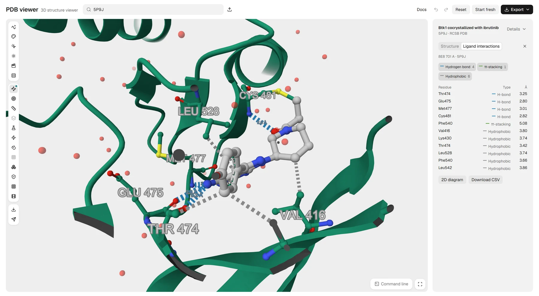
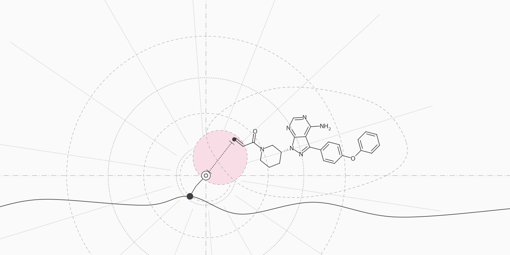
Figure 1 shows that crystal structure in the PDB Viewer. The acrylamide has already bonded to the Cys481 sulfur, 1.85 Å away in the deposited coordinates, and the drug makes four hydrogen bonds, one π-stacking contact and six hydrophobic contacts with the kinase.[15]
Ibrutinib is one of 36 approved small molecules that were designed to bond to one residue of one protein, and 19 of those 36 bond to a cysteine. Docking a covalent ligand is really three questions: where the ligand sits before it reacts, whether its warhead can reach the residue from there, and how fast the reaction then runs. Covalent docking programs build the bond into the search to answer the first two. None answers the third, and the tools differ mainly in when they apply the bond.
The two steps behind a covalent inhibitor
Modern covalent inhibitors bind in two steps. The molecule first docks reversibly, like any inhibitor, and the warhead reacts only if that pose puts it next to the right residue.[1] Two constants describe the process: KI, the affinity of the first step, and kinact, the rate of the second. Because the effect grows with time, a single IC50 depends on how long the assay ran, and Strelow argues that covalent inhibitors should be compared by kinact/KI instead.[2]
A docking score can speak, at most, to the first step. A docking score is not a binding affinity even for ordinary ligands, and a covalent ligand adds a reaction the function never sees. Leng and colleagues compared docking scores with measured pIC50 values for covalent inhibitors of 17 targets. The correlation was below |r| = 0.2 for most pairs of target and program, and several were clearly negative.[11]
The second constant depends on how reactive the warhead is and on how well the pose holds it beside the residue. Docking sees only the second part, which is why the check that matters most is geometric: can the warhead reach?
Docking without the bond
The simplest route is to dock the intact ligand with any non-covalent program and measure afterwards how far the warhead sits from the residue. It works better than one might expect. Scarpino and colleagues compared six covalent programs on a large set of covalent complexes and found 40 to 60% of top poses within 2 Å of the experimental pose, about what non-covalent tools achieve.[3]
Non-covalent docking into proteins with the cysteine mutated to alanine reproduced the binding modes only slightly less well, at much lower cost, so anchoring the ligand does not necessarily improve accuracy.
The catch is the one in the opening example. A pose can reproduce the scaffold and miss the warhead, because RMSD averages over every heavy atom and the warhead is a few of them at the end of the molecule. The worked example below measures how often that happens.
Docking with the bond built in
Anchored docking fixes the bond in advance and searches only for poses that satisfy it. AutoDock4 has two such methods, a two-point attractor and a flexible side chain method. The flexible side chain method recovered the crystal pose for 75% of 20 test complexes and failed for the largest inhibitors.[4] CovalentDock, also built on AutoDock, adds an energy term for forming the linkage and geometry constraints for the new bond.[5]
GNINA's command line does the same with two flags that name the receptor atom and a SMARTS pattern for the ligand atom, and it can optimize the ligand and residue complex with a force field afterwards.[12] GOLD, MOE, ICM-Pro and FITTED follow the same logic and were part of the Scarpino comparison.[3] Anchoring has a cost: the residue must be the right one and its side chain must be able to reach the ligand, and a wrong choice gives a confident wrong pose with a perfect bond length.
Docking in two steps
Two-step methods copy the mechanism: dock non-covalently, then form the bond and refine. CovDock from Schrödinger docks with Glide, forms the bond heuristically and refines the complex with Prime; on 38 complexes, 76% of poses were within 2 Å.[6]
Attracting Cavities reached 78% on 304 complexes against 66% for GOLD and 35% for AutoDock, and HCovDock reports 70.5% for the top pose and 93.2% within the top ten on 207 complexes.[8][9] Each method was tested on a set its authors assembled, so the rates cannot be ranked against one another.
DOCKovalent takes a different step to the same end. It pre-builds the adducts of each library compound, samples poses constrained to ideal bond lengths and angles, and scores them with DOCK's physics-based function, one screen per likely rotamer of the nucleophile. Used prospectively against AmpC β-lactamase, RSK2 kinase and JAK3 kinase, it found actives in 5 of 6, 5 of 8 and 9 of 15 compounds tested, and crystal structures of AmpC and RSK2 complexes confirmed the predicted poses.[7]
Co-folding with a bond constraint
Co-folding models predict the protein and the ligand together, and some accept a covalent bond as an input. On CoFD-Bench, 218 recently solved covalent complexes, AlphaFold 3, Chai-1 and Boltz-1x gave better ligand RMSD than AutoDock-GPU, CovDock and GNINA, but their accuracy fell for pocket and ligand pairs unlike their training data. The classical programs were steady and modest, and limited mainly by the receptor conformation they were given.[10]
The same study found that AlphaFold 3 can point to the native covalent residue even when no bond is specified.[10] Leng and colleagues found Boltz-2 strongest at reproducing poses on their 2,172-complex benchmark, and report it as an upper bound because overlap with its training data is likely.[11] The bond itself is an assumption you supply. The model will draw the adduct whether or not the reaction occurs, so a high confidence score describes a structure with the bond imposed, not covalent reactivity.
| Approach | Tools | How the bond is used | Access |
|---|---|---|---|
| Without the bond | AutoDock Vina, smina, GNINA, DiffDock | Not modeled; measure the warhead distance afterwards | Free; all four run on ProteinIQ |
| Bond built in | AutoDock4, CovalentDock, GNINA command line, GOLD, MOE, ICM-Pro | A named ligand atom is bonded to a named receptor atom during the search | AutoDock4 and GNINA are free; GOLD, MOE and ICM-Pro are commercial |
| Two steps | CovDock, Attracting Cavities, HCovDock, DOCKovalent | Non-covalent pose first, then bond formation and refinement; DOCKovalent samples pre-built adducts | Web servers with the papers for HCovDock and DOCKovalent; CovDock is in the Schrödinger Suite |
| Co-folding | Boltz-2, AlphaFold 3, Chai-1 | Bond given as an input constraint, or residue inferred from a non-covalent run | Boltz-2 is MIT-licensed and runs on ProteinIQ |
Five covalent drugs, docked without the bond
To see what the missing bond costs, we docked five approved covalent drugs into the crystal structures of their own covalent complexes with AutoDock Vina 1.2.7 (Table 2).[13][14] Each drug was built from its parent structure with the warhead intact, so the ligand could not know where it had reacted.
The receptor was chain A of the crystal with waters and ligands removed, except the GDP and magnesium of KRAS; the main protease (Mpro) is a dimer, and only the protomer with the cysteine was kept. The box was a 25 Å cube on the crystal ligand, with five seeds per drug and up to nine poses each.
We measured two things for every pose: the heavy-atom RMSD to the crystal ligand, without fitting and with the reacted warhead matched to the intact one, and the distance from the electrophilic carbon to the cysteine sulfur.
The sulfur and carbon van der Waals radii sum to 3.5 Å, so we call a warhead within reach at 4.0 Å or less. That threshold is ours, not a published criterion. The cysteine in these files already has the conformation that fits the adduct, the most favorable receptor a docking run can be given.
| Drug, target and structure | Warhead | Top pose RMSD (Å) | Warhead to sulfur (Å) | Both checks passed |
|---|---|---|---|---|
| Ibrutinib, BTK Cys481, 5P9J | Acrylamide | 1.80 to 1.85 | 6.14 to 6.41 | 0 of 5 |
| Afatinib, EGFR Cys797, 4G5J | Aminocrotonamide | 5.77 to 5.83 | 7.32 to 7.54 | 0 of 5 |
| Sotorasib, KRAS Cys12, 6OIM | Acrylamide | 1.31 to 2.06 | 3.70 to 4.51 | 0 of 5 |
| Adagrasib, KRAS Cys12, 6UT0 | Fluoroacrylamide | 2.25 to 5.99 | 4.13 to 5.36 | 0 of 5 |
| Nirmatrelvir, Mpro Cys145, 7SI9 | Nitrile | 1.60 to 1.66 | 3.79 to 3.87 | 5 of 5 |
Twelve of the 25 top poses pass the 2 Å rule: all five for ibrutinib, all five for nirmatrelvir and two for sotorasib. Only five of those twelve, all nirmatrelvir, also put the warhead within 4 Å of the sulfur. Ibrutinib is the sharpest case. Ten of its 45 poses are within 2 Å of the crystal pose, none has the warhead within 4 Å, and the closest approach of any pose is 4.89 Å, in a pose that is 4.1 Å off the crystal ligand.
The two checks can disagree in the other direction. For sotorasib, the two runs closest to the crystal pose, at 1.31 Å, held the acrylamide 4.4 to 4.5 Å from the sulfur. The three runs at 2.05 Å, just outside the rule, held it at 3.70 to 3.74 Å.
Adagrasib depended on the seed: three runs found a pose scoring about −10.9 kcal/mol that sat 2.25 Å from the crystal ligand with the warhead 4.1 to 4.2 Å from the sulfur, and two stayed with poses scoring about −8.0 kcal/mol that were 6.0 Å away. A single run would have told a different story each time.
Afatinib shows the plainest failure. No pose in any run came within 2 Å of the crystal ligand, and the top pose was 5.8 Å away with the warhead more than 7 Å from Cys797. Whether the cause is the low resolution of 4G5J, the flexible crotonamide chain or the pocket, the distance check catches it as clearly as the RMSD does.
Does building the bond in fix this? We reran the same five drugs with the covalent mode of GNINA 1.3.3, which bonds the warhead carbon to the cysteine sulfur during the search.[12] The runs used the same ligand conformers, receptors, box and exhaustiveness, three seeds per drug, on the CPU with the default CNN rescoring. The warhead sits 1.81 Å from the sulfur in every pose, because the program enforces it. The pose is the open question.
None of the 15 top-ranked poses was within 2 Å of the crystal ligand. Ibrutinib's were 9.4 to 9.5 Å away, adagrasib's 10.7 to 12.8 Å, nirmatrelvir's 4.2 to 5.1 Å and sotorasib's 5.6 to 5.7 Å, and afatinib's were 2.03 Å in one run and 7.1 Å in the other two.
The right pose was often in the list. For sotorasib, a pose at 1.43 to 1.45 Å appeared in all three runs, ranked second to fifth. Ranking by GNINA's empirical score instead of its CNN score put that pose first every time, and gave 3 of 15 top poses within 2 Å. The bond made the warhead reachable and left the pose undecided. This is a spot check of one program at its default settings, not a benchmark, and we did not tune it.
A workflow for covalent docking
Start from the question, not the program (Table 3). The residue comes first because every covalent method assumes it, and the distance check comes before any score is read.
| Question | Start with | Add | Check |
|---|---|---|---|
| Which residue reacts? | Mass spectrometry or a crystal structure of a related ligand | A non-covalent co-folding run to see where the ligand lands | Do not let docking choose; a wrong residue gives a confident wrong pose |
| Where does the ligand sit before it reacts? | Non-covalent docking with several seeds | A repeat with the cysteine in each plausible rotamer | Warhead to sulfur distance in every pose, not only the top one |
| Does the bonded complex fit the pocket? | Covalent docking, or co-folding with the bond | Refinement of the adduct | Compare with the non-covalent pose; check geometry and strain |
| Which compounds in a library? | A covalent method tested on known ligands of this target | A non-covalent control against the cysteine-to-alanine mutant | Order a few compounds early; hit rates are the only real test |
| How potent will it be? | A kinact/KI assay | Intact-protein mass spectrometry | Docking scores do not answer this |
Prepare the receptor as for any docking job, and then pay attention to the nucleophile. Prepare the protein with the cysteine's protonation state checked, since a cysteine reacts as the thiolate and its PROPKA pKa shows how likely that is. DOCKovalent holds the nucleophile fixed during sampling and recommends a separate screen for each likely rotamer.[7] A receptor taken from the covalent crystal, as ours were, has the rotamer that fits the adduct; an apo or predicted structure may not.
Redocking the crystal ligand into its own structure is still the first test. For a covalent ligand it needs the second column of the table too: report the warhead distance next to the RMSD, because the RMSD alone passed ibrutinib five times in five.
What each approach cannot see
Each approach fails where its model of the reaction stops, and each failure has a cheap check (Table 4).
| Approach | What it cannot see | Sign of trouble | Check |
|---|---|---|---|
| Without the bond | Whether the warhead can reach the residue | A low RMSD with the warhead far from the sulfur | Measure the distance in every pose |
| Bond built in | Whether the residue is the right one | A perfect bond length in a pose that is otherwise wrong | Dock without the bond and compare |
| Two steps | Reactions outside its parameters | Lower success for ring-opening and disulfide-forming warheads than for Michael additions | Test on known ligands with the same warhead |
| Co-folding with a bond | Whether the reaction occurs; targets unlike its training data | High confidence on every sample, with or without the bond | Compare samples and measure that the constraint holds |
| Any of them | kinact, warhead reactivity, glutathione stability | Scores that do not follow measured potency | Measure kinact/KI for the few compounds that matter |
A passed check proves less here than it appears to. A low RMSD says nothing about the warhead, and a clean PoseBusters report checks chemistry and geometry, not whether the right atoms are close. Run the distance check first. It takes a minute, and it is the test that ibrutinib's five best poses failed.
Covalent docking tools on ProteinIQ
ProteinIQ does not run covalent docking with a bond to a named residue: the hosted GNINA leaves its covalent options out, and Vina, smina and DiffDock have none. Boltz-2 accepts the bond as input. A practical order is to prepare and protonate the receptor, dock without the bond with several seeds, measure the warhead distance, co-fold with the bond to compare, and then check the pose (Table 5).
| Task | Tool |
|---|---|
| Inspect a crystal structure, its covalent link and its contacts | PDB Viewer |
| Repair a structure and add missing atoms before docking | PDBFixer |
| Estimate the pKa of the target cysteine | PROPKA |
| Set the protonation state of the ligand | Dimorphite-DL |
| Flag reactive groups in a compound library | Brenk filter |
| Dock the intact ligand without the bond | AutoDock Vina, GNINA, smina |
| Co-fold protein and ligand with a covalent bond constraint | Boltz-2 |
| Check that a pose is chemically and physically valid | PoseBusters |
| Measure a pose against the crystal ligand | RMSD Calculator |
| List the contacts a pose makes with the pocket | PLIP |
Frequently asked questions
What is covalent docking?
It is molecular docking in which the ligand is bonded to a named protein atom, for example a warhead carbon to a cysteine sulfur, and only poses that allow the bond are kept. Most covalent methods also mimic the two-step mechanism: a reversible docking step followed by bond formation.[1][6]
Can AutoDock Vina do covalent docking?
Vina has no covalent mode, so it docks every ligand as if it were not going to react, as in our five-drug test above. AutoDock4 has two covalent methods, a two-point attractor and a flexible side chain method, and GNINA's command line has covalent flags.[4][12]
Is covalent docking more accurate than regular docking?
Not necessarily. Across six covalent programs, 40 to 60% of top poses were within 2 Å of the experimental pose, similar to non-covalent tools, and docking into a cysteine-to-alanine mutant did almost as well.[3] The bond helps most when you need the warhead placed next to the residue.
Do I need to know the target residue?
For most covalent methods, yes, because the bond is an input. Co-folding is the exception to explore: in one benchmark AlphaFold 3 identified the native covalent residue through non-covalent runs, with a ligand RMSD comparable to covalent co-folding.[10]
Can a docking score predict covalent potency?
Rarely. Across 17 covalent targets, the correlation between docking scores and pIC50 was below |r| = 0.2 for most target and program pairs.[11] Covalent potency is better described by kinact/KI than by a single IC50.[2]
How do I dock a covalent inhibitor in GNINA?
On the command line, name the receptor atom with the covalent receptor atom flag, for example A:481:SG, and give a SMARTS pattern that matches the one ligand atom that forms the bond. The ProteinIQ version of GNINA does not offer these options.[12]