RMSD calculator
Calculate RMSD between protein structures with automatic alignment
Input
Output
Configure input settings on the left, then click "Calculate"
Calculate RMSD between protein structures with automatic alignment
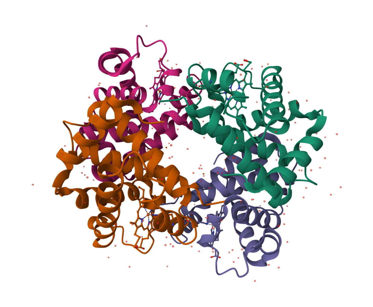
Assess docking model quality by comparing predicted complexes against native references. DockQ v2.1.3 supports protein, nucleic-acid, and supported small-molecule interfaces with faithful native metrics.
PoseBusters validates generated or docked molecular poses with chemically and structurally grounded quality checks for molecular geometry, intermolecular interactions, and optional reference-pose agreement.

Assign protein secondary structure using the DSSP algorithm. The gold standard for hydrogen bond-based structure assignment from coordinates.
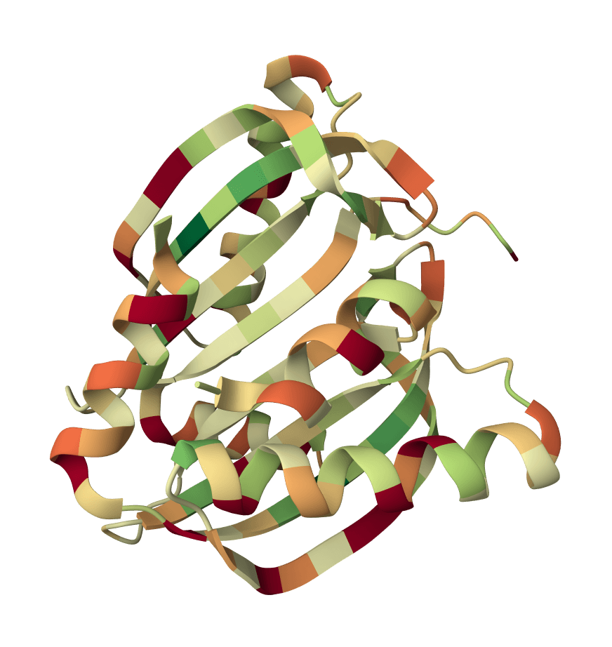
Validate protein structure quality with all-atom contact analysis, Ramachandran plots, rotamer assessment, and geometry checks.
Generate a downloadable PDBsum structural summary report archive for a single protein structure.
Calculate the radius of gyration (Rg) for protein structures from PDB files. Supports multiple chains and atom selection options.
Calculate Solvent Accessible Surface Area (SASA) for protein structures using the Shrake-Rupley algorithm.
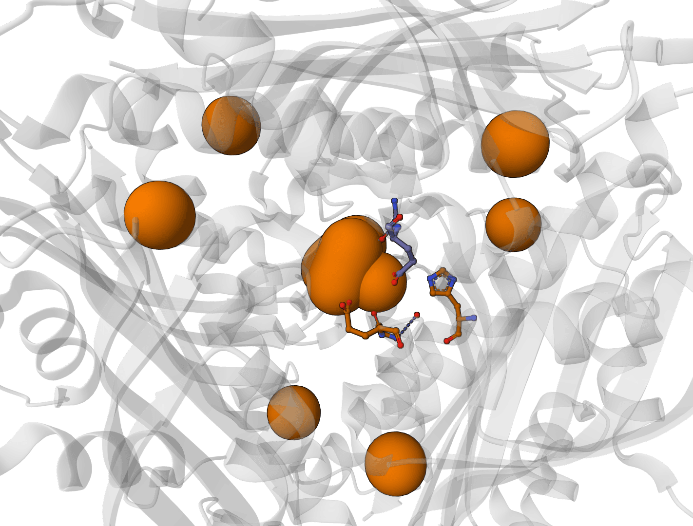
Predict metal and water binding sites in protein structures using 3D convolutional neural networks (AllMetal3D + Water3D).
Scoring function for interprotein interactions in AlphaFold2, AlphaFold3 and Boltz predictions. Calculates ipSAE, ipTM, pDockQ, pDockQ2, and LIS scores to assess protein-protein interface quality.
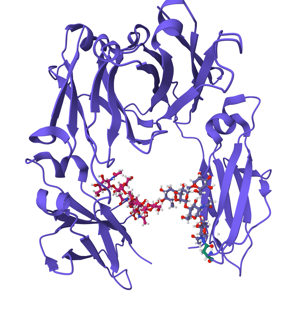
Geometric deep learning model for predicting protein binding sites directly from 3D structure. Identifies where proteins interact with other proteins, antibodies, or disordered proteins with high accuracy, including for novel protein folds.
Root Mean Square Deviation (RMSD) is the most widely used metric for quantifying structural similarity between proteins. It measures the average distance between corresponding atoms in two superimposed structures, expressed in Ångströms (Å).
An RMSD of 0 Å indicates identical structures. Values below 2 Å typically suggest high similarity, while values above 4 Å indicate significant structural differences. RMSD is essential for validating predicted structures against experimental data, tracking conformational changes in molecular dynamics, and assessing the diversity of structural models.
For more comprehensive structural comparison that includes TM-score and sequence identity, use USAlign. To search for structurally similar proteins in databases, try FoldSeek.
RMSD calculates the square root of the average squared distances between pairs of equivalent atoms:
where is the Euclidean distance between the -th pair of corresponding atoms after superposition.
Before calculating RMSD, structures must be optimally superimposed. This tool uses the Kabsch algorithm to find the rotation matrix that minimizes RMSD between the two structures.
The algorithm works in three steps. First, both structures are centered on their centroids. Second, the optimal rotation matrix is computed using singular value decomposition (SVD) of the covariance matrix. Third, one structure is rotated to best match the other before computing the final RMSD.
Disable automatic alignment only if your structures are already pre-aligned in the same coordinate frame.
The choice of which atoms to compare significantly affects the result.
Alpha carbons (CA) — The standard choice for backbone comparison. Using only Cα atoms (one per residue) provides a robust measure of overall fold similarity while being insensitive to side chain conformations.
Backbone atoms (N, CA, C, O) — Includes all main chain atoms. This gives a more detailed backbone comparison but increases sensitivity to local conformational differences.
All atoms — Compares every atom in the structure. This is most sensitive to differences but can be dominated by flexible side chains and may not reflect overall structural similarity.
Reference structure — The structure you want to compare against. Upload a PDB file or fetch from RCSB using a 4-letter PDB ID.
Comparison structures — One or more structures to compare with the reference. You can upload multiple PDB files or enter multiple PDB IDs separated by commas.
Structure — Outputs one RMSD value per comparison file. Atoms are matched across the entire structure by residue number.
Chain — Compares each chain in the reference against each chain in the comparison structure. This produces a matrix of all pairwise chain comparisons, useful for identifying which chains correspond between structures.
| Column | Description |
|---|---|
| Reference | The reference structure (or chain in chain mode), e.g., 5VF9_A |
| Comparison | The comparison structure/chain being evaluated |
| RMSD (Å) | Root mean square deviation in Ångströms |
| Atoms | Number of atom pairs used in the calculation |
| Skipped | Atoms present in one structure but not the other |
The significance of an RMSD value depends on protein size and the atoms being compared.
For Cα RMSD between similar-sized proteins, these ranges provide rough guidance. Values below 1 Å indicate nearly identical structures. Between 1–2 Å suggests high similarity with minor differences. Values of 2–4 Å show moderate similarity with significant local variations. Above 4 Å typically indicates different conformations or folds.
A high "Skipped" count suggests the structures have different residue numbering or missing regions, which can affect the reliability of the comparison.
RMSD has known limitations as a similarity metric.
It is sensitive to outliers. A single misaligned loop or flexible terminus can dramatically increase the global RMSD even when the core structures are nearly identical.
RMSD is size-dependent. An RMSD of 3 Å has different significance for a 500-residue protein versus a 50-residue protein. For size-independent comparison, TM-score (available in USAlign) is preferred.
Structures must have corresponding atoms. RMSD requires matching residue numbers between structures. Insertions, deletions, or different numbering schemes will result in skipped atoms.