Radius of gyration
Calculate the radius of gyration (Rg) for protein structures from PDB files.
Input
Output
Configure input settings on the left, then click "Generate"
Calculate the radius of gyration (Rg) for protein structures from PDB files.
Faithful static-mode Aggrescan3D tool for per-residue aggregation propensity analysis from a single protein structure.
Plot net charge vs pH for protein sequences. Visualize how protein charge changes across pH 0-14 and identify the isoelectric point (pI) where the net charge crosses zero.
Match experimental peptide masses against theoretical digest fragments of a protein sequence. Identify peptides from mass spectrometry data by peptide mass fingerprinting.
Generate Kyte-Doolittle hydropathy plots to visualize hydrophobic and hydrophilic regions along protein sequences. Identify transmembrane domains and surface-exposed regions.
Generate hydrophobicity plots using 24 different amino acid scales. Visualize hydrophobic and hydrophilic regions for protein analysis, epitope prediction, and membrane protein studies.
Isoelectric Point Calculator 2.0 - Predict protein/peptide isoelectric point (pI) using 18+ validated pKa scales, SVR models, and deep learning. Supports proteins, peptides, and comprehensive analysis.
Predict protease and chemical cleavage sites across a protein sequence for up to 39 enzymes simultaneously. Identify where each enzyme cuts, the cleavage residue, and context window around each site.
Cleave a protein sequence with a chosen protease and compute the masses of the resulting peptides. Supports multiple enzymes, missed cleavages, chemical modifications, and different ion types for mass spectrometry experiment planning.
Predict pKa values of ionizable groups in proteins and protein-ligand complexes from 3D structure. PROPKA calculates environment-driven pKa shifts for standard ionizable residues, terminal groups, and supported ligand atom types.
Calculate protein parameters, including molecular weight, theoretical pI, extinction coefficients, aromaticity, secondary structure fractions, atomic composition, estimated half-life, and several indices, including instability, aliphatic index, and GRAVY.
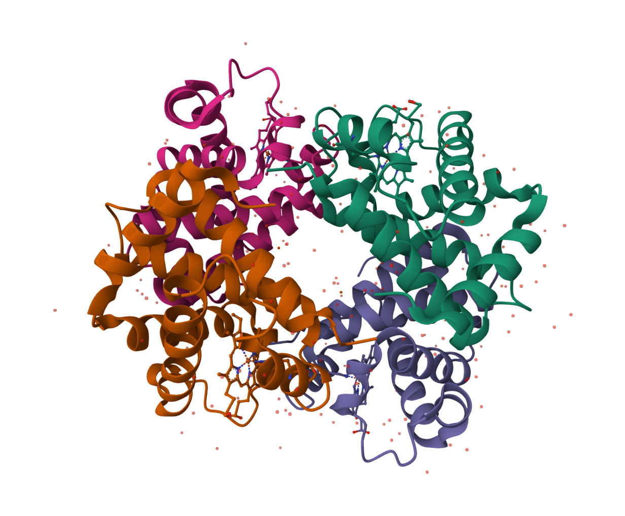
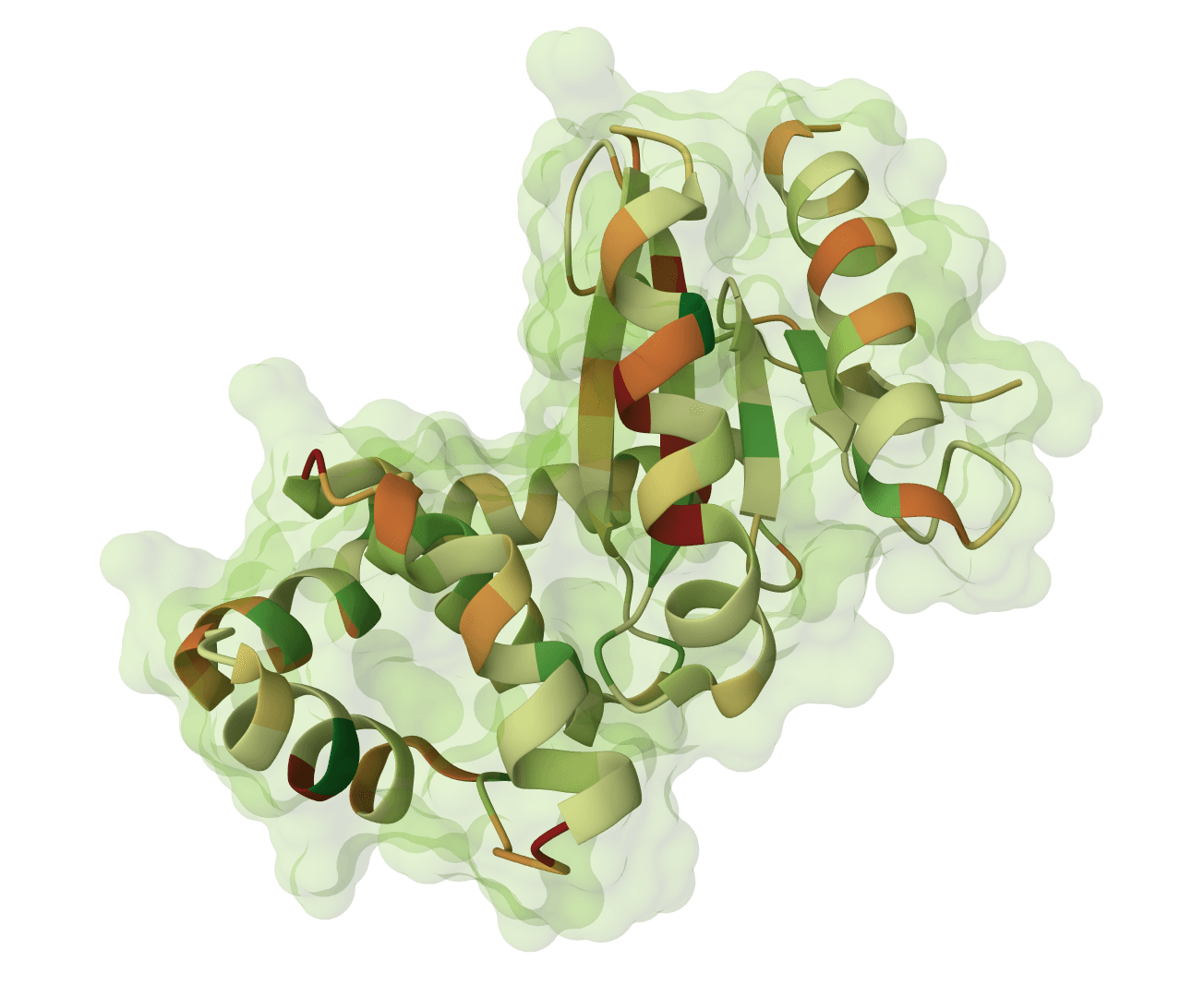
The radius of gyration () quantifies the compactness of a protein structure. It measures how mass is distributed around a protein's center of mass—a compact, well-folded protein has a small , while an extended or unfolded structure has a larger value.
is widely used in structural biology to assess folding states, compare conformational changes, and validate computationally predicted structures. Molecular dynamics simulations track over time to detect unfolding events or conformational transitions.
For sequence-based analysis of protein properties like composition and charge, see Protein Parameters. To visualize your structure alongside calculations, use the PDB Viewer.
The radius of gyration is the root-mean-square distance of all atoms from the protein's center of mass. The formula is:
Where:
The tool uses atomic masses to weight each atom's contribution. Heavier atoms like sulfur have more influence than lighter atoms like hydrogen.
Different atom selections provide complementary information:
Alpha carbon calculations are fastest and most reproducible across different structure sources (X-ray, NMR, computational models). Use all-atom calculations when side chain packing matters.
For globular proteins, follows a predictable relationship with chain length:
Where is the number of residues. A 100-residue globular protein typically has Å. Values significantly above this suggest extended conformations or multi-domain arrangements.
1UBQ)A or A,B). Leave empty to include all chains.The output table contains one row per chain or structure analyzed:
| Column | Description |
|---|---|
| ID | Structure/chain identifier |
| Num Atoms | Number of atoms included in the calculation |
| Rg (Å) | Radius of gyration in angstroms |
The absolute value depends on protein size, so compare against expectations for your protein's length:
When comparing structures, small differences (< 1 Å) are often within experimental or computational uncertainty. Larger changes (> 2–3 Å) typically indicate meaningful conformational differences.
Radius of gyration analysis helps answer several structural biology questions: