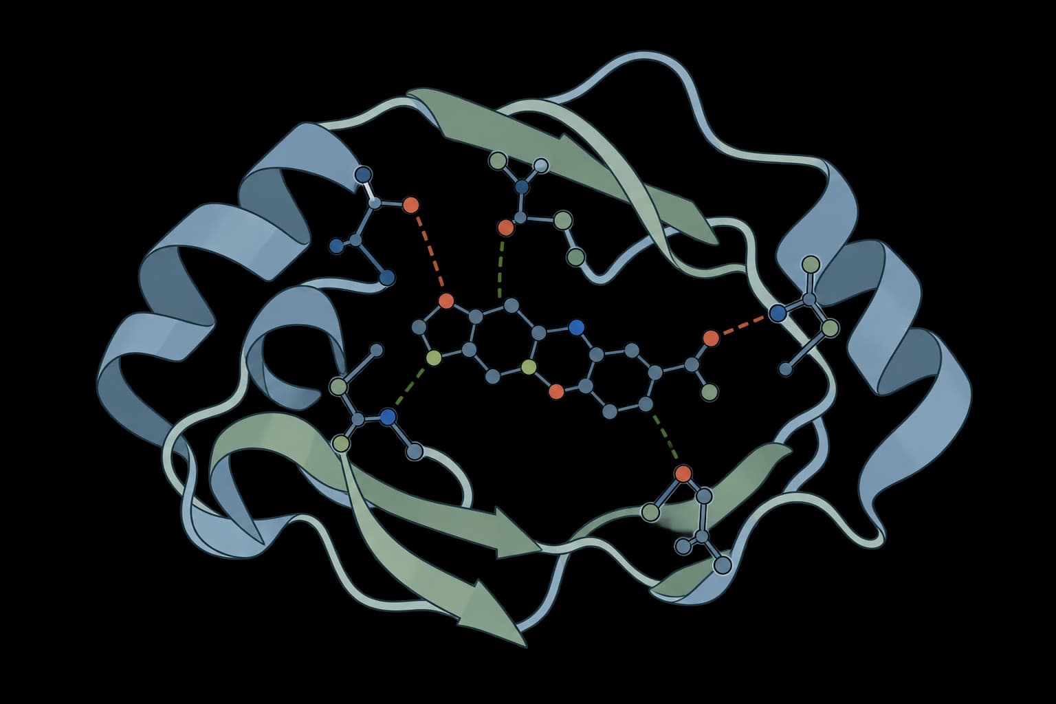
Profile protein-ligand interactions from a PDB complex structure.
Input
What is PLIP?
PLIP, the Protein-Ligand Interaction Profiler, detects noncovalent contacts in three-dimensional protein-ligand complexes. It applies geometric rules to identify hydrophobic contacts, hydrogen bonds, water bridges, salt bridges, pi stacking, pi-cation interactions, halogen bonds, and metal complexes.
PLIP describes interactions present in a submitted pose. It does not dock the ligand, calculate binding affinity, or prove that a contact is energetically favorable. Its main value is turning a complex structure into a reproducible interaction inventory that can be compared across crystal structures, docking poses, and designed complexes.
How to use PLIP online
PLIP 3.0.1 accepts a complex in PDB format or an RCSB PDB ID. Small-molecule mode requires protein ATOM records and a non-water ligand encoded as HETATM. Peptide, intra-chain, chain-group, and residue-region modes also support ligands represented by protein residues. ProteinIQ returns one row per detected interaction and the native reports, processed structures, and logs. PNG images, PyMOL sessions, and compressed reports are optional.
Pricing
A standard single-model complex costs 11 credits. The exact quote is calculated before submission. Multi-model files can increase the quote; thresholds, interaction mode, and optional output settings do not change the current calculation.
Input requirements
| Input | Requirements |
|---|---|
Complex structure | One .pdb or .ent file up to 50 MiB, or one RCSB PDB ID. Protein and ligand must already share the same coordinate frame. Multi-model PDB files are accepted. |
| Protein records | At least one protein atom represented by an ATOM record. |
| Ligand records | Small-molecule mode requires at least one non-water ligand represented by HETATM records. Other modes select protein chains or residue regions. |
A separate receptor file and ligand file cannot be submitted to this tool. They must first be combined into one valid complex PDB. Small molecules written as ATOM records may be interpreted as part of the protein and should be re-exported as HETATM.
Settings
| Parameter | Type | Default | Description |
|---|---|---|---|
Interaction mode | enum | small_molecule | small_molecule, peptide_ligand, intra_chain, chain_groups, or regions. |
Peptide or ligand chain IDs | string | conditional | Required in peptide mode; separate chain IDs with commas or spaces, for example B, C. |
Chain ID | string | conditional | Required in intra-chain mode, for example A. |
Chain groups | string | conditional | Required in chain-group mode; receptor chains first and ligand chains second, for example [['A'], ['B', 'C']]. |
Residue regions | string | conditional | Required in region mode; ligand region first and optional receptor region second, for example ({A: 1-20}, {B: 17-46}). A list of pairs is also supported. |
Advanced options
| Parameter | Type | Default | Description |
|---|---|---|---|
PDB model | integer | 1 | Model number, at least 1. PLIP falls back to model 1 when the requested model is absent. |
Skip hydrogen addition | boolean | false | Keep the supplied hydrogen model; otherwise PLIP adds polar hydrogens. |
Treat DNA/RNA as receptor | boolean | false | Include nucleic acids in the receptor instead of treating them as ligands. |
Keep covalently bound ligand fragments separate | boolean | false | Analyze covalently connected ligand fragments separately. |
Consider alternate atom locations | boolean | false | Include alternate atom locations. |
Disable PDB fixing | boolean | false | Disable PLIP's native PDB repair. |
Do not write fixed PDB files | boolean | false | Suppress writing fixed structures without disabling repair. |
Disable canonical PDB atom mapping | boolean | false | Disable ligand canonical-to-PDB atom mapping. PLIP also disables this mapping in peptide and intra-chain modes. |
Keep modified residues as ligands | boolean | false | Retain modified residues among ligand candidates. |
Interaction thresholds
Distances are in Å and must be greater than 0 and at most 10. Angle thresholds must be greater than 0 and less than 180°. The two angle-deviation controls follow PLIP's distinct rules described below. PLIP adjusts its binding-site search distance when a larger distance threshold requires it.
| Parameter | Type | Default | Description |
|---|---|---|---|
Aromatic ring planarity (°) | number | 5 | Maximum ring planarity deviation. |
Hydrophobic distance (Å) | number | 4 | Maximum hydrophobic contact distance. |
Hydrogen bond distance (Å) | number | 4.1 | Maximum donor-acceptor distance. |
Hydrogen bond donor angle (°) | number | 100 | Minimum donor angle. |
Pi stacking distance (Å) | number | 5.5 | Maximum aromatic ring-center distance. |
Pi stacking angle deviation (°) | number | 30 | Maximum deviation from parallel or perpendicular orientation; greater than 0 with no native upper bound. |
Pi stacking offset (Å) | number | 2 | Maximum ring offset. |
Pi-cation distance (Å) | number | 6 | Maximum charge-to-ring-center distance. |
Salt bridge distance (Å) | number | 5.5 | Maximum distance between opposite charge centers. |
Halogen bond distance (Å) | number | 4 | Maximum halogen-acceptor distance. |
Halogen acceptor angle (°) | number | 120 | Optimal acceptor angle; must exceed the angle deviation. |
Halogen donor angle (°) | number | 165 | Optimal donor angle; must exceed the angle deviation. |
Halogen angle deviation (°) | number | 30 | Greater than 0 and smaller than both optimal halogen angles. |
Water bridge minimum distance (Å) | number | 2.5 | Minimum water oxygen-polar atom distance; must be smaller than the maximum. |
Water bridge maximum distance (Å) | number | 4.1 | Maximum water oxygen-polar atom distance. |
Water bridge minimum omega (°) | number | 71 | Minimum acceptor-water oxygen-donor hydrogen angle; must be smaller than the maximum. |
Water bridge maximum omega (°) | number | 140 | Maximum acceptor-water oxygen-donor hydrogen angle. |
Water bridge minimum theta (°) | number | 100 | Minimum water oxygen-donor hydrogen-donor atom angle. |
Additional outputs
| Parameter | Type | Default | Description |
|---|---|---|---|
Create PNG images | boolean | false | Generate native PLIP interaction pictures with open-source PyMOL. |
Create PyMOL sessions | boolean | false | Generate native .pse sessions for interactive inspection in PyMOL. |
Compress XML and text reports | boolean | false | Return .xml.gz and .txt.gz instead of uncompressed reports; the interaction table remains available. |
Hydrogen placement influences donor and acceptor geometry. Protonation states, missing atoms, alternate locations, and unresolved waters can therefore change the interaction profile even when the heavy-atom pose is unchanged.
Results
The interaction table begins with the complex and binding-site identity, then includes fields specific to each contact type.
| Field | Meaning |
|---|---|
interaction_type | PLIP contact class, such as hydrogen_bond, hydrophobic_interaction, or pi_stack. |
binding_site_uid | Combined identifier for the PDB entry and ligand site. |
ligand_hetid, ligand_chain, ligand_position | Ligand residue identity in the PDB complex. |
resnr, restype, reschain | Interacting protein residue number, residue type, and chain when reported for that interaction. |
distance_angstrom | Principal atom or group distance in native Å units when the PLIP report supplies one. |
| Interaction-specific fields | Donor and acceptor indices, angles, ring geometry, charge centers, water atoms, or metal coordination details. |
The exact columns vary because hydrogen bonds, pi interactions, and metal complexes require different geometric descriptions. The Files tab preserves the complete XML and text reports, processed PDB files, optional PNG images and PyMOL sessions, and full logs. Compression changes the downloadable report containers without changing the interaction table or report contents.
How PLIP detects interactions
PLIP assigns functional groups and then evaluates distance and angle rules for each interaction class. Hydrogen bonds use donor-acceptor geometry, pi stacking uses aromatic ring centers, plane angles, and offsets, and salt bridges use the distance between opposite charge centers. Water bridges require one water molecule positioned between compatible protein and ligand donor-acceptor groups.
Hydrophobic contacts receive additional reduction steps. When several nearby atoms describe essentially the same residue-level contact, PLIP keeps representative shortest contacts so that large hydrophobic groups do not overwhelm the interaction list.
Interpreting a PLIP profile
Contact counts are best used as descriptors, not as a binding score. A pose with more contacts is not automatically better: one well-oriented hydrogen bond or metal coordination may be more meaningful than several marginal hydrophobic contacts, and strained ligand geometry can still produce an attractive-looking profile.
Practical review should include:
- Residue identity: Check whether contacts involve known catalytic, recognition, or resistance residues.
- Geometry: Inspect distances and angles near method cutoffs rather than treating all detected contacts as equally strong.
- Conservation across poses: Contacts repeated across independently generated poses are more informative than a contact unique to one orientation.
- Structure preparation: Confirm protonation, cofactors, metals, waters, alternate conformations, and missing side chains.
ProLIF is better suited to compact interaction fingerprints across many poses. PLIP is better when a detailed, human-readable interaction report for a complex is needed. Before interpreting a generated pose, PoseBusters can check whether its ligand chemistry and receptor placement are physically plausible.
Related tools

ProLIF
Calculate protein-ligand interaction fingerprints from 3D structures.

PoseBusters
Validate generated and docked ligand poses with structure, chemistry, and reference-pose checks.

ScanNet
Predict protein binding sites using geometric deep learning on 3D structures.

DeepRank-GNN-esm
Score every protein interface with ESM-2 and a graph neural network

DockQ
Assess docking quality between model and native structures

IPSAE
Score interprotein interactions in AlphaFold and Boltz predictions

StaB-ddG
Predict mutation effects on protein binding

AllMetal3D
Predict metal and water binding sites in proteins with 3D CNN models.

DSSP
Assign helices, sheets, turns, bends, and PPII structure from atomic coordinates.

LocScale
Physics-informed local sharpening for cryo-EM density maps.