Input
5000 credits
Output
Configure input settings on the left, then click "Submit job"
Run alchemical free energy calculations for drug discovery using Open Free Energy. Supports Absolute Hydration Free Energy (AHFE) and Relative Binding Free Energy (RBFE) calculations with GPU-accelerated OpenMM simulations.
Predict molecular energies, atomic forces, atomic charges, stress tensors, and Hessians from coordinate-bearing molecular structures with AIMNet2 neural network potentials.
Run GPU-accelerated molecular dynamics simulations using OpenMM. Simulate protein and protein-ligand complex dynamics with industry-standard force fields (AMBER, CHARMM) and OpenFF ligand parameterization.
GPU-accelerated molecular docking using the AutoDock4 force field. Up to 56x faster than serial AutoDock via CUDA parallelization of the Lamarckian Genetic Algorithm.
AutoDock Vina is a widely-used molecular docking tool that predicts protein-ligand binding modes using physics-based force fields. Fast, reliable, and the gold standard for structure-based drug discovery.
Analyze molecular dynamics trajectories using a ProteinIQ tool pinned to MDAnalysis 2.9.0. Calculate RMSD, residue-aggregated RMSF, radius of gyration, distance tracking, and additional trajectory observables from standard topology and trajectory files.
ORB v3 is a universal interatomic potential (machine learning force field) that predicts energies, forces, and stress tensors for atomic systems. Supports both molecular and materials structures with geometry optimization using conservative and direct model variants.
Open-source molecular docking platform using physics-based scoring functions. CPU-optimized algorithms achieve sub-angstrom accuracy (0.014A RMSD) without GPU requirements.
SMINA is a fork of AutoDock Vina with enhanced scoring functions, custom scoring support, and 10-20x faster minimization. Ideal for scoring function development, pose refinement, and high-performance docking workflows.
Boltz-2 is a biomolecular foundation model for structure and binding affinity prediction. Supports proteins, ligands, DNA, and RNA in multi-component complexes. Automatically scales GPU resources for large complexes. Predicts binding affinity with near-FEP accuracy at 1000x faster speed.
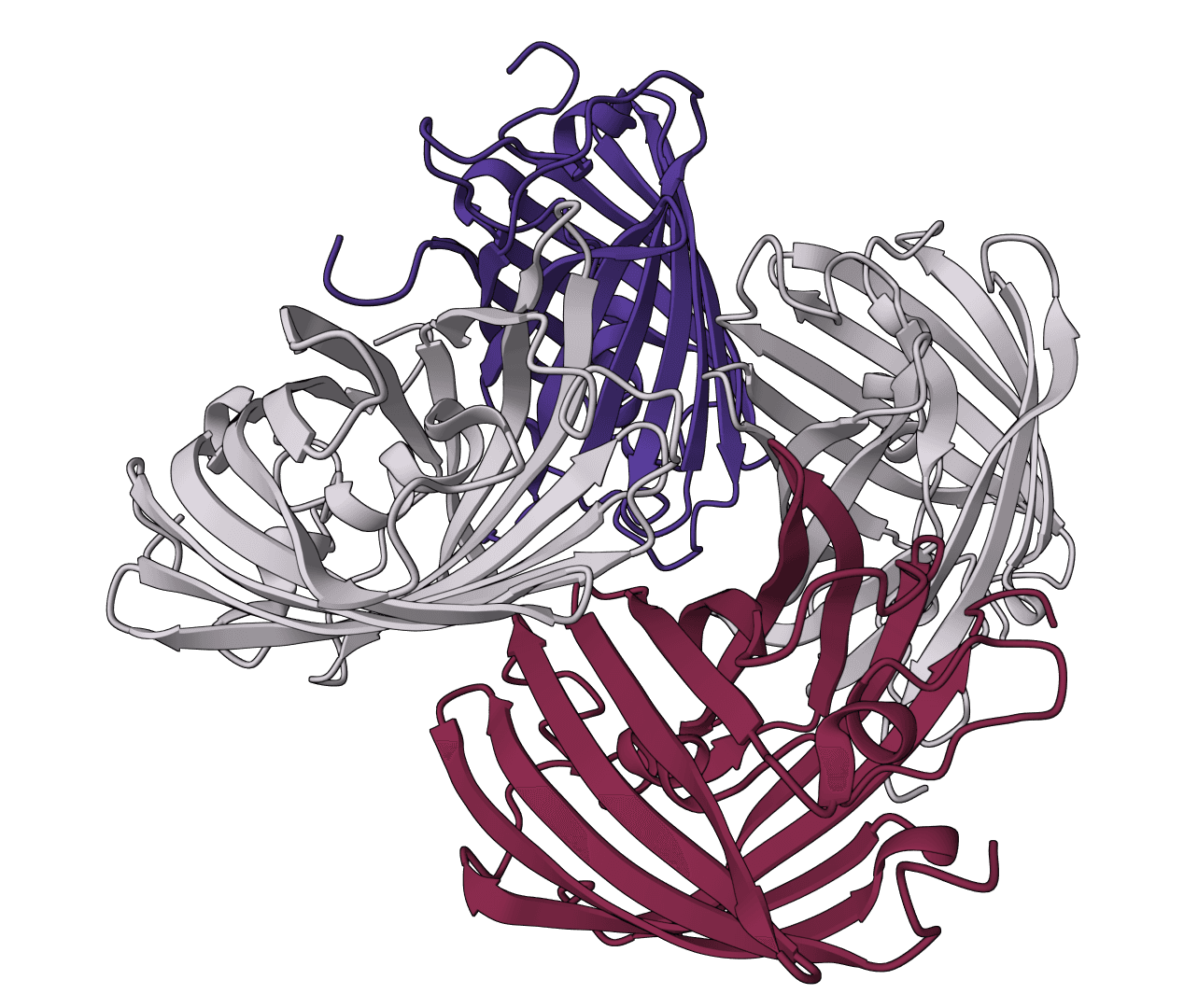
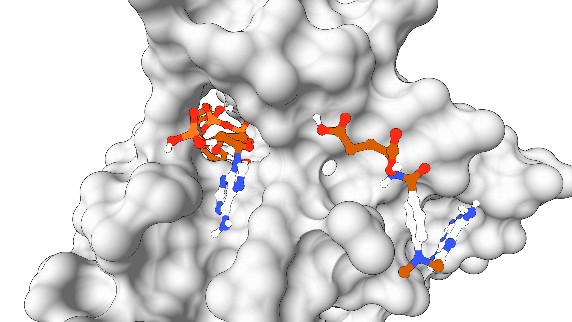
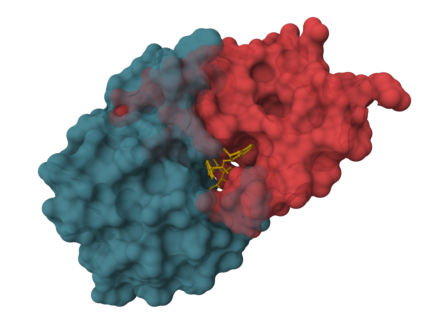
gmx_MMPBSA calculates binding free energies from molecular dynamics trajectories using MM/PBSA (Molecular Mechanics/Poisson-Boltzmann Surface Area) and MM/GBSA (Molecular Mechanics/Generalized Born Surface Area) methods. Developed by Valdés-Tresanco and colleagues, it bridges GROMACS simulations with AMBER's MMPBSA.py engine, enabling end-state free energy calculations for protein-ligand, protein-protein, and protein-nucleic acid complexes.
These methods estimate binding affinity by calculating the free energy difference between bound and unbound states. The calculation combines molecular mechanics energies (van der Waals, electrostatic) with implicit solvation models, offering a balance between the speed of empirical scoring functions and the accuracy of rigorous alchemical methods.
Binding free energy () is computed from snapshots extracted from an MD trajectory:
Each term comprises several energy components:
| Component | Description |
|---|---|
| Van der Waals interactions (Lennard-Jones potential) | |
| Coulombic electrostatic interactions | |
MM/PBSA solves the Poisson-Boltzmann equation for electrostatic solvation, providing higher accuracy but at greater computational cost. MM/GBSA uses analytical Generalized Born approximations, running faster while maintaining reasonable accuracy for ranking compounds.
| Model | Description |
|---|---|
GB-HCT (igb=1) | Hawkins-Cramer-Truhlar model, fast but less accurate |
GB-OBC1 (igb=2) | Onufriev-Bashford-Case model, variant 1 |
GB-OBC2 (igb=5) | OBC variant 2, recommended for most applications |
GB-Neck (igb=7) | Improved treatment of interstitial regions |
GB-Neck2 (igb=8) | Further refinements to neck region calculations |
The OBC2 model (igb=5) provides a good balance of accuracy and speed for protein-ligand systems.
ProteinIQ provides cloud-based access to gmx_MMPBSA, eliminating the need to configure AMBER, GROMACS, or compute environments locally.
| Input | Description |
|---|---|
MD Trajectory file | GROMACS trajectory (.xtc, .trr) or multi-model PDB. Contains atomic coordinates across simulation frames. |
Topology file | GROMACS topology (.tpr or .top). Defines force field parameters and system topology. |
Index file | Optional GROMACS index file (.ndx). Specifies receptor and ligand atom groups. Auto-detected if omitted. |
Input parameter file | Optional gmx_MMPBSA input file. Overrides settings with custom parameters. |
| Setting | Description |
|---|---|
Calculation method | MM/GBSA (faster, suitable for screening), MM/PBSA (more accurate), or Both for comparison |
Energy decomposition | When enabled, calculates per-residue energy contributions to identify key binding site residues |
| Setting | Description |
|---|---|
Start frame | First frame to analyze (1-indexed) |
End frame | Last frame to analyze. Use -1 to include all frames |
Frame interval | Analyze every nth frame. Set to 2 or higher to reduce computation for long trajectories |
| Setting | Description |
|---|---|
Salt concentration | Ionic strength in molar (default 0.15 M, physiological conditions) |
GB model | Generalized Born variant for MM/GBSA. GB-OBC2 (igb=5) recommended |
Results include total binding free energy with component breakdown:
| Column | Description |
|---|---|
DELTA TOTAL | Net binding free energy (kcal/mol). More negative indicates stronger binding. |
VDWAALS | Van der Waals contribution |
EEL | Electrostatic energy (gas phase) |
EGB or EPB | Polar solvation (GB or PB method) |
ESURF or ENPOLAR | Nonpolar solvation (surface area term) |
When energy decomposition is enabled, per-residue contributions identify which amino acids drive binding.
| (kcal/mol) | Interpretation |
|---|---|
| < −15 | Very strong binding |
| −10 to −15 | Strong binding (nanomolar range) |
| −5 to −10 | Moderate binding |
| > −5 | Weak binding |
These values should be interpreted as relative rankings rather than absolute affinities. MM/PBSA and MM/GBSA excel at comparing similar ligands binding to the same target, but absolute values carry significant uncertainty.
The single-trajectory approach used here assumes identical conformations for complex, receptor, and ligand. This reduces noise but misses conformational reorganization effects. Results depend heavily on the quality and length of the MD simulation—short simulations with poor sampling produce unreliable energies.
Entropy contributions are not calculated by default due to computational cost and large uncertainties. For charged ligands, electrostatic and polar solvation terms often exhibit significant cancellation, amplifying small errors. Results should guide compound prioritization rather than predict absolute binding constants.
| Polar solvation energy (PB or GB model) |
| Nonpolar solvation, proportional to solvent-accessible surface area |
| Conformational entropy (optional, computationally expensive) |