Input
100 credits
Output
Configure input settings on the left, then click "Submit job"orLoad an example (it's free)
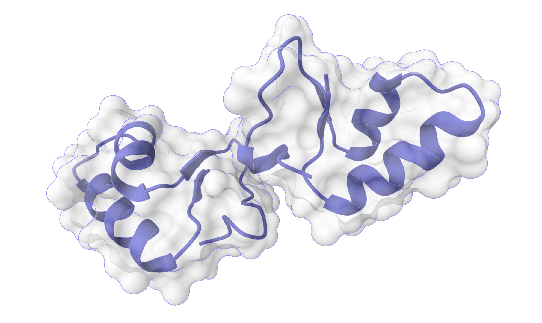
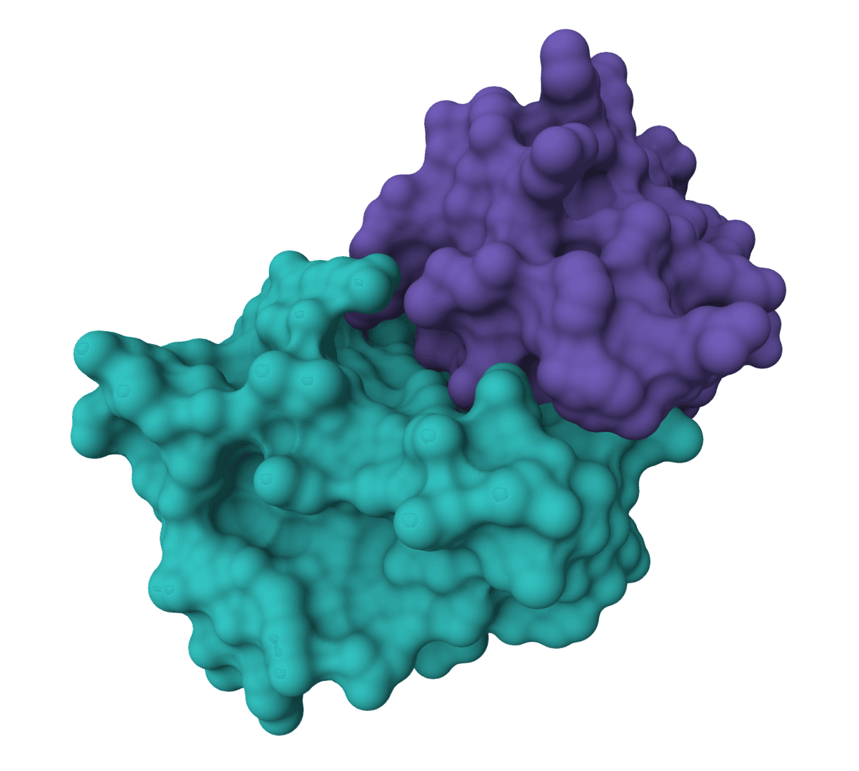
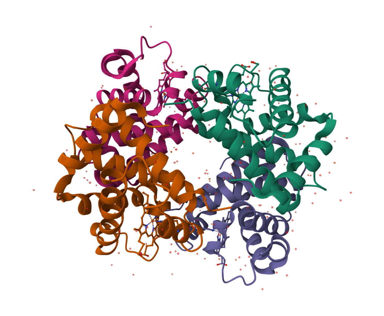
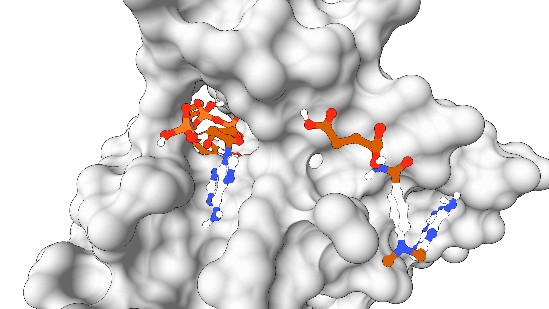
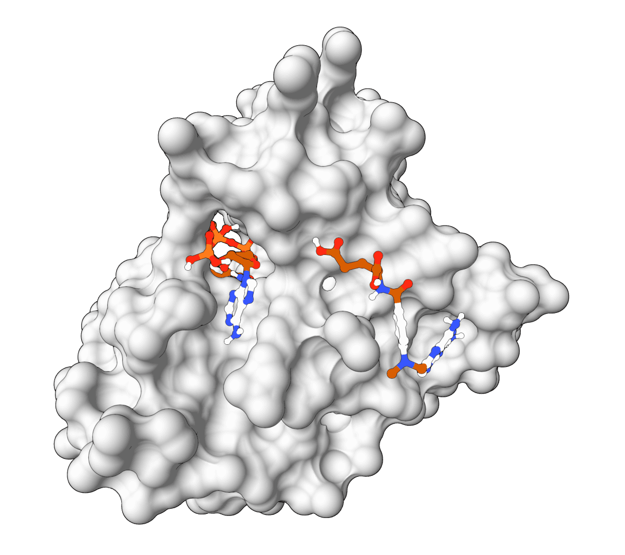
Quick protein-protein docking
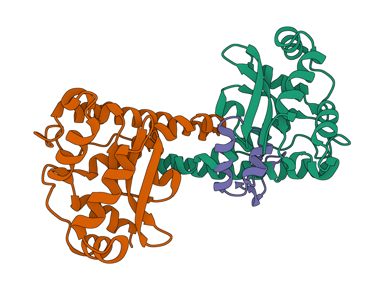
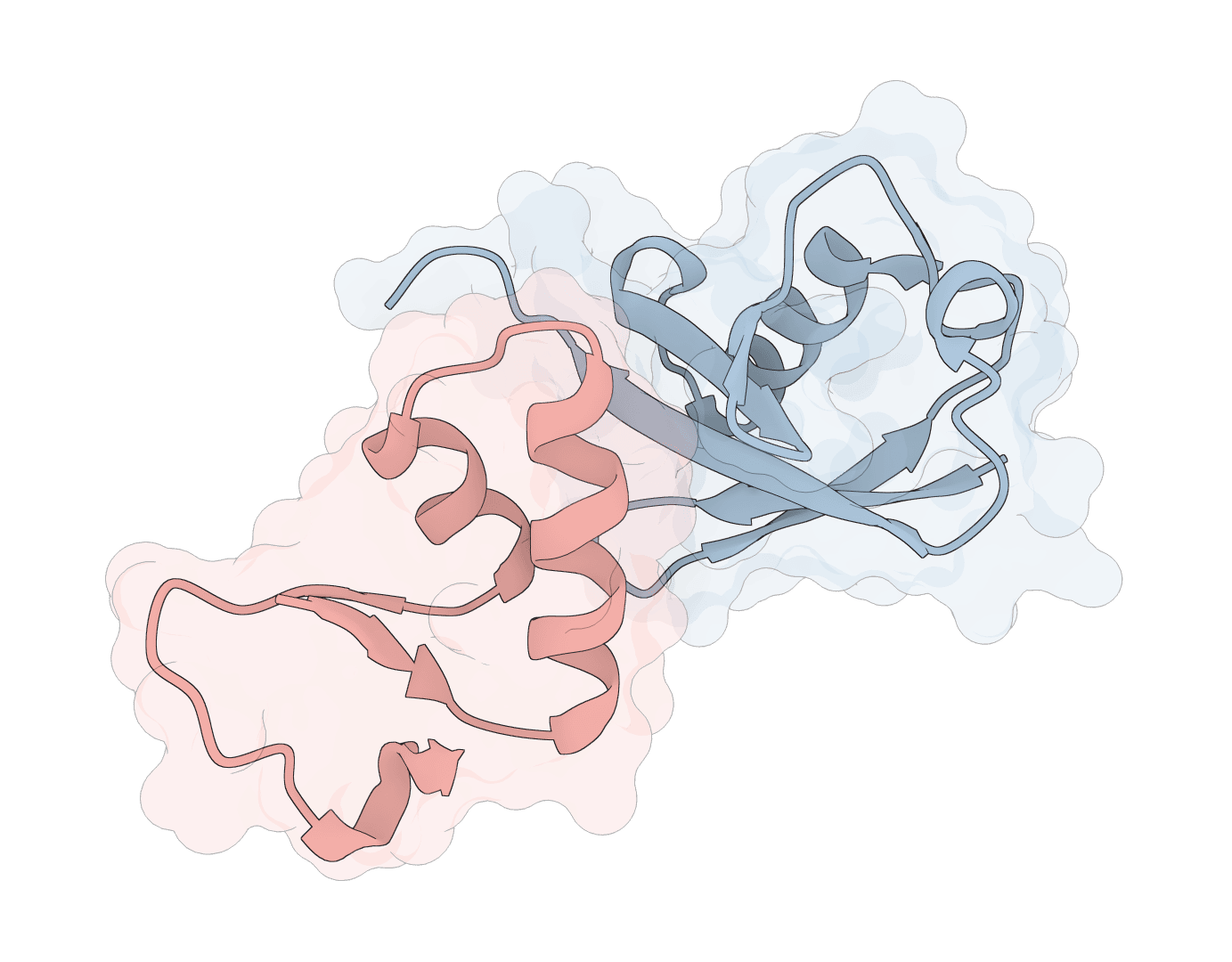
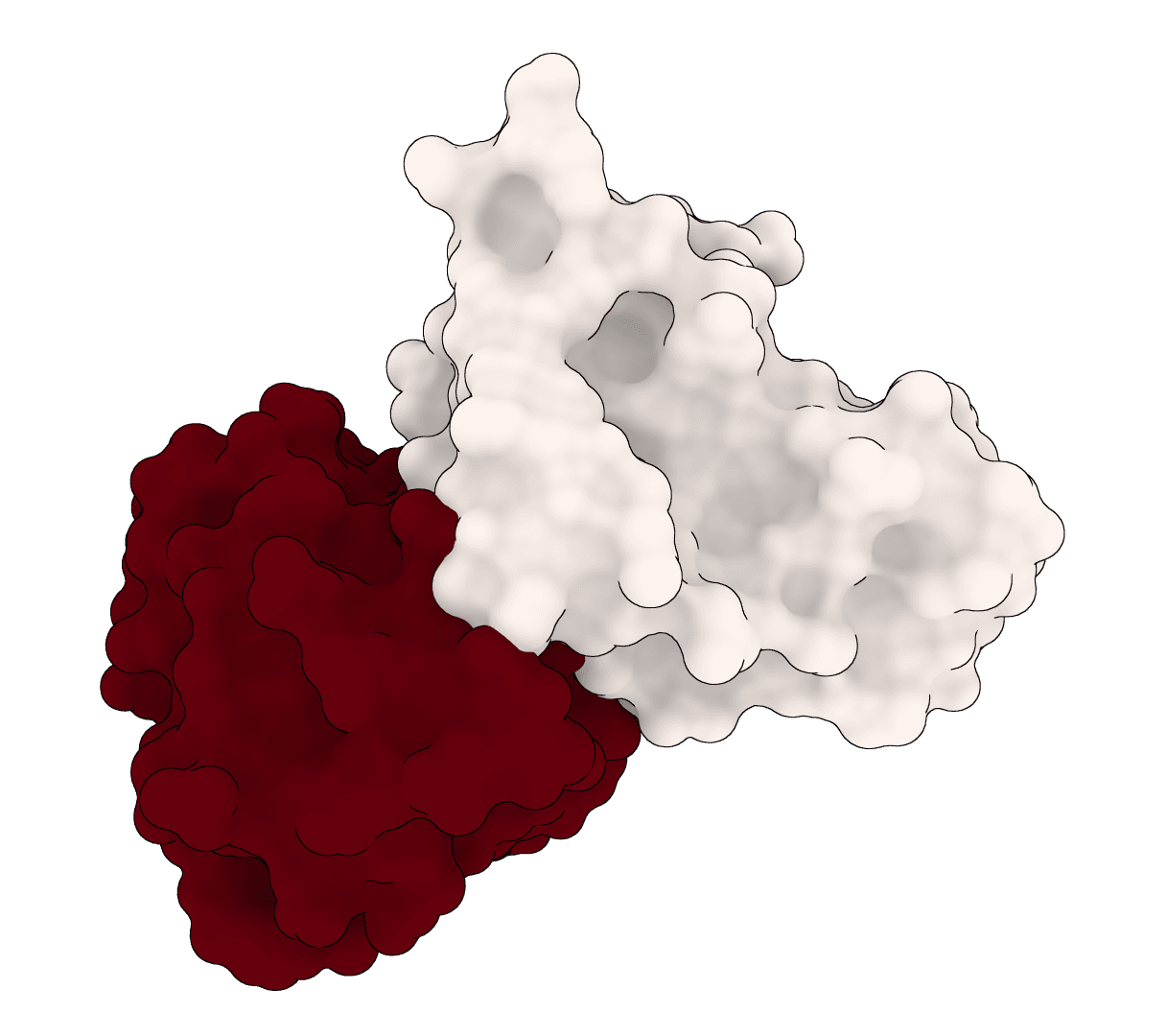
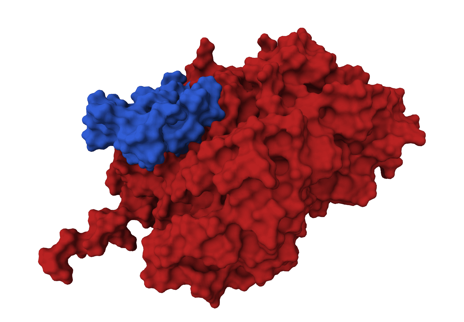
Data-driven docking with interface residues
Quick protein-protein docking
Data-driven docking with interface residues
LightDock is a protein-protein, protein-peptide, and protein-DNA docking framework using Glowworm Swarm Optimization (GSO). It predicts macromolecular binding modes and interfaces for biological complexes.
AF2Dock adapts AlphaFold2-style co-folding for structure-based protein-protein docking. It docks receptor and ligand protein structures with flow-matching refinement and ranks sampled complexes by iPTM.
ColabDock is a protein-protein docking framework that uses AlphaFold2 to predict complex structures guided by experimental restraints from cross-linking mass spectrometry, NMR, or other sources.
DFMDock (Denoising Force Matching Dock) is a diffusion model that unifies sampling and ranking for protein-protein docking within a single framework. It predicts docked poses for protein-protein complexes from unbound structures using denoising score matching with optional clash force guidance.
EquiDock is an SE(3)-equivariant graph neural network for rigid protein-protein docking. It predicts a binding pose for a protein-protein complex from unbound structures using geometric deep learning, with DIPS and DB5 pretrained checkpoints from the native release.
GeoDock predicts flexible protein-protein docking complexes from two separate protein structures using a multi-track iterative transformer and the DIPS 0.3 checkpoint from the Gray Lab release.
ParaSurf is a state-of-the-art surface-based deep learning model for predicting interactions between antibodies and antigens. It identifies paratope binding sites on antibody structures with high accuracy across multiple benchmark datasets.
GPU-accelerated molecular docking using the AutoDock4 force field. Up to 56x faster than serial AutoDock via CUDA parallelization of the Lamarckian Genetic Algorithm.
AutoDock Vina is a widely-used molecular docking tool that predicts protein-ligand binding modes using physics-based force fields. Fast, reliable, and the gold standard for structure-based drug discovery.
Open-source molecular docking platform using physics-based scoring functions. CPU-optimized algorithms achieve sub-angstrom accuracy (0.014A RMSD) without GPU requirements.
HADDOCK3 (High Ambiguity Driven protein-protein DOCKing) is an integrative modeling platform for predicting the structure of biomolecular complexes. Unlike docking methods that rely solely on shape complementarity, HADDOCK3 incorporates experimental or bioinformatic data as restraints to guide the search. This makes it particularly effective when partial interface information is available from mutagenesis, NMR chemical shift perturbations, cross-linking mass spectrometry, or epitope mapping.
HADDOCK3 is developed by the Bonvin Lab at Utrecht University and represents a complete redesign of the original HADDOCK platform with a fully modular workflow.
Upload two protein structures (PDB or mmCIF format, or enter a PDB ID) as the receptor and ligand, optionally provide interface residue numbers for each partner, and submit the job. ProteinIQ returns ranked docked complexes with HADDOCK scores, individual energy components (van der Waals, electrostatics, desolvation, AIR violations, buried surface area), and cluster assignments, all without local installation or CNS configuration.
| Input | Format | Notes |
|---|---|---|
Receptor | PDB, ENT, CIF, mmCIF, or PDB ID | Protein atoms required. mmCIF inputs are converted to PDB automatically. |
Ligand | PDB, ENT, CIF, mmCIF, or PDB ID | The second docking partner, typically the smaller protein. |
Active residues | Comma-separated numbers or ranges (e.g., 38,40,45 or 38-45) | Residues directly involved at the interface. |
Passive residues | Same format | Surface residues near active residues that may contribute to binding. |
Chain ID | Single letter | Leave blank to use the first detected chain after preprocessing. |
| Setting | Default | Description |
|---|---|---|
Number of models | 50 | Rigid-body sampling count. Start with 50; use 100-200 with reliable restraints. |
Top models to return | 10 | Final complexes returned after clustering and refinement. |
Ab initio mode | Center-of-mass | Fallback guidance when no restraints are provided. cmrest applies center-of-mass restraints; ranair uses random AIRs; none docks freely. |
CPU cores | 12 | Parallelization for CNS jobs. Higher values speed up large runs. |
Rigid body tolerance | 5 | Percentage of failed rigidbody models accepted before the run aborts (default). |
Flexref tolerance | 5 | Same as above for the semi-flexible refinement stage. |
EM refinement tolerance | 5 | Same for energy minimization. |
Select top models before refinement | 200 | Rigid-body models passed to flexref. Capped at sampling count automatically. |
Minimum cluster population | 1 | Clusters with fewer members than this are discarded. Set low for small sampling counts. |
Each returned complex includes a HADDOCK score and the individual energy terms that compose it. Cluster information is included when clustering finds more than one group.
| Column | Description |
|---|---|
rank | Global rank by HADDOCK score (lower is better). |
score | Weighted HADDOCK score in arbitrary units. |
vdw | Intermolecular van der Waals energy (kcal/mol). |
elec | Intermolecular electrostatic energy (kcal/mol). |
desolv | Empirical desolvation energy (kcal/mol). |
air | Ambiguous interaction restraint violation energy (kcal/mol). |
bsa | Buried surface area at the interface (Ų). |
cluster | Cluster identifier. Models in the same cluster adopt similar interface conformations. |
The core concept is the Ambiguous Interaction Restraint. An AIR encodes experimental uncertainty without requiring exact atomic contacts: it defines a distance restraint between any atom of an active residue on one protein and any atom on the active or passive residues of the partner. This flexibility accommodates the fact that most experimental techniques identify interface-proximal residues, not specific atomic contacts.
Active residues incur an energy penalty if they remain solvent-exposed in the docked complex. Passive residues are allowed but not required to participate. Random removal of a fraction of restraints during each docking trial ensures the search samples a range of conformations even when some restraints are incorrect.
The ProteinIQ implementation runs a five-stage pipeline:
Stage 1 - Topology generation (topoaa): Generates CNS-format all-atom topology for both proteins. Histidine protonation states are assigned automatically.
Stage 2 - Rigid body docking (rigidbody): Proteins are randomized in orientation and subjected to rigid-body energy minimization guided by AIRs or ab-initio restraints. The Number of models setting controls how many structures are generated here.
Stage 3 - Semi-flexible refinement (flexref): Top-scoring rigid-body models undergo simulated annealing in torsion angle space. Side chains and backbone atoms near the interface are free to move, optimizing local contacts without perturbing the global fold.
Stage 4 - Energy minimization refinement (emref): Selected models are refined by Cartesian energy minimization with electrostatics, van der Waals forces, and desolvation terms. This produces the final scored coordinates.
Stage 5 - Clustering and evaluation (clustfcc, seletopclusts, caprieval): Models are grouped by fraction of common contacts (FCC). Top models per cluster are selected, then scored by CAPRI evaluation, which computes the per-model energy breakdown and global ranking used in the results table.
The final HADDOCK score is a weighted sum of energy terms evaluated at the emref stage:
The heavy weight on van der Waals and desolvation reflects their central role in protein-protein recognition. Electrostatics is downweighted because its long-range character and sensitivity to dielectric treatment make it less reliable as a discriminator. Individual term values are returned alongside the composite score so that models can be compared by specific interaction type.
Typical scores for crystallographically validated complexes fall between -100 and -200. Absolute values scale with interface size, so ranking within a run is more informative than comparing scores across different protein pairs.
Both receptor and ligand accept multi-chain PDB files. HADDOCK3's rigidbody CNS script internally indexes chain segments rather than molecules, which causes it to reference mol_fix_origin_N variables beyond what the module defines when more than two chain segments are present. This is a confirmed native bug in HADDOCK3 2025.11.0: a two-chain receptor plus a one-chain ligand creates three segments, and the third variable is undefined, aborting the run.
ProteinIQ works around this by collapsing each upload into a single chain before passing it to HADDOCK3. Chain A is used for the receptor body and chain B for the ligand body. If interface residues are specified per chain, residue numbers are remapped onto the collapsed structure before restraint generation. When no restraints are provided for a multi-chain input, center-of-mass restraints (cmrest) are enabled automatically to maintain intermolecular guidance during rigid-body docking.
For single-chain inputs, overlapping chain IDs between receptor and ligand are remapped automatically (for example, if both use chain A, the ligand is reassigned to chain C before docking).
The vdw term is usually the most reliable discriminator between good and poor models. A very positive value (above +50) suggests steric clashes that were not resolved during refinement. The desolv term captures burial of hydrophobic surface; large negative values indicate a well-packed hydrophobic core. An air value significantly above zero means active residues are not forming the expected contacts, which can indicate incorrect restraints or a poorly sampled binding mode.
The buried surface area (bsa) is not part of the score formula but correlates with interface size. Values below 500 Ų typically indicate poor packing; values above 1500 Ų suggest large, well-formed interfaces.
Clusters with high population and low average score represent binding modes that are both energetically favorable and structurally consistent across multiple independent trials. When the top cluster contains most models, the docking has converged on a single binding mode. Fragmented clusters with similar scores suggest an underdefined interface, where increasing sampling or adding restraints would help.
If restraints are reliable, the top-ranked model within the largest cluster is most likely to approximate the native binding mode. For completely unknown interfaces (ab initio mode), the top 2-3 clusters should be examined since multiple modes may be plausible.
Data-driven docking uses active and passive residues to focus the search on known interface regions. Success rates exceed 70% in benchmark studies when restraints are correctly assigned. Convergence is faster because the rigid-body sampling is not exploring the full translational and rotational space.
Ab initio docking runs when no restraints are provided. HADDOCK3 generates random AIRs automatically (ranair), or restricts sampling using center-of-mass restraints (cmrest). Success rates drop to 30-40%, but the mode is useful when no interface information is available or when testing whether experimental observations are consistent with a proposed binding site.
Center-of-mass restraints (cmrest, the default) represent the best trade-off for ab initio runs: they ensure the proteins come into proximity without biasing which surface forms the interface.
HADDOCK3 is the right choice when some experimental interface information is available. The data-driven mode consistently outperforms purely computational approaches for these cases. It handles protein-protein complexes of arbitrary size, including antibody-antigen, enzyme-substrate, and homo-oligomeric systems.
LightDock is faster for exploratory runs with no interface data, using swarm intelligence to sample a wide range of docking poses. It does not support AIRs natively, so it loses the advantage HADDOCK3 has when restraints are available.
ColabDock and EquiDock are deep learning approaches that require no restraints and run faster, but cannot incorporate experimental data the way HADDOCK3 can.
For protein-ligand docking (small molecule into a protein binding site), use AutoDock Vina, GNINA, or DiffDock instead. HADDOCK3 is designed for macromolecular interfaces.
NMR-based complex modeling: Chemical shift perturbations identify residues affected by binding. Map perturbed residues to active, surface-adjacent residues to passive, and submit. HADDOCK3's accuracy in this regime is well established across hundreds of benchmark complexes.
Mutagenesis-guided docking: Mutations that disrupt binding localize the interface. Alanine scan results map directly to active residues. Surrounding exposed surface residues serve as passive restraints.
Antibody-antigen docking: Predicted CDR loops (from tools like AbLang-2 or ImmuneBUilder) can be used as active residues for the antibody partner. Experimentally identified epitope residues serve as active restraints for the antigen. Multi-chain antibody structures (VH + VL) are preprocessed into a single rigid body, with distance restraints maintaining chain connectivity.
Cross-linking mass spectrometry integration: Distance constraints from cross-linked residue pairs can be treated as unambiguous restraints in HADDOCK3. These supplement AIRs and further narrow the conformational search.
The method assumes proteins maintain their unbound conformations. Semi-flexible refinement allows local side-chain and backbone adjustments at the interface, but large conformational changes upon binding are not captured. If the complex involves intrinsic disorder, coiled-coil rearrangements, or domain-level conformational shifts, accuracy will be limited regardless of restraint quality.
Computational time scales with sampling count. A run with 50 rigid-body models typically completes in 15-30 minutes; 200 models with full refinement can take 1-2 hours.
HADDOCK3 requires ATOM records and standard protein residues. Non-standard ligands, cofactors, and glycans require additional topology and parameter files not currently available through ProteinIQ. Clean structures using PDB Fixer before submission.
TIMEOUT_ERROR: Job exceeded runtime limits. Reduce Number of models or lower tolerance settings.NO_COMPLEXES_GENERATED: HADDOCK3 finished but produced no models. Validate input PDB quality with PDB Fixer and consider adding interface restraints.INVALID_RESTRAINT_SELECTION: Chain or residue IDs in restraint fields do not match the preprocessed structures. Leave chain fields blank for automatic selection, or provide valid IDs from the input PDB.MODULE_OUTPUT_GENERATION_ERROR: A specific workflow stage (usually rigidbody or flexref) failed to produce any models within the tolerance. For multi-chain inputs, check whether chains were processed correctly. For single-chain inputs with restraints, verify residue numbers match the PDB exactly.HADDOCK_EXECUTION_ERROR: Generic CNS runtime failure. Retry with safer defaults; contact support if it repeats.