OpenMM is a high-performance molecular dynamics simulation toolkit that enables GPU-accelerated simulations of biomolecular systems. Developed by the Pande and Chodera labs at Stanford and MSKCC, OpenMM provides a flexible platform for studying protein dynamics, conformational changes, and ligand-bound complexes with industry-standard force fields like AMBER, CHARMM, and OpenFF small-molecule parameters.
Molecular dynamics (MD) simulations model atomic motion over time by numerically integrating Newton's equations of motion. Each atom experiences forces from bonded interactions (bonds, angles, dihedrals) and non-bonded interactions (electrostatics, van der Waals). By tracking these forces over femtosecond timesteps, MD reveals how proteins move, fold, and interact with their environment on nanosecond to microsecond timescales.
Our implementation uses OpenMM 8, which combines classical force fields with optional machine learning potentials for enhanced accuracy. The simulations run on GPU hardware, achieving speeds of 50-500 ns/day depending on system size—fast enough for routine equilibration checks or extended sampling of conformational transitions.
How does molecular dynamics work?
The physics of motion
At each timestep, OpenMM calculates the potential energy of the system and derives forces from the gradient:
Fi=−∇iU(r1,r2,…,rN)
where U is the potential energy function and ri are atomic coordinates. Forces accelerate atoms according to Newton's second law (F=ma), and velocities are integrated to update positions.
The potential energy function (force field) decomposes into bonded and non-bonded terms:
U=Ubonds+Uang
Time integration
OpenMM uses Langevin dynamics, which adds friction and random forces to simulate coupling with an implicit heat bath:
midt2d
The friction coefficient γ and temperature T control thermal equilibration. Random forces R(t) sample from a Gaussian distribution to maintain the target temperature.
Force fields
AMBER14-SB is the default force field, offering well-validated parameters for proteins based on extensive benchmarking against NMR data and quantum calculations. It uses fixed partial charges and Lennard-Jones potentials for non-bonded interactions.
CHARMM36 provides an alternative with different charge assignments and dihedral parameterization. It offers superior lipid parameters if you plan membrane simulations or protein-lipid interaction studies.
Both force fields assume a classical treatment of electron density—atoms are point masses with fixed charges. This approximation works well for most protein simulations but breaks down for processes involving electronic polarization, charge transfer, or bond breaking.
Solvation models
Explicit water (TIP3P) surrounds the protein with thousands of discrete water molecules. Each water has fixed geometry and partial charges on oxygen and hydrogen. Explicit solvent captures hydrogen bonding, hydrophobic effects, and solvent-mediated interactions with high fidelity.
The computational cost scales with system size. A 100-residue protein in a typical water box requires ~30,000 total atoms, with water molecules comprising ~90% of the atoms.
Implicit solvent (GBn2) replaces discrete water molecules with a continuum dielectric. The generalized Born model approximates electrostatic screening by surrounding atoms with a polarizable medium characterized by dielectric constant and Born radii.
Implicit solvent reduces system size dramatically (protein atoms only) and eliminates water equilibration time. However, it sacrifices detailed hydrogen bonding and can distort conformational preferences for surface-exposed residues.
Input parameters
Simulation settings
Simulation duration controls how long the production simulation runs. Longer simulations sample more conformational space but cost more credits and time.
1 ns (quick test) — Verify the system is stable and check for obvious problems
10 ns (equilibration) — Standard equilibration check; sufficient to detect major issues
50 ns (standard) — Production-quality sampling for small conformational changes
100 ns (production) — Extended sampling for larger motions or binding events
Solvation model determines how solvent is treated. Use Explicit water (TIP3P) for accurate dynamics including hydrogen bonding and realistic solvent behavior. Use Implicit solvent (GBn2) for faster sampling when detailed solvent interactions aren't critical.
Force field selects the energy function parameters. AMBER14-SB is recommended for most protein work. Choose CHARMM36 if planning membrane simulations or if your workflow uses CHARMM-derived parameters downstream.
Ligand file is optional and is used only to parameterize a small organic ligand that is already present in the uploaded structure as non-water HETATM coordinates. The ligand file does not place or dock a separate molecule into the protein. Metal complexes and other ligands requiring custom parameters are not supported by this tool.
Environment
Temperature sets the simulation temperature in Kelvin. 300 K (27°C) is standard physiological temperature. Use higher temperatures (350-400 K) for enhanced sampling of slow processes, or lower temperatures for cold-adapted proteins.
Pressure controls the barostat target in bar. 1.0 bar is standard atmospheric pressure. Most simulations use NPT ensemble (constant number, pressure, temperature) to allow box volume fluctuations.
Ionic strength specifies NaCl concentration in molar. 0.15 M matches physiological conditions. The system is first neutralized with counterions, then excess salt is added to reach the target concentration.
pH determines protonation states of titratable residues (histidine, aspartate, glutamate, lysine, arginine). Standard protonation at pH 7.0 is appropriate for most cytoplasmic proteins. Adjust for proteins in acidic compartments (lysosomes, pH 4.5-5.0) or extracellular environments.
Output options
Save interval controls trajectory frame frequency. 50 ps produces manageable file sizes while capturing relevant dynamics. Use 10 ps for detailed analysis of fast motions or transition states. Use 100 ps for long simulations where disk space is limited.
Remove water from output strips solvent molecules from the trajectory file. This reduces file size by ~90% while retaining all protein coordinates. Enable this unless you specifically need to analyze water dynamics or hydration shells.
Advanced settings
Timestep is the integration step size. 2 fs is standard when hydrogen bonds are constrained. 4 fs (HMR) uses hydrogen mass repartitioning—hydrogen masses are increased while heavy atom masses decrease to maintain total mass. This allows larger timesteps without instability but requires validation for your specific system.
Minimization steps controls energy minimization before dynamics. Minimization removes bad contacts and steric clashes from the initial structure. 1000 steps suffice for most structures. Increase for structures with severe clashes or added loops.
Equilibration time sets the equilibration phase duration before production data collection. Equilibration includes gradual heating (NVT) and density relaxation (NPT). 0.5 ns is typically sufficient; increase for larger systems or membrane simulations.
Bond constraints determines which bonds are held rigid:
All bonds — Constrain all bonds, required for 4 fs timesteps
None — No constraints, requires 0.5-1 fs timesteps (rarely needed)
Understanding the results
Trajectory output
OpenMM produces a trajectory file containing atomic coordinates at each save interval. The trajectory can be visualized in molecular viewers (VMD, PyMOL, Chimera) to observe protein motion over time.
Key observations from trajectory analysis:
RMSD — Root-mean-square deviation from the starting structure indicates overall conformational stability
RMSF — Root-mean-square fluctuation per residue reveals flexible regions
Radius of gyration — Measures protein compactness; changes indicate unfolding or large conformational shifts
Secondary structure — Track helix/sheet content over time to detect folding/unfolding events
Energy monitoring
The simulation reports potential energy, kinetic energy, and temperature at regular intervals. Stable simulations show:
Potential energy fluctuating around a constant value (not drifting)
Temperature fluctuating around the target value (300 K ± 5 K typical)
No sudden energy spikes indicating steric clashes or numerical instabilities
Common issues
Structure explodes — Usually indicates bad starting contacts or missing parameters. Run energy minimization first with PDB Fixer. Ensure all atoms have appropriate force field parameters.
Temperature drifts — Check that the thermostat is functioning correctly. Very large systems may need longer equilibration.
Protein unfolds — May indicate force field issues with your specific protein or unrealistic starting conditions. Verify the structure quality with MolProbity before simulation.
When to use OpenMM
Use molecular dynamics when
Studying protein flexibility and conformational changes
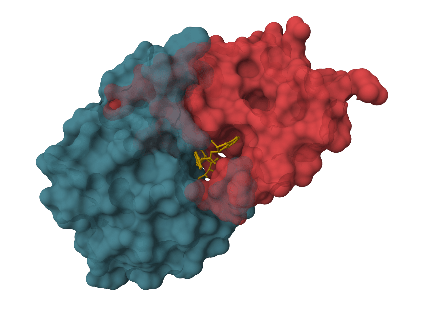
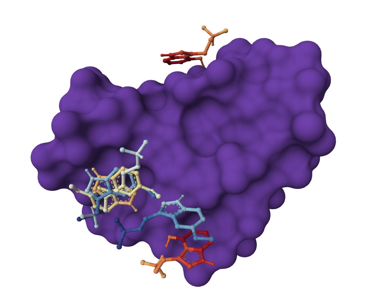
Investigating binding site dynamics and ligand accommodation
Run MD on docking complexes to assess pose stability
Frequently asked questions
Is OpenMM free to use?
Yes. ProteinIQ provides OpenMM simulations at no cost within free tier credit limits. OpenMM itself is open-source software (MIT/LGPL license) developed by the academic community.
How long do simulations take?
Simulation time depends on system size, solvation model, and duration. A 100-residue protein with explicit water for 10 ns typically completes in 30-60 minutes. Implicit solvent simulations run 3-5x faster. The 2-hour maximum timeout accommodates 100 ns explicit solvent runs for most proteins.
What's the difference between explicit and implicit solvent?
Explicit solvent adds discrete water molecules that interact realistically with the protein—hydrogen bonding, hydrophobic effects, and electrostatic screening are captured physically. Implicit solvent replaces water with a continuum approximation, running faster but sacrificing detailed solvent interactions. Use explicit solvent for production simulations; use implicit for quick conformational sampling or when water isn't relevant to your question.
Should I use AMBER or CHARMM force fields?
For standard protein simulations, AMBER14-SB is recommended and well-validated. CHARMM36 offers advantages for membrane simulations due to superior lipid parameters. Both produce reasonable results for most proteins—the differences are typically smaller than uncertainties from other sources (sampling, starting structure quality).
How do I know if my simulation is stable?
Check these indicators:
Energy stays within normal fluctuation range (no spikes or drift)
Temperature fluctuates around target value
RMSD plateaus rather than continuously increasing
Visual inspection shows no unfolding or unrealistic motions
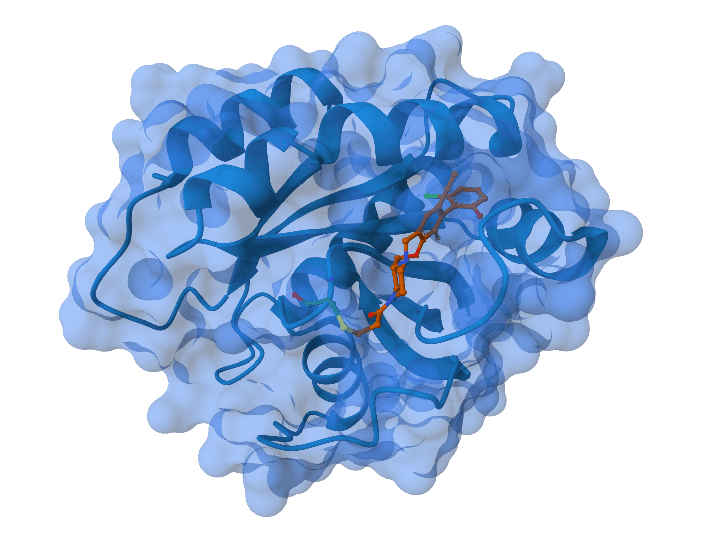
Can I simulate protein-ligand complexes?
Yes, when the uploaded PDB/mmCIF already contains both protein and ligand coordinates. The optional ligand slot accepts an SDF or MOL2 file for the same embedded ligand so OpenMM can generate OpenFF parameters for common organic small molecules. It is not a docking or placement step, and metal-containing ligands or coordination complexes require custom force-field parameters outside this tool.
What should I do if the simulation crashes?
Most crashes result from bad starting structures. Try:
Ensure all atoms have valid force field parameters
For ligand complexes, confirm the ligand is embedded in the structure and matches the uploaded SDF/MOL2 file
How much conformational sampling do I need?
This depends on your question:
Stability check: 1-10 ns suffices to detect major problems
Local flexibility: 10-50 ns captures side chain rotations and loop motions
Domain motions: 50-100 ns may capture larger conformational changes
Rare events: Enhanced sampling methods may be needed for processes slower than 100 ns
Can I continue a simulation from where it left off?
Not currently through the web interface. Each simulation starts fresh from the uploaded structure. For extended sampling, run successive simulations starting from the final frame of previous runs.
References
Eastman, P., Galvelis, R., et al. (2024). OpenMM 8: Molecular Dynamics Simulation with Machine Learning Potentials. J. Phys. Chem. B, 128(1), 109-116. DOI: 10.1021/acs.jpcb.3c06662
Maier, J.A., et al. (2015). ff14SB: Improving the Accuracy of Protein Side Chain and Backbone Parameters from ff99SB. J. Chem. Theory Comput., 11(8), 3696-3713.
Huang, J., et al. (2017). CHARMM36m: an improved force field for folded and intrinsically disordered proteins. Nat. Methods, 14, 71-73.