How do nanobodies, scFvs and Fabs differ from antibodies?
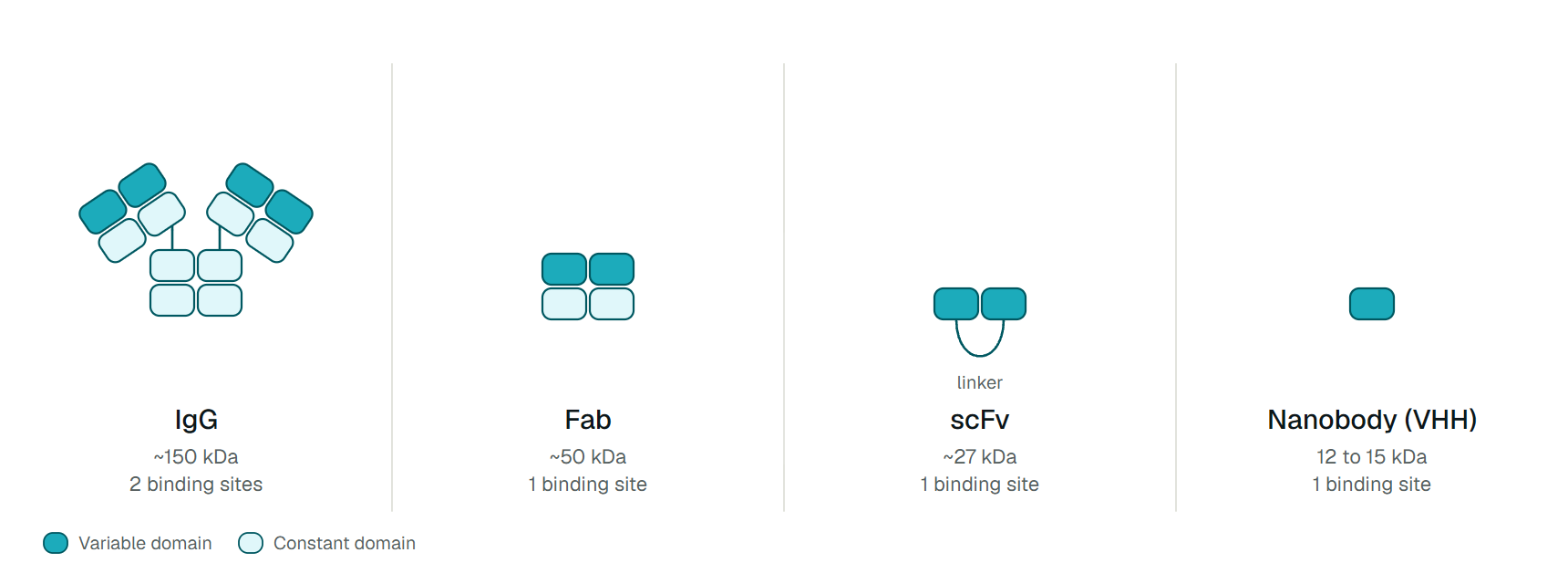
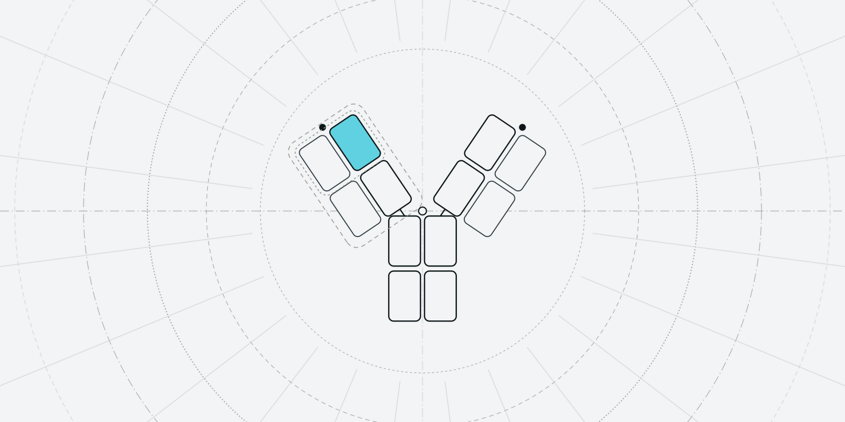
A full IgG antibody has two binding arms and an Fc stem, and weighs about 150 kDa. A Fab keeps one arm (about 50 kDa), an scFv keeps only the two variable domains joined by a peptide linker (about 27 kDa), and a nanobody is the single variable domain of a camelid heavy-chain antibody, called a VHH, at 12 to 15 kDa.[1] The masses are the easy part. Each cut also removes something the full antibody supplied without anyone noticing.
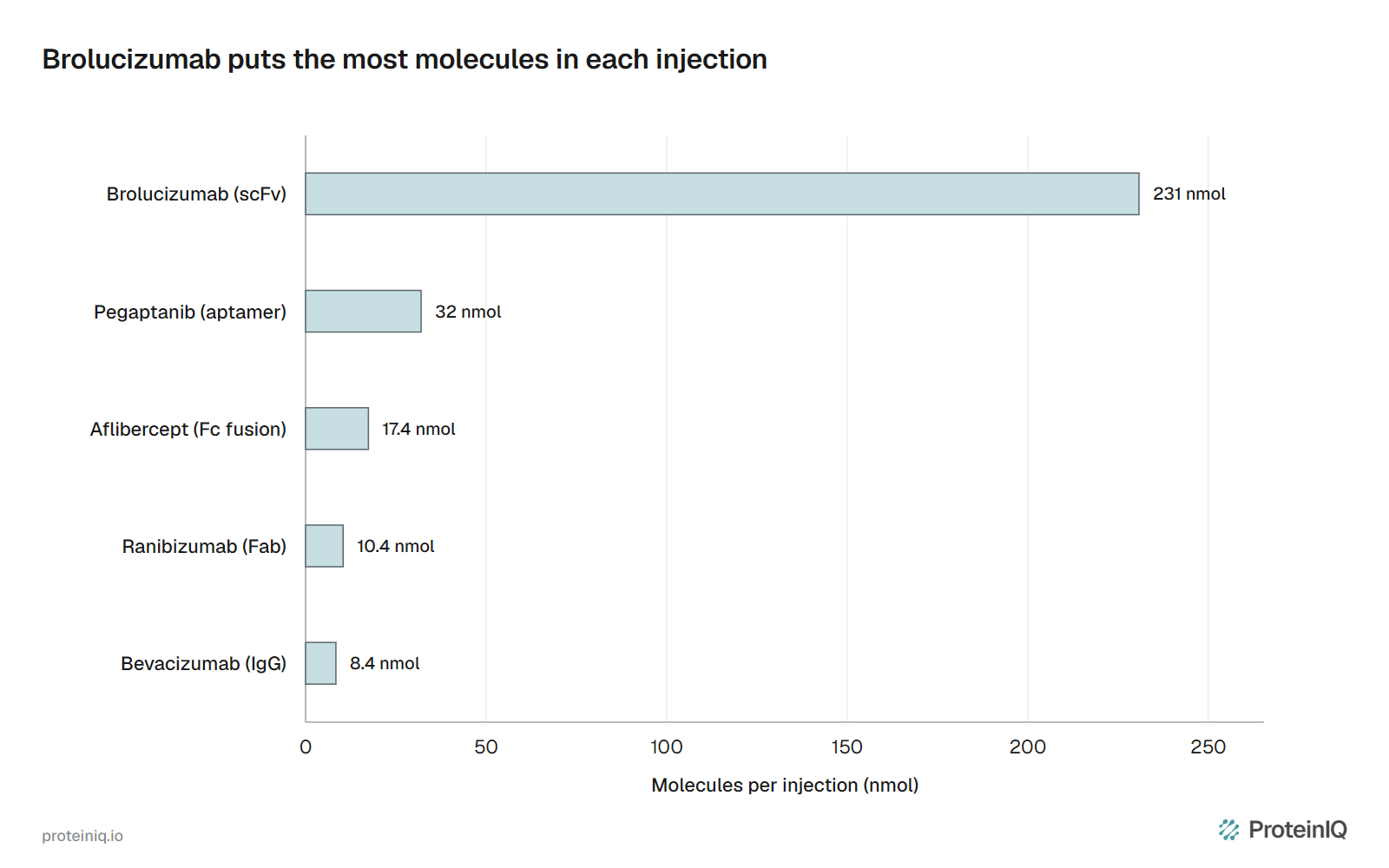
The price shows up in the eye. VEGF-A is blocked in clinics by an IgG (bevacizumab, used off-label), a Fab (ranibizumab), an scFv (brolucizumab) and a PEGylated aptamer (pegaptanib). A 6 mg dose of brolucizumab holds about 22 times as many molecules as a 0.5 mg dose of ranibizumab, and more than half of the eyes in its phase 3 trials stayed on injections every 12 weeks.[19] Its label also warns of retinal vasculitis and vascular occlusion.[14]
Five properties decide between the formats: how many molecules a dose carries, how long they stay in the body, how tightly a binder holds on (avidity), whether the fragment stays folded without its partner domains, and how the immune system reacts to it. Aptamers, which are nucleic acids, face the same five tests.

What a full antibody supplies that fragments lose
The first loss is time. IgG1 lasts about 21 days in humans because its Fc binds the FcRn receptor and is recycled.[7] A Fab or scFv has no Fc and loses that recycling. A nanobody sits below the roughly 60 kDa cutoff of the kidney's filter and clears with a half-life of about 2 hours.[2][1] Twenty-one days is 504 hours, so the gap is roughly 250-fold, though the two values come from different studies.
Half-life is a design variable, not a fixed property of the format. Ozoralizumab, approved in Japan in 2022, is a 38 kDa nanobody drug with two TNF-binding domains and a third that binds serum albumin, and it is dosed every four weeks.[6] Fusing an albumin-binding domain can raise a fragment's half-life toward that of albumin, 19 days.[1]
The second loss is avidity. An IgG grips an antigen with two arms, while a Fab, scFv or VHH has one site. In one study reviewed by Harmsen and De Haard, a bivalent anti-TNF VHH was 500-fold more potent than the monovalent domain.[2] That is why many fragment drugs are multimers: caplacizumab, the first approved nanobody drug, joins two VHH domains against von Willebrand factor.[1]
The Fc also carries effector function, which recruits complement and killer cells (see the Fab and Fc regions). Where the goal is only to block a target, losing it is a gain. Fragments are not glycosylated and can be made in microbes, whereas full-length antibodies generally need mammalian cells.[1]
Fab versus scFv
A Fab holds the light chain and the first two heavy-chain domains, so VH and VL are anchored by their constant partners (CH1 and CL). Bates and Power attribute the Fab's greater stability to this mutual stabilization.[1] An scFv drops the constant domains and relies on one linker, which has to span about 3.5 nm without distorting the binding site. scFvs show lower thermostability and a greater tendency to aggregate than the parent antibody, which raises immunogenicity risk.[1]
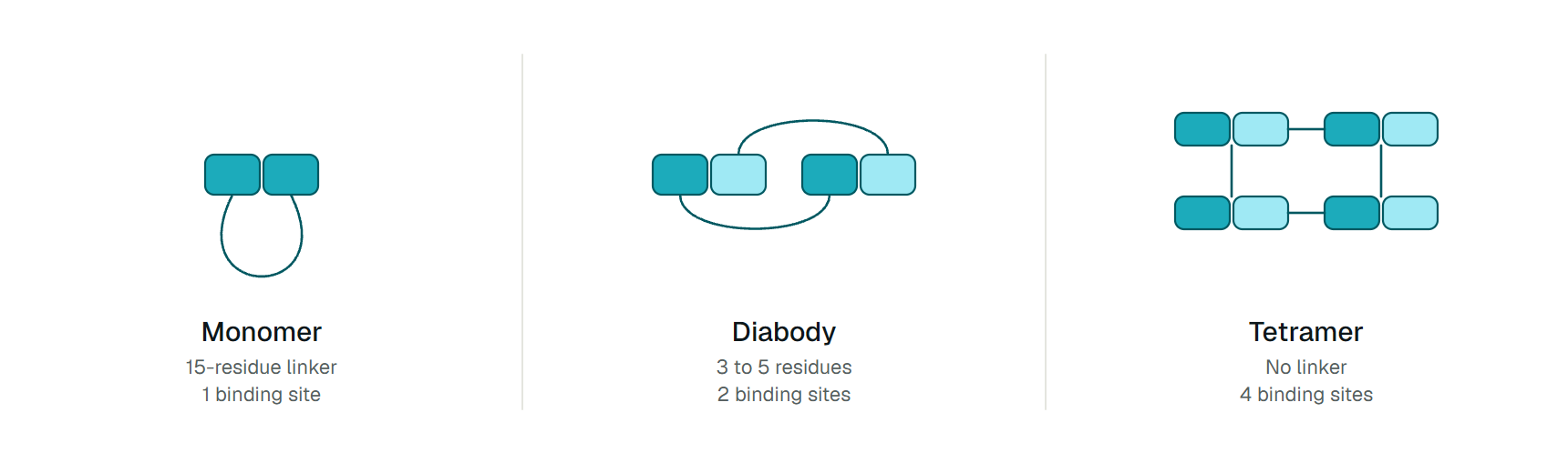
The linker also decides what the molecule is. In the NC10 antibody, with the domains in VL to VH order, a 15-residue linker gave mostly monomers, 3 to 5 residues gave diabodies and a direct fusion gave mainly tetramers.[4] In anti-TAG-72 fragments, oligomeric state and stability varied dramatically with linker, domain order and even the purification method.[5] "scFv" names a family of related molecules, and a result for one construct need not carry over to the next.
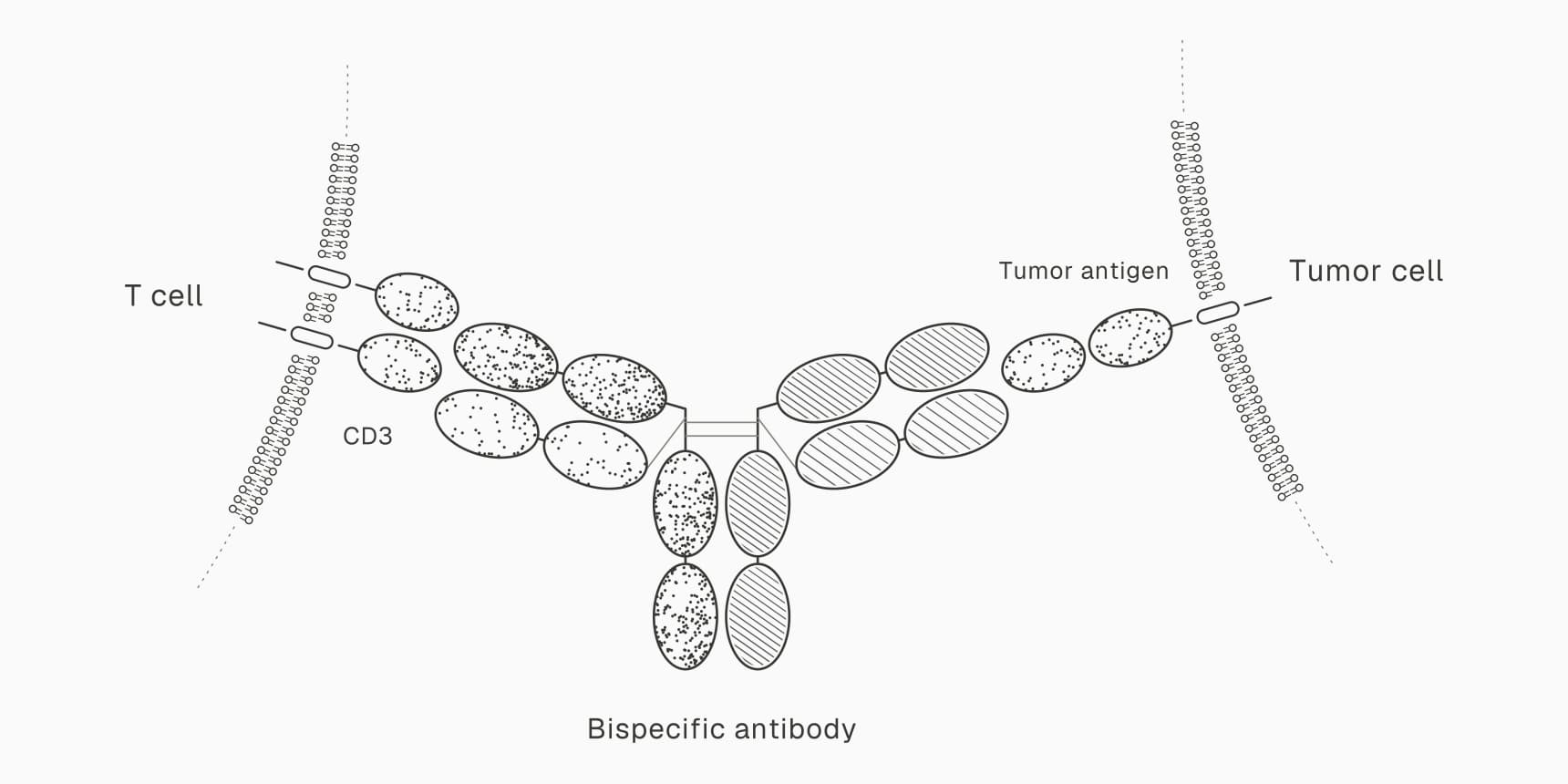
The clinical record favors the Fab. Bates and Power count abciximab, idarucizumab and ranibizumab as FDA-approved Fabs, and certolizumab pegol is a PEGylated one.[1] The scFv has brolucizumab, approved in 2019 and produced in E. coli.[14] Two scFvs joined end to end form the bispecific T-cell engager blinatumomab.
Nanobodies versus scFvs
A nanobody needs no partner, so it escapes the scFv's pairing problem. In a conventional VH, four framework 2 positions (37, 44, 45 and 47 in Kabat numbering) form the hydrophobic interface with VL. In a VHH those positions carry substitutions that make the domain more hydrophilic, and a human VH given the same changes was more stable than the original.[2] Camelid heavy-chain-only antibodies have no light chain, so the single domain is the whole binding site.
The result behaves well in the lab. VHHs have stayed functional at 90 degrees Celsius, mainly because they refold efficiently after denaturation.[2] Llama VHH fragments have given yields of hundreds of milligrams per litre in yeast, though Bates and Power note that these traits are not present in all nanobodies.[1]
Binding differs too. Many dromedary VHHs have an extended CDR3 loop that can reach enzyme active sites and other cryptic epitopes, and VHH affinities are generally comparable to those of conventional fragments.[2] Haptens and peptides that sit in the groove between VH and VL are the exception, where a single domain may be at a disadvantage.[2]
Immunogenicity is usually low. Camelid VH sequences resemble the human IGHV3 family, and most clinical trials have found minimal anti-drug antibodies. Two potent single-domain drugs, the tetrameric DR5-binding TAS266 and a TNFR1-binding domain, were exceptions that show why preexisting antibodies must be tested for, and humanizing a VHH can trade foreign sequence for low-level aggregation.[3]
Antibodies versus aptamers
An aptamer is a short single-stranded DNA or RNA molecule, selected in vitro by SELEX, that folds into a shape binding its target with picomolar to low nanomolar affinity.[8] It is made by chemical synthesis, is easy to modify and can be denatured and refolded reversibly.[9] Those are real advantages over an antibody that has to be grown in cells.
The catch is the one fragments face, in harsher form. Unmodified aptamers are metabolically unstable and rapidly filtered by the kidney, so nearly all human trials used modified aptamers, and PEGylation is the main way to extend half-life.[8] Only two aptamer drugs have FDA approval, pegaptanib in 2004 and avacincaptad pegol in 2023, set against the monoclonal antibodies the FDA has approved.[9] The oligonucleotide drug list places them among antisense and siRNA drugs.
Pegaptanib shows the problem in numbers. It is a 28-nucleotide oligonucleotide carrying two 20 kDa PEG units, about 50 kDa in all.[11] By subtraction the aptamer is about 10 kDa, so four-fifths of the drug is PEG added to slow filtration, the job an albumin binder does for a nanobody.
One target in five formats
VEGF-A has drugs in several formats, and their label doses can be converted to a common unit. Table 1 lists them, with molecules per dose calculated from the label mass and dose.
| Drug | Format | Mass (kDa) | Dose | Molecules per dose (nmol) | Interval |
|---|---|---|---|---|---|
| Bevacizumab | IgG | 149 | 1.25 mg | 8.4 | Off-label in the eye |
| Ranibizumab | Fab | 48 | 0.5 mg | 10.4 | Monthly |
| Aflibercept | Receptor Fc fusion | 115 | 2 mg | 17.4 | Every 8 weeks after 3 monthly doses |
| Pegaptanib | PEGylated aptamer | 50 | 1.6 mg | 32 | Every 6 weeks |
| Brolucizumab | scFv | 26 | 6 mg | 231 | Every 8 to 12 weeks after 3 monthly doses |
The small format made the high dose possible. Tadayoni and colleagues describe brolucizumab's 26 kDa size as allowing a concentrated molar dose in one injection.[17] It is about 22 times ranibizumab, 13 times aflibercept and 28 times bevacizumab. In human aqueous humor its half-life was 9.0 days against 2.88 days for aflibercept,[18] which fits intervals of up to 12 weeks.
The dose may also be part of the safety problem. Retinal vasculitis or vascular occlusion may follow the first injection, and the label contraindicates the drug in active intraocular inflammation.[14] Real-world cohorts found intraocular inflammation in about 4 to 10% of treated eyes, often within three months.[19]
Anti-drug antibodies are a prerequisite, since high titers were required to produce inflammation in monkeys, but only 2.1% of antibody-positive patients developed vasculitis or occlusion, so other factors must matter.[20] A root cause analysis named several: a linear epitope shared with bacteria, non-natural surfaces from the scFv format, drug that turns non-native over time in the eye, and immune complexes whose formation depends on dose intensity.[21] Both studies came from the manufacturer, which is worth knowing when weighing them.
Pegaptanib went the other way. It binds the VEGF165 isoform, and in the label's two pivotal trials 73% and 67% of treated patients lost fewer than 15 letters of vision, against 60% and 53% on sham injection.[11] Direct comparisons with ranibizumab are not possible across separate trials, but the antibody drugs restored some lost vision while the aptamer mainly slowed disease.[10]
Within the antibodies, format changed little at a similar molar dose. Monthly bevacizumab and ranibizumab gained 8.0 and 8.5 letters in the CATT trial, from doses of 8.4 and 10.4 nmol.[16] Format mattered when it moved the dose by an order of magnitude or exposed the immune system to new surfaces.
Which format should you start with?
Table 2 maps a goal to a starting format and the property most likely to go wrong.
| If you need | Start with | Check first |
|---|---|---|
| A drug that lasts weeks and recruits immune cells | Full IgG | Mammalian production cost and Fc effector function |
| A monovalent blocker that reaches tissue | Fab | Clearance, and whether one site is enough |
| A binder for a cleft or an enzyme active site, or a module for multispecifics | Nanobody | Preexisting anti-drug antibodies, humanization and a half-life extender |
| A small fragment made in microbes from an existing antibody | scFv | Linker length, oligomers, aggregation and exposed surfaces |
| A chemically synthesized, easily modified binder | Aptamer | Nuclease stability, kidney filtration and PEG |
Screen candidates early for developability, and check that an scFv or nanobody carries no aggregation-prone patches before committing to a construct.
Antibody, nanobody and scFv tools on ProteinIQ
The order is usually to number and check the sequence, humanize it if needed, screen it for developability, predict its structure, and only then design or redesign binders.
- ANARCI: number antibody and nanobody variable domains
- BioPhi: humanize a sequence and score its humanness
- TAP2: compare developability against clinical-stage antibodies
- ImmuneBuilder: predict antibody and nanobody structures
- RFantibody and BoltzGen: design antibody or nanobody binders against a target
Frequently asked questions
Is a nanobody the same as a VHH?
In practice, yes. VHH is the technical name for the single variable domain of a camelid heavy-chain antibody, and Nanobody is a trade name for the same format.[6] A VHH is 12 to 15 kDa.[1]
Which is smaller, an scFv or a Fab?
An scFv, at about 27 kDa against about 50 kDa for a Fab. A nanobody is smaller still.[1]
Why do nanobodies clear from the blood so fast?
They sit below the roughly 60 kDa cutoff of the kidney's filter, so a typical half-life is about 2 hours.[2][1] An albumin-binding domain fixes this: ozoralizumab is dosed every four weeks.[6]
Why choose an aptamer over an antibody?
Aptamers are chemically synthesized, easy to modify and can refold after denaturation.[9] They need modification or PEGylation to survive nucleases and kidney filtration, and only two have reached FDA approval.[8][9]
Has any fragment drug been approved?
Yes. Approved Fabs include abciximab, idarucizumab and ranibizumab, brolucizumab is an scFv, and caplacizumab and ozoralizumab are nanobody drugs.[1][14][6]