TL;DR
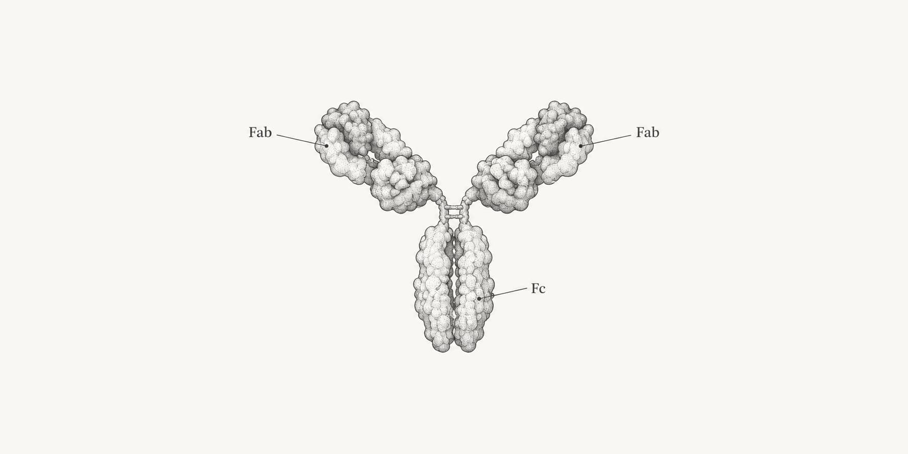
- A typical IgG contains two Fab regions and one Fc region, joined by a flexible hinge.
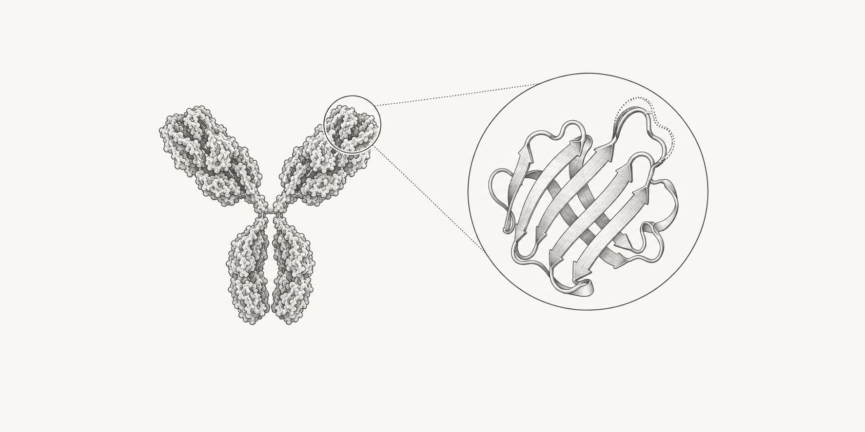
- Each Fab contains one complete light chain plus the VH and CH1 domains of one heavy chain; its paired VH and VL domains form one antigen-binding site.
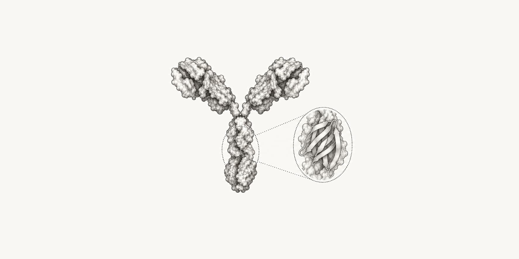
- The IgG Fc contains the paired CH2 and CH3 domains of the heavy chains and interacts with Fc receptors, C1q, and FcRn.
- Fab is not synonymous with the variable region: Fab includes both variable and constant domains, while Fv contains only VH and VL.
What are the Fab and Fc regions of an antibody?
The Fab regions are the two arms of an immunoglobulin G (IgG) antibody that bind antigen. The Fc region is the stem that interacts with immune receptors and other proteins. One IgG antibody has two Fab regions and one Fc region.[1][4][5]
| Feature | Fab region | Fc region |
|---|---|---|
| Position in IgG | The two arms of the Y | The stem of the Y |
| Composition | One complete light chain plus VH and CH1 from one heavy chain | Paired CH2 and CH3 domains from the two heavy chains |
| Main binding partners | Antigen epitopes | Fc receptors, C1q, FcRn, and other Fc-binding proteins |
| Antigen-binding sites | One per Fab | None in an unmodified antibody |
| Main role | Antigen recognition and direct blocking or neutralization | Immune-cell recruitment, complement activation, transport, and IgG recycling |
| Typical IgG fragment mass | About 50 kDa | About 50 kDa |
Fab means fragment antigen-binding. Fc means fragment crystallizable. Fab contains both variable and constant domains, while Fc contains only heavy-chain constant domains.[1][2]
What does the Fab region do?
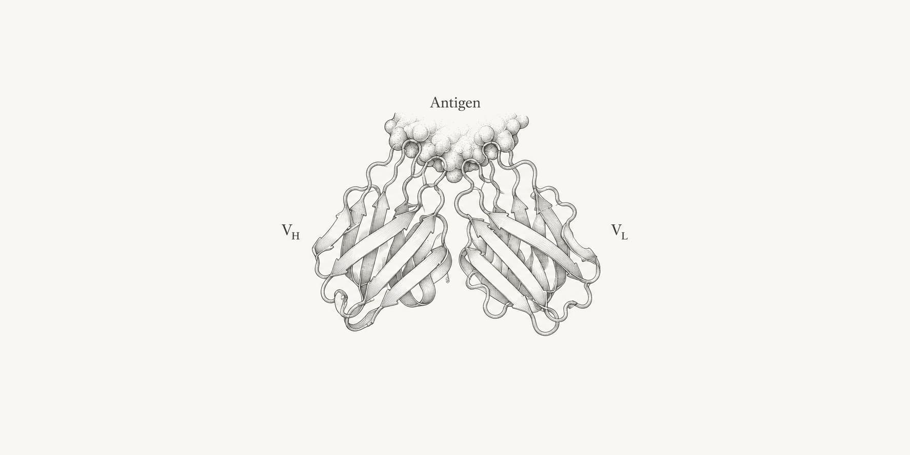
The Fab region recognizes and binds an epitope on an antigen. Each Fab arm contains one complete light chain—its variable (VL) and constant (CL) domains—plus the VH and CH1 domains of one heavy chain. VH and VL meet at the tip to form the antigen-binding surface, or paratope.[1]
An intact IgG therefore has two equivalent Fab arms and is usually bivalent: it can engage two copies of the same epitope. A Fab fragment contains only one arm and is monovalent, even though it preserves the parent antibody's antigen specificity.[1][2]
Fab is not the same as the antibody variable region. The Fv region contains only VH and VL, whereas Fab also includes CH1 and CL. The six complementarity-determining regions (CDRs) within VH and VL make many of the direct contacts with the antigen.
An antigen does not have a Fab or Fc region. It presents an epitope, which is recognized by the antibody's paratope.
What does the Fc region do?
The Fc region recruits immune functions after the Fab arms bind their target. It interacts with Fc receptors, complement protein C1q, and FcRn, which influence immune-cell activity, complement activation, transport, and IgG recycling.[4][5][6]
In IgG, Fc consists of the paired CH2 and CH3 domains of the two heavy chains. The CH3 domains associate closely, while glycans separate the CH2 domains and help maintain their structure.[1][4] These interactions vary by antibody class and subclass, so changing the constant region can change biological activity without changing antigen specificity.[6]
What binds to the Fc region?
Different Fc-binding partners contact different surfaces:
| Fc partner | Principal IgG interaction region | Functional consequence |
|---|---|---|
| Fcγ receptors | Lower hinge and CH2 surfaces | Activating or inhibitory immune-cell signaling; phagocytosis and cytotoxicity |
| C1q | Primarily the CH2-facing Fc surface in clustered IgG | Initiation of the classical complement pathway |
| FcRn | CH2–CH3 interface | pH-dependent IgG recycling and transport |
FcRn binds at acidic endosomal pH, rescues IgG from degradation, and releases it again near neutral pH.[4][5][7]
Human IgG carries an N-linked glycan at Asn297 in each CH2 domain (EU numbering). These glycans influence Fc structure and Fcγ-receptor binding, so glycosylation can change Fc activity.[4]
What does the antibody hinge do?
The IgG hinge lies between CH1 and CH2. It contains inter-heavy-chain disulfide bonds and acts as a flexible tether, allowing the two Fab arms to change their angle and distance from the Fc.[1][6] This flexibility helps both arms engage differently spaced epitopes.
Whether a few hinge residues are assigned to Fab or Fc depends on the cleavage site, antibody subclass, and naming convention. Fab and Fc are historically defined proteolytic fragments; VH, CH1, CH2, and CH3 are sequence domains with more explicit boundaries.
How are Fab and Fc fragments made?
The terms Fab and Fc come from protease-digestion experiments. Papain and pepsin cut IgG on different sides of the inter-heavy-chain disulfide bonds in the hinge.[1][2][3]
| Molecule or fragment | Antigen-binding sites | Fc retained? | Typical IgG mass | How it is obtained |
|---|---|---|---|---|
| Intact IgG | 2 | Yes | ~150 kDa | Complete antibody |
| Fab | 1 | No | ~50 kDa | Papain cleavage above the hinge disulfides or recombinant expression |
| F(ab')2 | 2 | No | ~110 kDa | Pepsin cleavage below the hinge disulfides; the Fab arms remain linked |
| Fc | 0 | It is the Fc | ~50 kDa | Papain cleavage leaves the paired heavy-chain stem |
Papain and pepsin fragment boundaries vary slightly with antibody sequence and digestion conditions, so the masses are approximate.[1][3][8]
These are rounded values. Sequence, subclass, cleavage site, glycosylation, and conjugated molecules change the exact mass. See the antibody molecular-weight guide for details.
Which antibody format should be used?
| Format | Useful when | Main limitation |
|---|---|---|
| Intact IgG | Bivalent binding, Fc-mediated activity, or FcRn-supported persistence is required | Fc-receptor interactions can create background or unwanted effector activity |
| Fab | Monovalent binding, structural studies, epitope mapping, or reduced Fc-receptor interference is required | Loses Fc function, bivalent avidity, and FcRn recycling |
| F(ab')2 | Bivalent antigen binding is needed without an intact Fc | Still lacks Fc-mediated effector function and recycling |
| Fc | Studying receptor/complement interactions, engineering effector function, or building Fc fusions | Has no native antigen-binding site |
Removing Fc changes molecular size, valency, biodistribution, and persistence as well as immune-cell recruitment.[7][8] A Fab result is therefore not automatically equivalent to a result from the same binder in intact IgG format.
In antibody engineering, the two regions are often optimized separately. Fab engineering changes affinity, specificity, or cross-reactivity. Fc engineering changes Fc-receptor engagement, complement activity, glycosylation, stability, or FcRn-mediated half-life.[7]
How are Fab and Fc regions analyzed?
ANARCI numbers VH and VL variable domains, while ABodyBuilder3 and ImmuneBuilder model paired antibody variable domains. These tools do not produce a complete Fc or a definitive intact-IgG orientation. The Molecular Weight Calculator estimates sequence mass, and the PDB Viewer can inspect domains and chain assignments in a supplied structure.
For intact IgG, hinge flexibility means that one static structure represents only one possible Fab–Fc arrangement.[6][7] See the antibody structure-prediction workflow for model selection and validation.