TL;DR
- Antibodies are antigen-binding proteins made by B cells; immunoglobulin is the name of the protein family.
- A conventional antibody contains two heavy chains and two light chains, with variable regions for antigen recognition and constant regions that determine class and immune activity.
- Antibodies bind specific sites called epitopes and can neutralize targets, promote phagocytosis, activate complement, or recruit immune cells.
- Humans have five main antibody classes: IgG, IgA, IgM, IgD, and IgE.
What are antibodies?
Antibodies are proteins made by B cells that recognize and bind specific molecular sites called epitopes. After binding, an antibody can block its target directly or recruit other parts of the immune system to respond to it.[1][2][8]
The antibody protein family is called immunoglobulin, abbreviated Ig. B cells display membrane-bound immunoglobulin as their antigen receptor, while plasma cells secrete the soluble form. Antibody and immunoglobulin are therefore often used interchangeably.[1]
What is the difference between an antibody and an antigen?
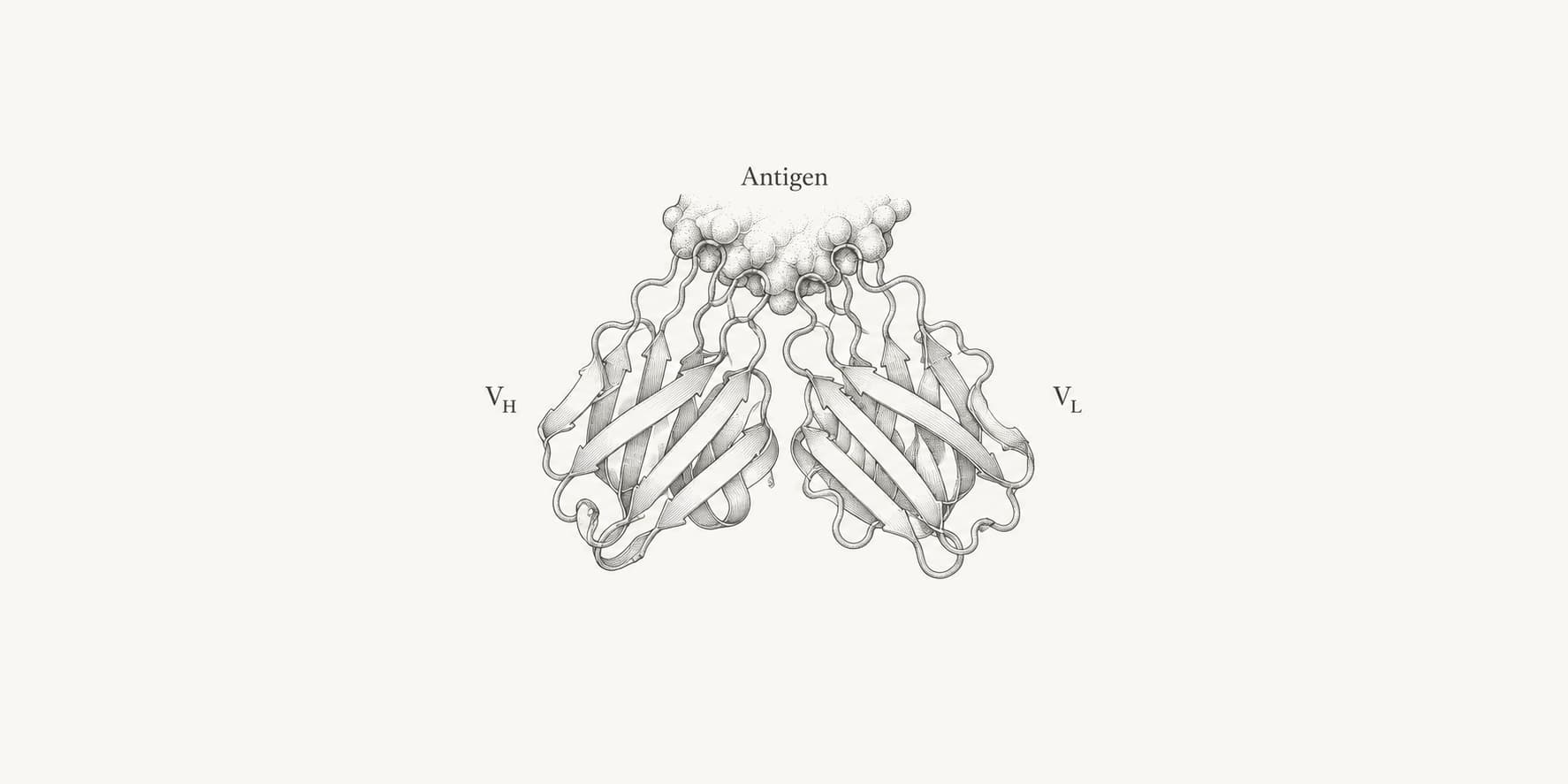
An antibody is the binding protein. An antigen is a molecule or molecular structure that can be recognized by an antibody or another adaptive immune receptor. The precise part of an antigen contacted by an antibody is its epitope; the complementary binding surface on the antibody is its paratope.[3][6]
An antigen is not necessarily a pathogen or even foreign. Proteins, carbohydrates, lipids, small molecules, and self-components can act as antigens. One antigen can also present many overlapping epitopes, allowing different antibodies to bind the same target.
What are antibodies made of?
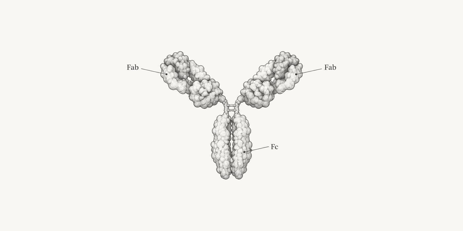
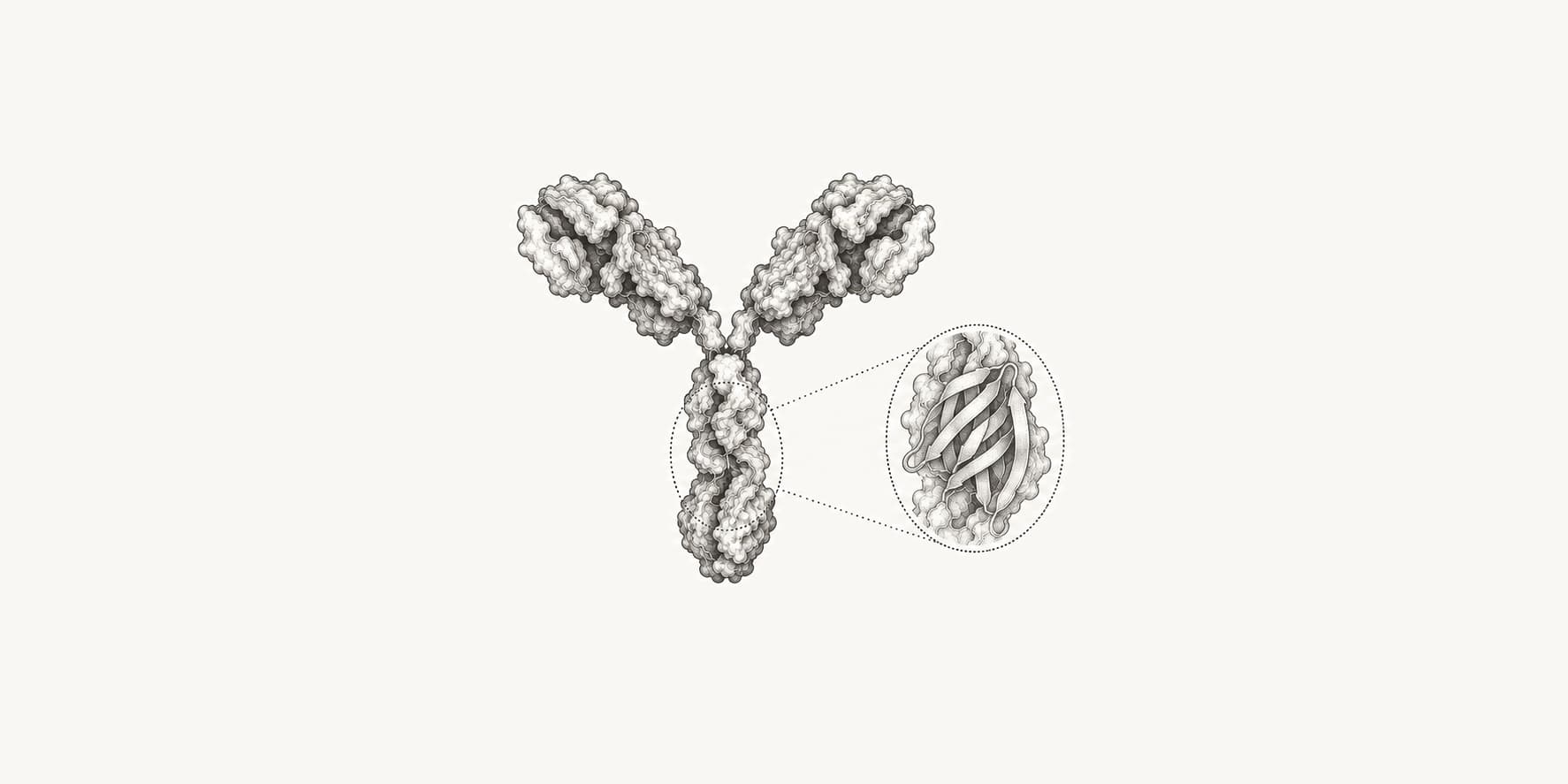
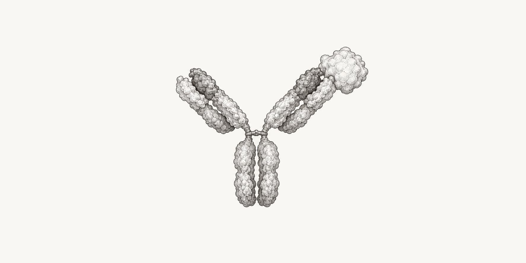
A conventional antibody unit contains two identical heavy chains and two identical light chains joined by disulfide bonds and noncovalent interactions. The familiar Y-shaped diagram usually represents an IgG molecule, which has a typical molecular weight of about 150 kDa.[1][2]
The two Fab arms contain the variable regions that recognize antigen. The Fc stem interacts with Fc receptors, complement proteins, and transport receptors, while the hinge allows the Fab arms to adopt different orientations.
The dedicated guides explain the antibody variable regions and Fab and Fc regions in detail. Not every secreted antibody is a single Y-shaped monomer: IgM and some forms of IgA assemble into larger complexes.
How do antibodies recognize antigens?
In a conventional antibody, the binding site is formed mainly by six complementarity-determining region loops: three from VH and three from VL. Their shape and chemical properties create a paratope complementary to an antigen epitope.[3][6]
Antibody-antigen binding is reversible and noncovalent. Hydrogen bonds, electrostatic interactions, van der Waals contacts, and hydrophobic effects collectively stabilize the complex. Affinity describes the strength of one binding interaction; avidity describes the combined strength of multiple simultaneous interactions.[3]
Specificity is not absolute. An antibody can cross-react when another molecule presents a sufficiently similar chemical surface, and strong binding does not automatically mean that the antibody neutralizes the target or works in every assay.
Where and how are antibodies produced?
B cells generate antibody diversity before antigen exposure by rearranging variable (V), diversity (D), and joining (J) gene segments. Heavy-chain variable regions use V, D, and J segments, while light-chain variable regions use V and J segments.[4]
When a B cell receives the appropriate antigen-dependent activation signals, it proliferates into a clone. Some descendants become plasma cells that secrete antibodies; others become memory B cells that support a faster response after later exposure.
Activated B cells can further modify their antibodies in germinal centers. Somatic hypermutation and selection produce affinity maturation, while class-switch recombination replaces the heavy-chain constant region to change antibody class and effector function without replacing the rearranged antigen-binding variable region.[4]
How do antibodies work?
Antibody binding can block a target directly or recruit immune mechanisms through the antibody constant region. The result depends on the epitope, antibody class, Fc structure, target location, and biological context.[5]
- Neutralization blocks a toxin, pathogen, ligand, or receptor interaction.
- Opsonization allows Fc receptors on phagocytes to recognize and engulf an antibody-coated target.
- Complement activation promotes inflammation, opsonization, or membrane damage.
- Antibody-dependent cellular cytotoxicity recruits immune cells that kill an antibody-coated target cell.
- Cross-linking groups targets into immune complexes that can be cleared.
Antibodies do not always protect the organism and usually do not “destroy” an antigen by themselves. Autoantibodies can bind self-components, IgE responses can drive allergy, and some antibody-antigen interactions can contribute to inflammation or disease. Binding, neutralization, and protection are therefore different experimental measurements.
What are the five antibody classes?
Humans have five main immunoglobulin classes, determined by the constant region of the heavy chain. The classes share the same general antigen-binding plan but differ in assembly, distribution, receptors, and immune functions.[2]
- IgG is the major serum class and supports neutralization, opsonization, complement activity, and placental transfer.
- IgA is found as a monomer in serum and commonly as a dimer in mucosal secretions.
- IgM is displayed as a monomer on B cells and commonly secreted as a pentamer during early primary responses.
- IgD is mainly a membrane-bound receptor on mature naïve B cells.
- IgE binds Fc receptors on mast cells and basophils and participates in allergy and responses to parasites.
IgG and IgA also have subclasses. The guide to antibody classes and subclasses explains what counts as an antibody “type.”
How do polyclonal, monoclonal, and recombinant antibodies differ?
These terms describe different properties of an antibody preparation. Polyclonal and monoclonal describe the diversity of the antibody population, while recombinant describes how an antibody is produced.[7]
| Term | Meaning |
|---|---|
| Polyclonal antibodies | A mixture produced by multiple B-cell clones, usually recognizing several epitopes |
| Monoclonal antibody | A defined antibody population derived from one selected clone or recombinant sequence |
| Recombinant antibody | An antibody expressed from cloned genes; it can be monoclonal, multispecific, or a designed fragment |
The monoclonal-antibody guide covers hybridoma and recombinant production, molecular heterogeneity, and validation.
How are antibodies analyzed and engineered?
Antibody analysis separates questions about sequence, numbering, structure, binding, and biological function. No single computational result establishes all of these properties.
- IgBLAST assigns germline V, D, and J genes and annotates variable-domain junctions.
- ANARCII standardizes variable-domain positions and CDR boundaries across antibody sequences and structures.
- ABodyBuilder3 and ImmuneBuilder predict three-dimensional antibody structures with confidence or error estimates.
- RFantibody performs structure-based de novo antibody and nanobody design.
- BioPhi evaluates antibody humanness and proposes humanization changes.
Sequence annotation and structure prediction must be followed by experimental tests of expression, folding, affinity, specificity, cross-reactivity, and biological activity. Those computational workflows sit within the broader field of antibody engineering.