What is the molecular weight of an antibody?
A typical IgG antibody has a molecular weight of about 150 kDa. IgA is about 160 kDa as a serum monomer, while pentameric IgM is about 970 kDa.
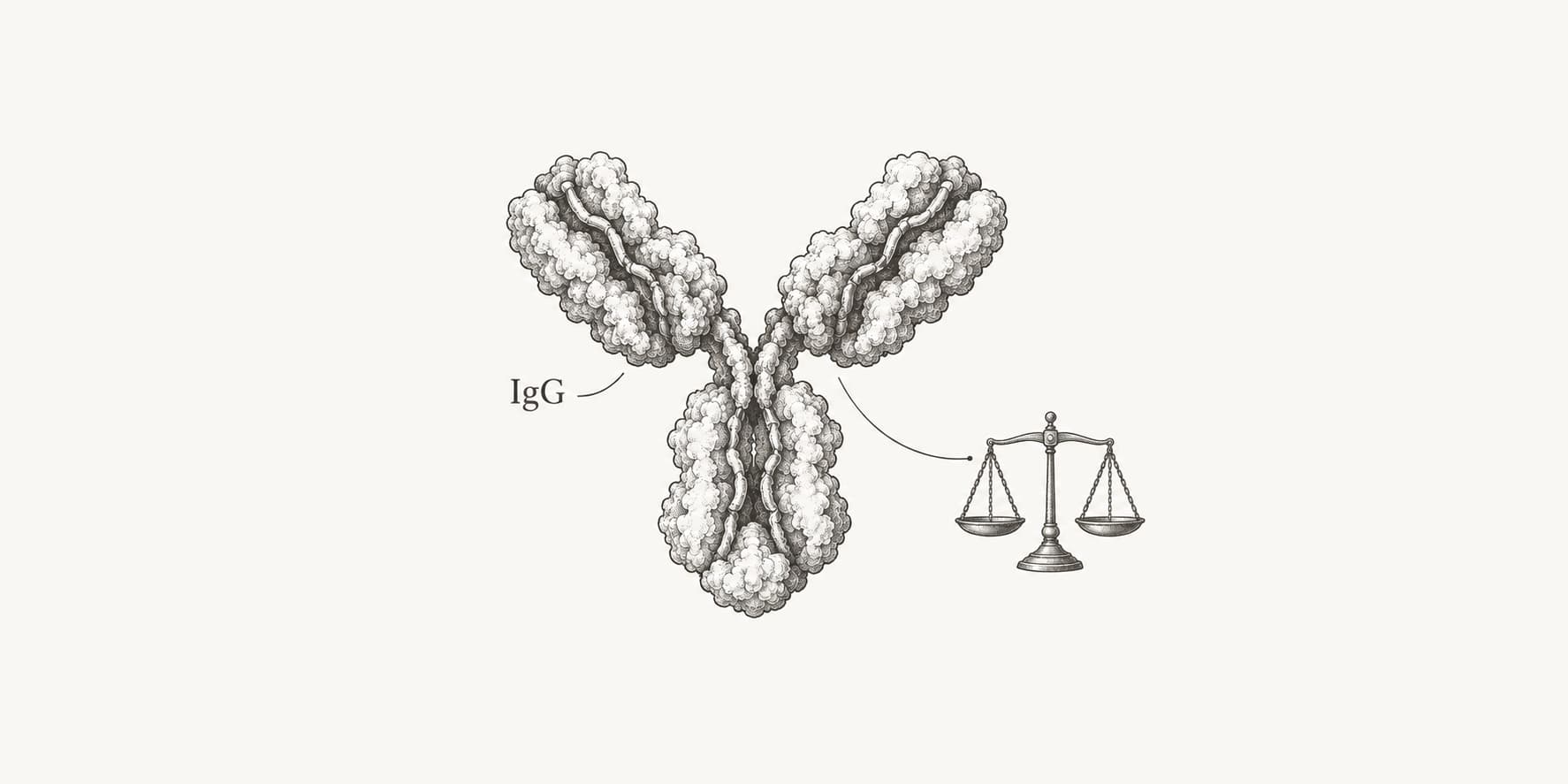
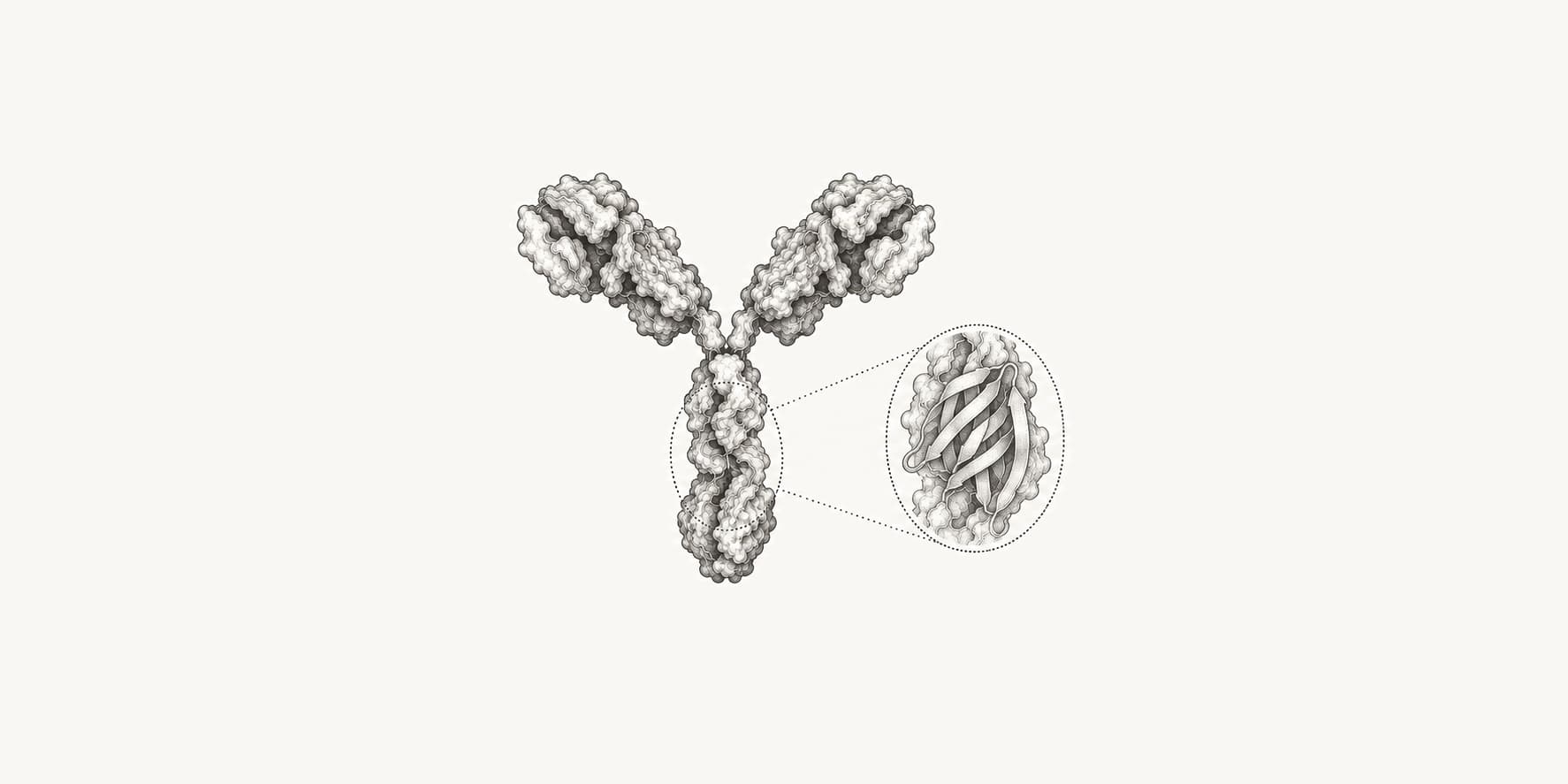
A typical antibody has a molecular weight of about 150 kilodaltons (kDa), or 150,000 daltons. This familiar figure refers to immunoglobulin G (IgG), the antibody form used for most research and therapeutic monoclonal antibodies.
Other antibody classes can be much larger. Secreted IgM is about 970 kDa, more than six times the mass of IgG, while serum IgA is about 160 kDa.
What is the molecular weight of an antibody?
The molecular weight of a typical IgG antibody is approximately 150 kDa.[1]
An IgG molecule contains two heavy chains of about 50 kDa each and two light chains of about 25 kDa each. Adding the four chains gives the standard estimate:
2 × 50 kDa + 2 × 25 kDa = 150 kDa.
The chains are held together by disulfide bonds and noncovalent interactions. The same four-chain plan appears across the five human antibody classes, although their heavy chains and assembled forms differ.
“Molecular weight” is the common laboratory term. Because a dalton and a gram per mole have the same numerical value for molecular mass, 150 kDa also corresponds to a molar mass of about 150,000 g/mol.
What are the molecular weights of IgG, IgM, and IgA?
IgG is about 146 to 150 kDa, monomeric IgA is about 160 kDa, and secreted pentameric IgM is about 970 kDa.[1][2]
IgA needs a second number because its form changes by location. Serum IgA is mainly a 160 kDa monomer. Secretory IgA found at mucosal surfaces is a larger complex of about 385 kDa. IgD and IgE are monomers of about 184 kDa and 188 kDa, respectively.[2]
The chart compares typical human antibody forms, not every subclass or glycoform. The 146 kDa value for IgG is often rounded to 150 kDa in laboratory use. IgM is also frequently rounded to 900 kDa, but the cited reference value is 970 kDa.[2]
What are the molecular weights of Fab and Fc fragments?
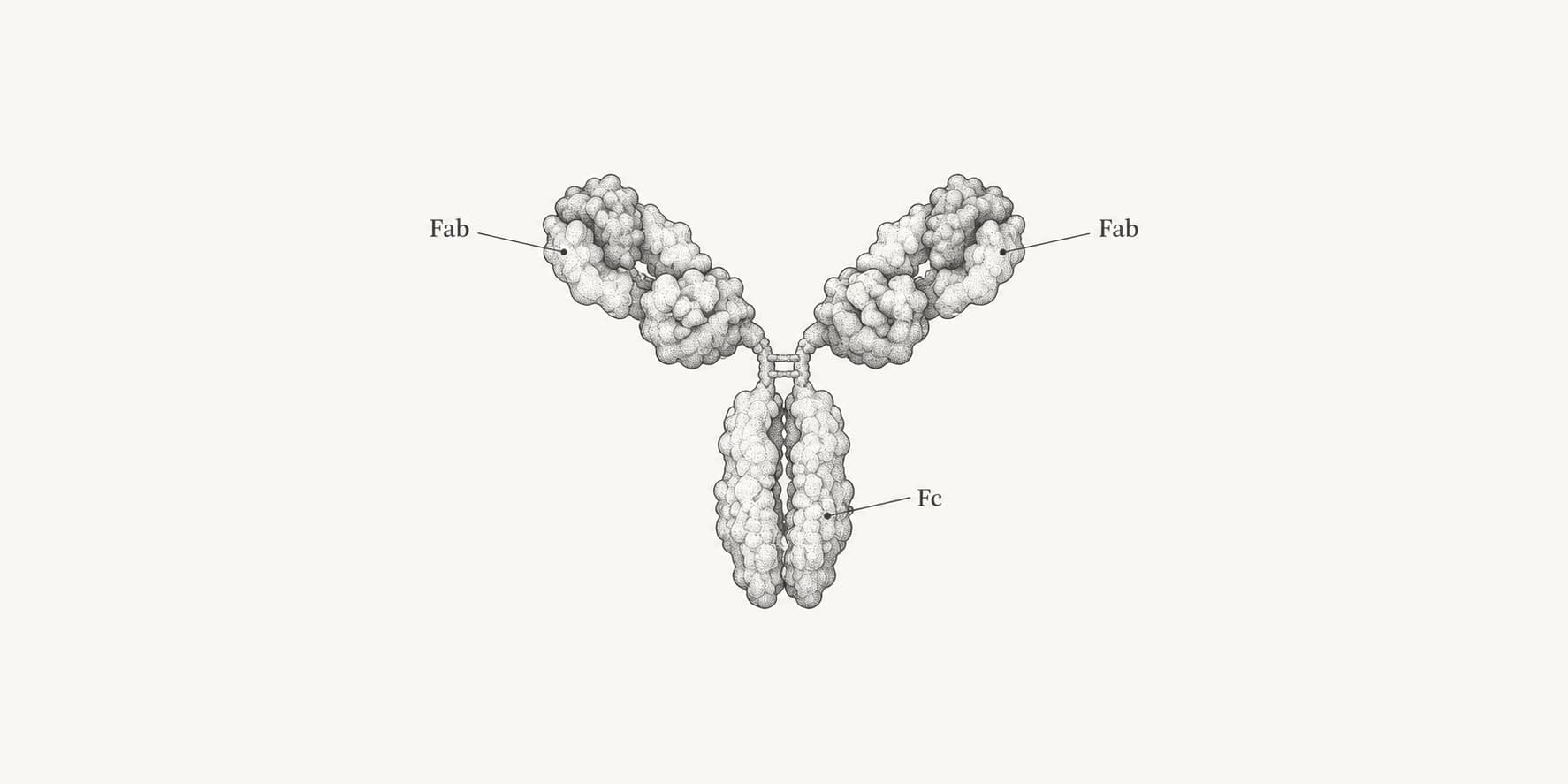
The Fab and Fc regions of IgG each produce fragments with a molecular weight of about 50 kDa after papain cleavage.[3][4]
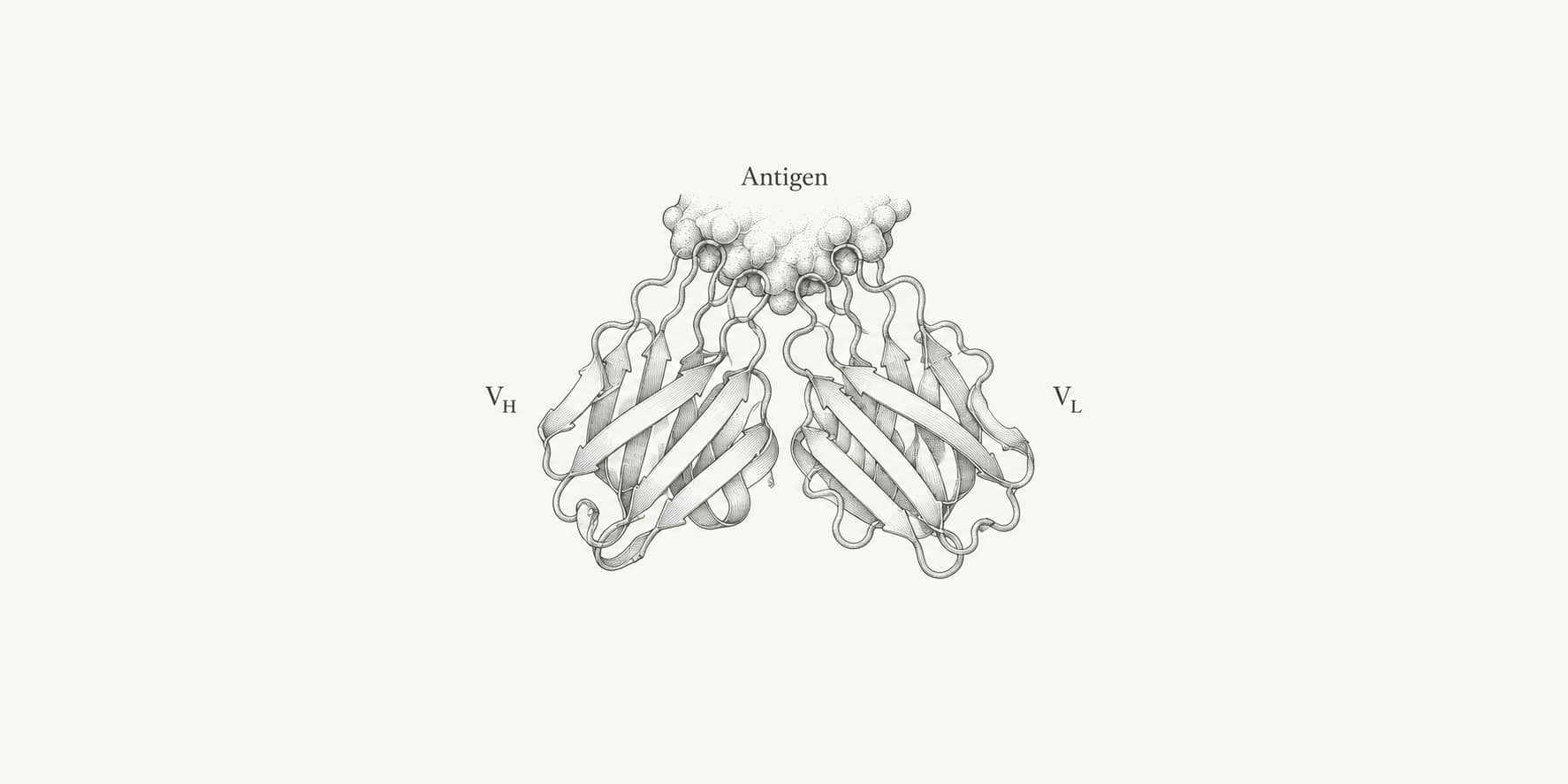
Fab contains one complete light chain plus the variable domain and first constant domain of one heavy chain. It binds antigen but has only one binding site. The Fc fragment contains the paired lower portions of the two heavy chains and binds Fc receptors and other immune-system partners.
An F(ab')2 fragment retains both antigen-binding arms and the hinge that joins them, giving it a molecular weight of about 110 kDa.[3]
These are rounded reference values. A Fab from the NIST monoclonal antibody reference material had a measured mass of 47,628 Da, not exactly 50,000 Da.[6]
Why does an antibody's exact molecular weight vary?
An antibody's exact molecular weight depends on its amino acid sequence, class, subclass, glycosylation, assembly state, and any engineered or chemical additions.
IgG antibodies are glycoproteins. Carbohydrates typically contribute about 1% to 5% of a glycosylated monoclonal antibody's roughly 150 kDa mass, and different attached glycans produce closely related glycoforms with different exact masses.[5]
Variable-region sequences also differ between antibodies. Therapeutic formats may add linkers, extra binding domains, polyethylene glycol, drugs, or other payloads. Aggregation raises the apparent size, while proteolysis lowers it.
This distinction matters in the lab. Under reducing SDS-PAGE conditions, an IgG separates into bands near 50 kDa and 25 kDa because its heavy and light chains are no longer linked. Under nonreducing conditions, intact IgG runs near 150 kDa.
For a sequence-specific estimate, a molecular weight calculator can calculate each heavy or light polypeptide chain and account for disulfide bonds. Glycans and conjugated payloads must then be added from the known composition or measured directly by mass spectrometry.