TL;DR
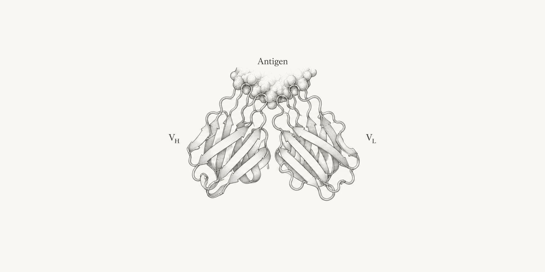
- In a conventional antibody, each antigen-binding site contains one heavy-chain variable domain (VH) paired with one light-chain variable domain (VL).
- VH and VL each contain three complementarity-determining regions (CDRs) separated by four framework regions (FRs).
- The six CDR loops form most of the antigen-binding surface, while the frameworks support the fold and influence VH–VL orientation.
- Heavy-chain variable regions are assembled from V, D, and J gene segments; light-chain variable regions use V and J segments.
What are antibody variable regions?
Antibody variable regions are the amino-terminal domains that form the antigen-binding sites. In a conventional antibody, each site contains one heavy-chain variable domain (VH) paired with one light-chain variable domain (VL). Together, VH and VL are called the fragment variable, or Fv, region.[1]
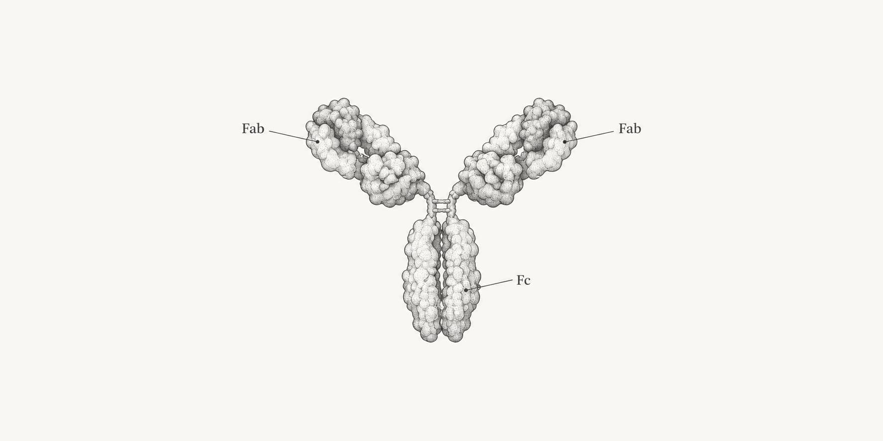
An intact IgG has two identical antigen-binding sites and therefore contains two VH–VL pairs. The variable region is only the tip of each antibody arm; it is not the entire Fab region, which also contains the CH1 and CL constant domains.
What are VH and VL?
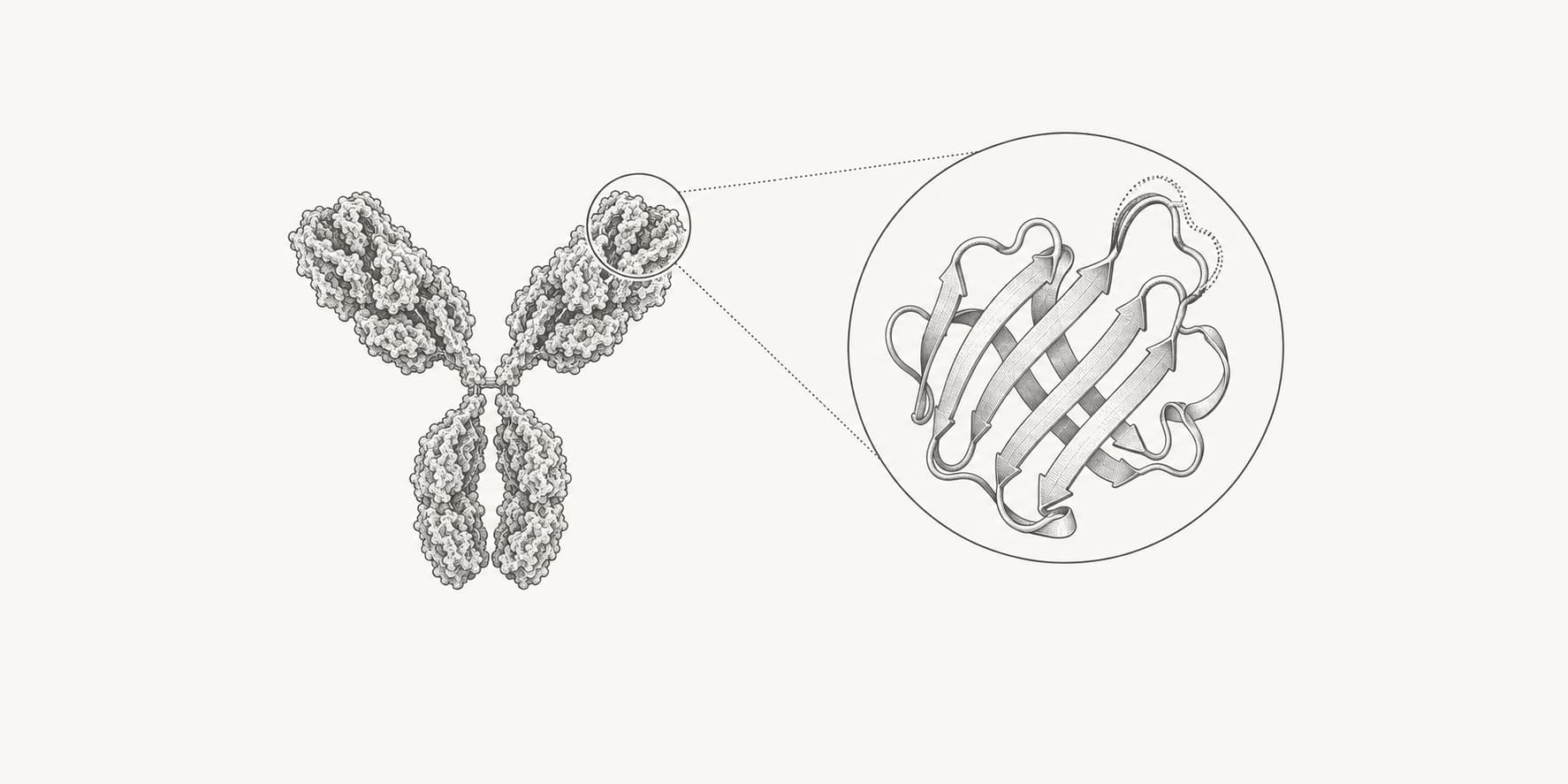
VH is the variable domain of an antibody heavy chain. VL is the variable domain of a light chain, which can be either kappa or lambda in conventional human antibodies. Each domain is approximately 110 amino acids long and adopts an immunoglobulin fold built from two beta sheets linked by a disulfide bond.[1][4]
| Term | Components | What it contributes |
|---|---|---|
| VH | Heavy-chain variable domain | Three heavy-chain CDRs and the heavy-chain side of the VH–VL interface |
| VL | Kappa or lambda light-chain variable domain | Three light-chain CDRs and the light-chain side of the VH–VL interface |
| Fv | One VH paired with one VL | One complete antigen-binding site |
| Fab | Fv plus CH1 and CL | One antigen-binding arm with variable and constant domains |
VH and VL associate noncovalently. Their relative orientation helps determine the shape of the binding surface, so the same CDR sequences cannot always be interpreted independently of their frameworks and chain partner.[5][6]
The VH–VL pair is not universal. Camelid heavy-chain-only antibodies lack a light chain and use one autonomous VHH domain to bind antigen. Engineered single-domain antibodies can likewise function without a paired VL.[9]
What are CDRs and framework regions?
Each VH and VL domain contains three complementarity-determining regions—CDR1, CDR2, and CDR3—separated by four framework regions, FR1 through FR4. A paired Fv therefore contains six CDRs and eight framework regions.[2][4]
The CDRs form exposed loops that create most of the paratope, the antibody surface that contacts an antigen epitope. The frameworks form much of the beta-sheet scaffold that supports those loops. Framework residues can also influence CDR conformation, VH–VL packing, stability, and, in some antibody–antigen complexes, direct antigen contact.[2][5]
CDR-H3 is usually the most diverse of the six CDRs in both sequence and length. It spans the heavy-chain V–D–J junction, making it especially variable and generally more difficult to model than the other CDR loops.[3][6]
| Variable-domain segment | Main structural role | Main source of sequence diversity |
|---|---|---|
| FR1 | Begins the immunoglobulin-fold scaffold | Germline V gene and somatic mutation |
| CDR1 | Antigen-binding loop | Germline V gene and somatic mutation |
| FR2 | Supports the domain and VH–VL interface | Germline V gene and somatic mutation |
| CDR2 | Antigen-binding loop | Germline V gene and somatic mutation |
| FR3 | Supports the fold and positions CDR3 | Germline V gene and somatic mutation |
| CDR3 | Central antigen-binding loop in many antibodies | V–J or V–D–J joining, junctional diversity, and somatic mutation |
| FR4 | Completes the variable-domain scaffold | Primarily the J gene segment |
How do variable and constant regions differ?
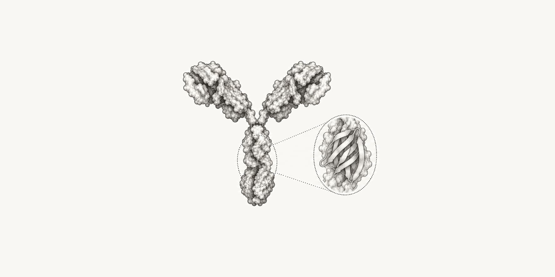
The variable region determines what the antibody recognizes. The constant region determines the antibody class and many of its interactions with immune receptors, complement proteins, and transport systems.[1]
| Feature | Variable region | Constant region |
|---|---|---|
| Position | Amino terminus of each heavy and light chain | Remainder of each chain toward the carboxyl terminus |
| Domains in one IgG arm | VH and VL | CH1 and CL, followed by heavy-chain Fc domains |
| Primary role | Antigen recognition | Structural support and class-dependent effector functions |
| Main diversity mechanisms | V(D)J recombination, junctional diversity, and somatic hypermutation | Different heavy- and light-chain constant genes; heavy-chain class switching |
| Determines antibody class? | No | Yes, through the heavy-chain constant region |
Class-switch recombination can replace the heavy-chain constant region while retaining the assembled variable region. The antibody can therefore preserve its antigen specificity while changing class and effector behavior.[3] See how many types of antibodies there are for the differences among IgM, IgD, IgG, IgA, and IgE.
How are antibody variable regions generated?
Variable-region diversity is created in stages:[3]
- During B-cell development, one V, one D, and one J gene segment are joined to encode VH. A light-chain variable region is assembled from one V and one J segment; light chains do not use a D segment.
- Imprecise joining removes or adds nucleotides at the segment boundaries. This junctional diversity has its largest effect on CDR3.
- Different heavy and light chains pair, creating further combinations of antigen-binding surfaces.
- After antigen stimulation, somatic hypermutation introduces additional changes into the assembled variable-region genes. Selection of B cells with improved binding produces affinity maturation.
V(D)J recombination occurs in DNA. It is different from splicing a rearranged variable-region transcript to a constant-region transcript, and it is different from class-switch recombination.
Why do numbering schemes change CDR boundaries?
Antibody variable regions differ in length, especially in their CDR loops. A residue's raw sequence index therefore does not reliably identify the equivalent structural position across antibodies. Numbering schemes provide a shared coordinate system for comparing sequences and defining CDR and framework boundaries.[4][8]
IMGT, Kabat, Chothia, Martin, and AHo do not place every CDR boundary at the same residue. A mutation described only as “in CDR1” is ambiguous unless the numbering scheme and chain are stated. For reproducible work, record the scheme, numbered sequence, chain type, and any insertion codes with the result.
What can an antibody variable-region sequence tell you?
| Question | What sequence analysis can establish | Important limitation |
|---|---|---|
| Is this VH, kappa VL, or lambda VL? | Domain and chain classification | Heavily engineered or incomplete sequences can be difficult to classify |
| Where are the CDRs and frameworks? | Scheme-specific boundaries and residue numbers | Boundaries differ among numbering schemes |
| Which germline genes are closest? | V, D, and J assignments and sequence identity | D-gene calls can be ambiguous; nucleotide input is more informative than protein input |
| Is the rearrangement productive? | Reading frame, stop codons, and junction details from nucleotide sequence | A productive sequence does not prove expression, folding, or antigen binding |
| What structure might VH and VL adopt? | A predicted paired variable-domain structure and confidence estimates | VH–VL orientation and CDR-H3 remain important uncertainty sources |
| Which antigen does it bind? | Not established from sequence annotation alone | Antigen specificity and affinity require binding evidence or a separately validated predictive method |
Heavy- and light-chain pairing should be preserved whenever possible. An unpaired VH or VL sequence omits half of a conventional antibody-binding site and cannot recover the original VH–VL interface or complete paratope.
How are antibody variable regions analyzed?
- Use IgBLAST for germline V, D, and J assignments, junction analysis, productivity, and CDR/framework annotation. Nucleotide input is required for the full rearrangement analysis; protein input provides more limited gene assignment.[7]
- Use ANARCII to classify and number antibody variable domains under IMGT, Kabat, Chothia, Martin, or AHo schemes. It can also renumber antibody structures.[10]
- Use ABodyBuilder3 to predict a paired VH–VL structure from heavy- and light-chain sequences. The output is a variable-domain model, not a complete Fab, Fc, or intact IgG structure.
- Review uncertain loops, chain pairing, germline calls, and sequence quality before using annotations in humanization, mutation design, or antibody structure prediction.
These analyses describe the sequence and predicted structure of the binding region. They do not by themselves establish antigen specificity, affinity, biological activity, or developability.