Antibody vs antigen, epitope vs paratope
Where the clean definitions stop working

Matic Broz, PhD12 min
What is the difference between an antibody and an antigen?
An antibody is an immunoglobulin made by B-lineage cells that binds one target with high specificity. An antigen is the substance the immune system recognizes and responds to, which is the thing an antibody binds.[1][3] The two words name roles in a binding event, not two kinds of molecule, so one protein can be either.
The antibody D1.3 shows it. D1.3 binds hen egg-white lysozyme, and in the crystal structure 1VFB 18 of its residues have an atom within 4 Å of the enzyme (our calculation).[6]
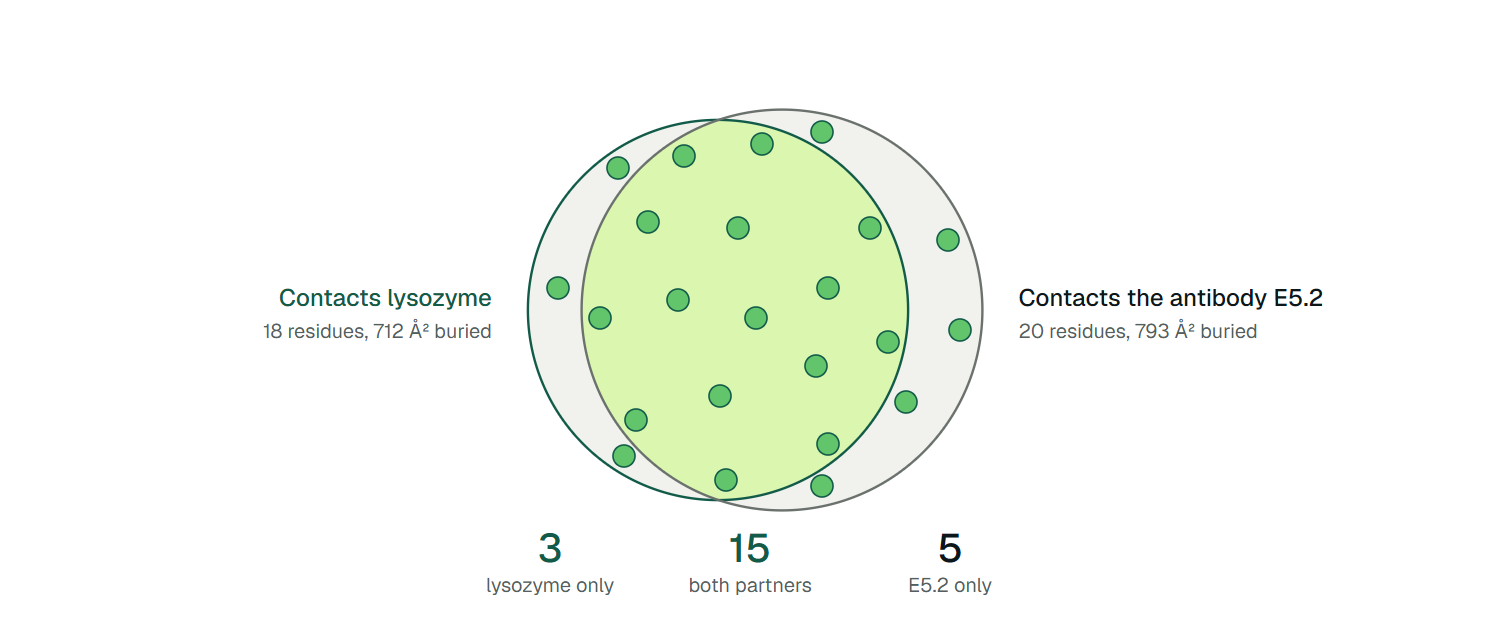
An anti-idiotypic antibody, E5.2, recognizes D1.3 itself. In the structure 1DVF, 20 residues of D1.3 touch E5.2, and 15 of them are among the 18 that touch lysozyme. The authors of that structure summarized it in one phrase: E5.2 "contacts substantially the same residues of D1.3 as lysozyme."[5]
Three pairs of terms get tangled around this. Antibody and antigen name the two sides of a binding event. Immunoglobulin and antibody name a protein family and one of its functions. Epitope and paratope name the two faces of a single contact. What decides between the words each time is the partner: you cannot say which one applies without saying what binds what.

An antibody can also be an antigen
The D1.3 structures let us count how completely one antibody plays both roles. Figure 1 compares D1.3 bound to lysozyme (1VFB, 1.8 Å) with D1.3 bound to E5.2 (1DVF, 1.9 Å), using the same distance rule and the same residue numbering in both files. Most of the surface that D1.3 uses to grip lysozyme is the surface that E5.2 grips.
Three D1.3 residues touch lysozyme only (light chain 91 and 93, heavy chain 102) and five touch E5.2 only (heavy chain 33, 56, 57, 58 and 98). The mimicry is real and partial: Braden and colleagues found that six of the 14 hydrogen bonds between D1.3 and E5.2 are structurally equivalent to hydrogen bonds between D1.3 and lysozyme, and that the match does not extend to the shape of the nonpolar surfaces.[5] An anti-idiotype copies the polar contacts better than the topography.
The same role reversal happens in patients. A therapeutic antibody is a protein, and the immune system can make antibodies against it. A meta-analysis of 46 studies of the six biologics approved for asthma found anti-drug antibodies in 2.91% of patients over follow-up (95% CI 1.60 to 4.55), ranging from 0.00% for omalizumab to 8.35% for benralizumab.[9]
Early work showing that rodent antibodies are immunogenic in humans is one reason humanized and fully human antibodies exist.[4] For a drug antibody, "antigen" therefore applies twice: to the target it was made for and to the drug itself.
How immunoglobulin and antibody differ
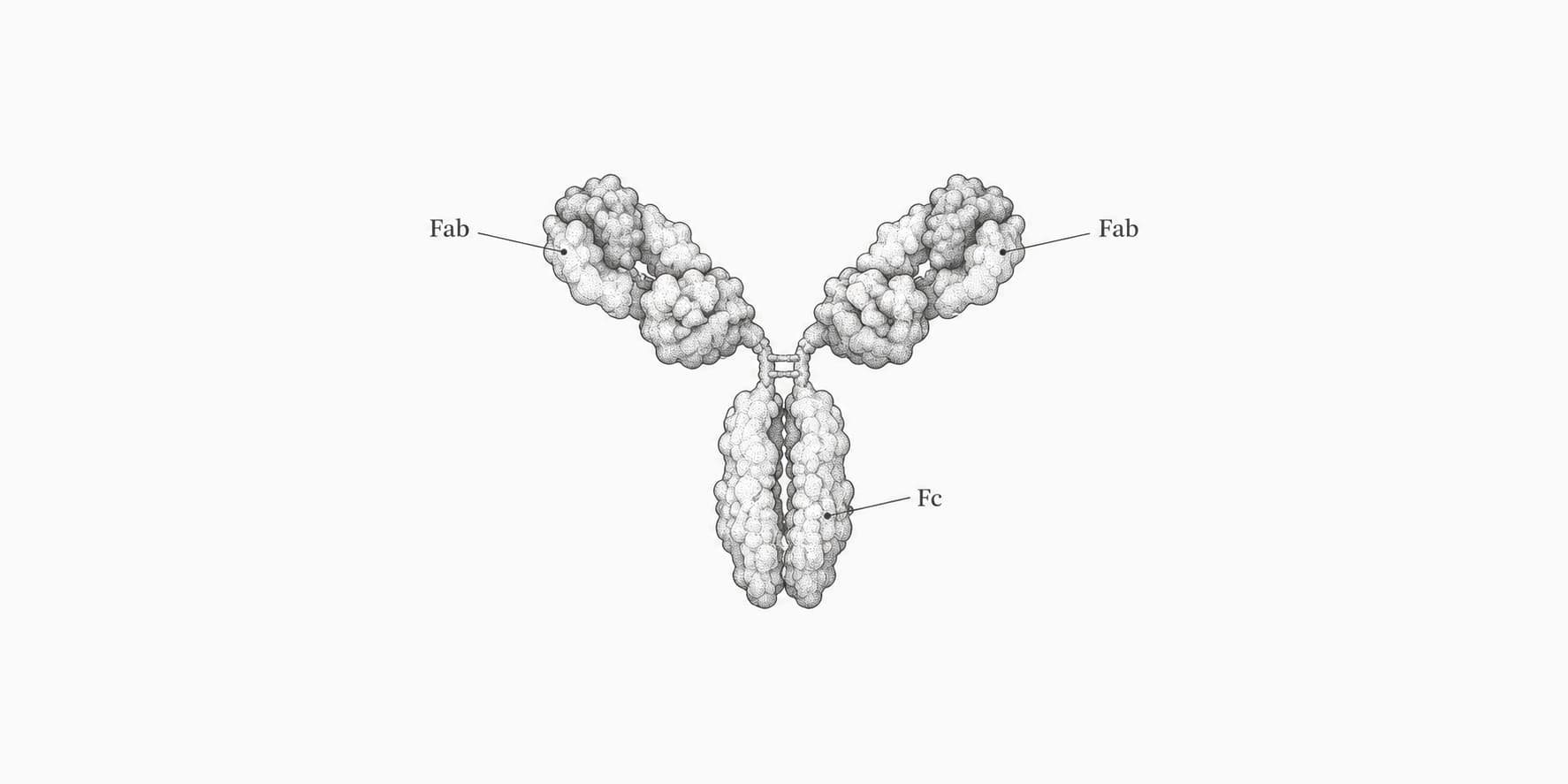
Immunoglobulin names the protein family and antibody names a function. The National Library of Medicine's MeSH vocabulary defines immunoglobulins as multi-subunit proteins made by B lymphocytes from immunoglobulin genes, built from two heavy and two light chains, and occurring as transmembrane forms (B-cell antigen receptors) or secreted forms (antibodies).[2]
It defines an antibody as an immunoglobulin with a specific sequence by virtue of which it interacts only with the antigen that induced its synthesis, or one of very similar shape.[1] The word therefore carries a claim about the target that immunoglobulin does not.
For the secreted proteins in serum the two words swap freely, and most papers use them that way. The difference shows in three places.
The receptor on a B cell is an immunoglobulin but is not a secreted antibody. Class names such as IgG, IgA and IgM label immunoglobulins by their heavy chain, which is why the five classes are classes of immunoglobulin.[11] And the family is wider than the two-heavy, two-light picture: camelids make IgG-like molecules built from heavy-chain dimers with no light chains, which still have an extensive antigen-binding repertoire.[10]
On MeSH's definition, an immunoglobulin sequenced from a patient's B cells, with no identified target, is an immunoglobulin until someone shows what it binds. The habit that follows is to write immunoglobulin for the family, class, gene or receptor, and antibody when you mean a molecule with a known target.
How an epitope and a paratope differ
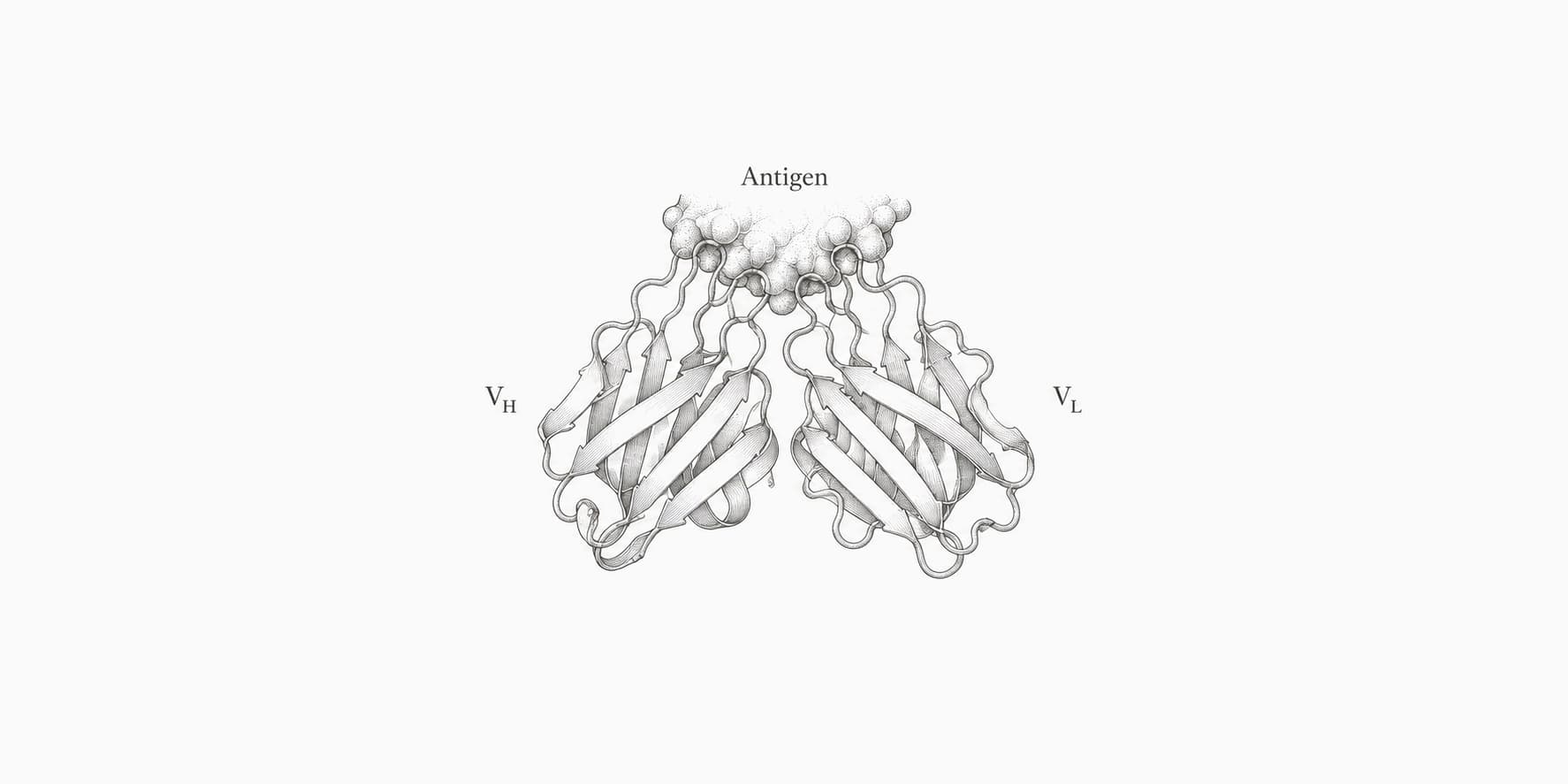
The epitope is the binding site on the antigen, and the paratope is the binding site on the antibody.[4] Each is a list of residues that touch the other, so each is defined by the pair. An antigen is the whole molecule; an epitope is one antibody's footprint on it.
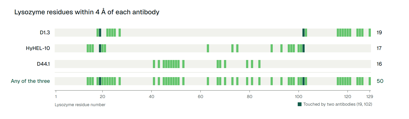
Lysozyme makes the point. It is 129 residues long, and dozens of antibodies are known to bind it.[4] Table 1 shows three of them, with the epitope each one leaves on the same protein.
| Antibody | Structure (resolution) | Epitope residues on lysozyme | Paratope residues on antibody | Epitope residues shared with another antibody |
|---|---|---|---|---|
| D1.3 | 1VFB (1.8 Å) | 19 | 18 | 2, both with HyHEL-10 |
| HyHEL-10 | 3HFM (3.0 Å) | 17 | 20 | 2, both with D1.3 |
| D44.1 | 1MLC (2.5 Å) | 16 | 19 | 0 |
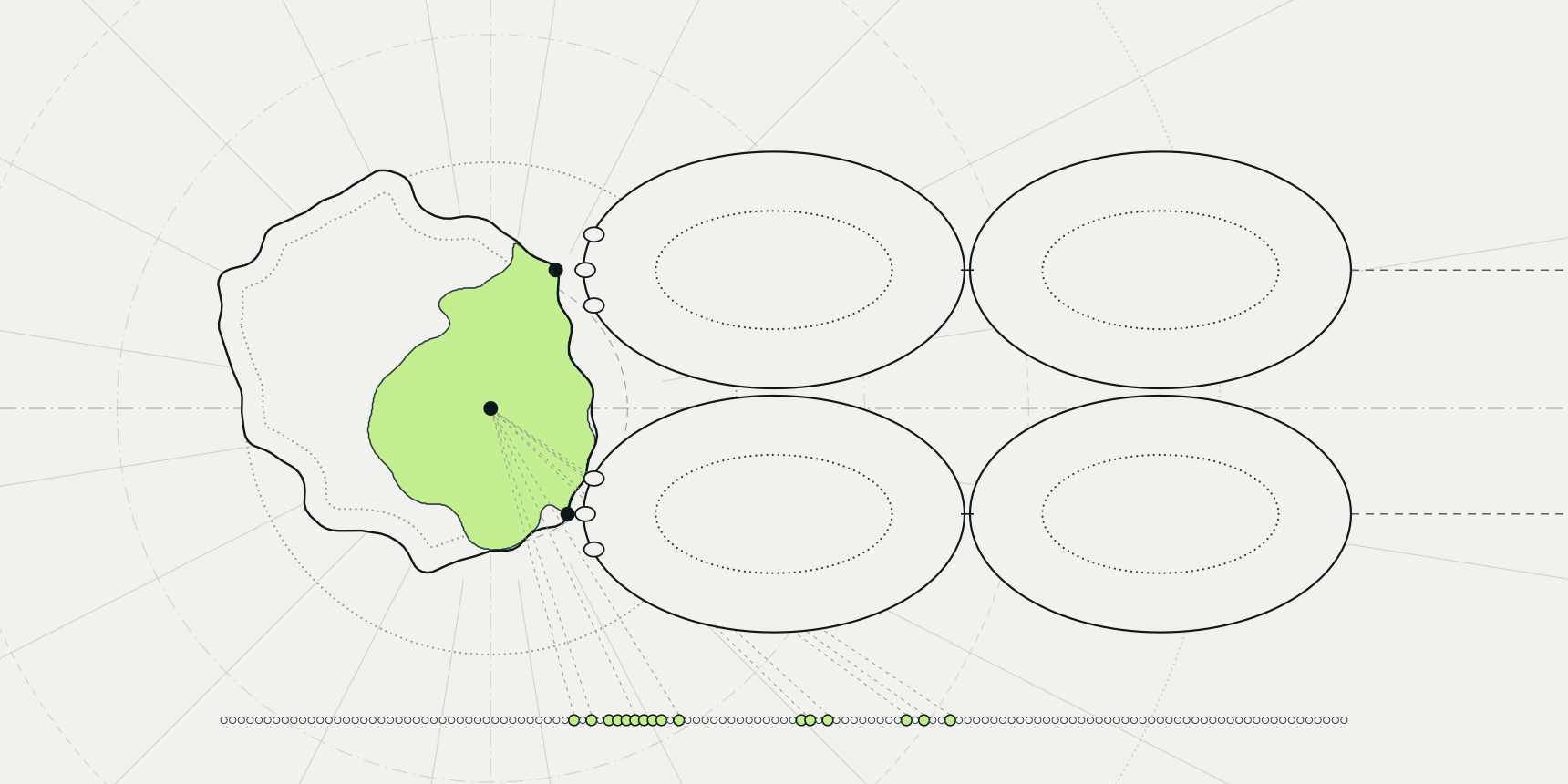
Together the three antibodies touch 50 distinct residues, 39% of the protein, and the footprints of D1.3 and HyHEL-10 overlap at only two residues. Figure 2 lays the footprints along the sequence: D44.1 shares no residue with either of the others.
Sela-Culang and colleagues add that the amino acid composition of epitopes is essentially indistinguishable from other surface residues, and that most of the lysozyme surface belongs to the epitope of at least one antibody.[4]
So "the epitope of lysozyme" is not a meaningful phrase; "the epitope of D44.1 on lysozyme" is. This is also why B-cell epitope prediction has no single right answer for an antigen. A residue is a true positive for one antibody and a false positive for the next.
Epitope and paratope size depends on the definition
Ask how large the D1.3 interface is and the answer changes with the rule. We applied five definitions to D1.3 bound to lysozyme (1VFB).[6] Residues with any atom within 4 Šof the partner number 19 on lysozyme and 18 on D1.3; a 5 Šcutoff gives 23 and 20. Residues that lose at least 1 Ų of accessible surface on binding also number 23 and 20, and a 10 Ų threshold gives 16 and 17.
The structural definitions agree within a factor of 1.5, from 16 to 23 residues. The fifth definition does not. The paratope is often equated with the six complementarity-determining regions (CDRs), but D1.3's six Kabat CDRs, located in the sequence without the antigen, hold 56 residues. Only 16 of them touch lysozyme (29%), and two contacting residues sit outside the CDRs altogether, light chain 49 and heavy chain 30.
That fits the general picture Sela-Culang and colleagues describe, in which an early analysis found that only 20 to 33% of CDR residues take part in binding, and CDR definitions miss about 20% of the residues that do.[4]
Contact counts also say nothing about energy. The same review describes the hotspot hypothesis, in which a few interface residues supply most of the binding energy. A paratope drawn from a contact list is a map of where atoms touch, not a list of what pays for the binding.
Constant domains can also change both affinity and epitope: the HyHEL-10 Fv binds lysozyme with a dissociation constant an order of magnitude lower than its Fab, and two anti-HIV antibodies with identical variable regions, one IgG1 and one IgA2, recognize only partly overlapping epitopes.[4]
Every structural number in the figures and tables above is ours. We took coordinates from the RCSB Protein Data Bank, removed waters and other heteroatoms, and used Biopython 1.85 for neighbor searches and for Shrake-Rupley surface areas (1.4 Å probe, 960 points); buried area is a chain set's accessible surface alone minus its surface in the complex. The script reproduces every count and area.
Which word should you use?
Each word is right for one job and leaves something unsaid that the sentence has to supply. These are writing habits drawn from the sections above, not results from the cited work.
- For the molecule an antibody binds, say antigen. The word does not say whether the molecule induces a response, because MeSH folds recognition and induction into one definition.[3] Name the antibody or the clone.
- For a binding protein with a known target, say antibody. It does not say that the protein binds only that target, so state the target and test cross-reactivity.
- For the protein family, a class, a gene or a B-cell receptor, say immunoglobulin. It says nothing about what the protein recognizes, so name the class (IgG, IgA, IgM, IgD or IgE).
- For the antigen surface an antibody touches, say the epitope of that antibody. It tells the reader nothing about the antigen alone, so give the antibody, the structure ID and the cutoff.
- For the antibody surface that touches the antigen, say paratope. It does not equal the CDRs (56 residues against 18 in D1.3), so report contact residues, not the CDR list.
- For the size of a binding site, give a count with its definition. A bare 19 or 23 does not show that they describe the same interface, so say the distance or area rule.
A quick test catches most mistakes: replace the word with "the thing that binds" or "the thing that is bound" and see whether the sentence still names the pair. If the next sentence needs the partner to make sense, the partner belongs in the first one.
Antibody and epitope tools on ProteinIQ
Start with a structure, either deposited or predicted from sequence, then number the variable domains and mark the binding residues on each side. When no complex has been solved, dock the antibody to the antigen, then measure the interface.
| Task | Tool |
|---|---|
| Predict an antibody Fv structure from heavy and light chain sequences | ABodyBuilder3 |
| Number variable domains and mark CDR boundaries in Kabat, Chothia, IMGT, Martin or AHo schemes | ANARCII |
| Assign germline V, D and J genes | IgBLAST |
| Predict paratope residues from an antibody structure alone | ParaSurf |
| Predict antibody-binding residues on an antigen structure | ScanNet |
| Dock an antibody to an antigen, with optional residue restraints | LightDock |
| Calculate per-residue accessible surface, alone and in a complex, to get buried area | SASA calculator |
| Review interaction diagrams and summaries for a deposited complex | PDBsum |
Predicted paratopes and epitopes are rankings of residues, not measured contacts, so compare them with a structure or a mutagenesis result when one exists. To reproduce the buried-area method behind Figure 1 in the SASA calculator, split the complex into its two partners, run each partner alone and inside the complex, and subtract.
 Measure the buried surface of an interfacePer-residue accessible surface for each partner alone and in the complex.Open tool
Frequently asked questions
What is the main difference between an antibody and an antigen?
An antibody is the immunoglobulin that binds, and an antigen is the substance it binds.[1][3] The words describe roles, so an antibody can itself be an antigen: the antibody D1.3 binds lysozyme, and the anti-idiotype antibody E5.2 binds D1.3 using substantially the same D1.3 residues.[5]
Is an immunoglobulin the same as an antibody?
For secreted proteins in serum the terms are used interchangeably. MeSH defines immunoglobulins as the protein family, with secreted forms called antibodies and transmembrane forms called B-cell antigen receptors, and it defines an antibody by its specific interaction with the antigen that induced it.[1][2]
What is the difference between an epitope and a paratope?
The epitope is the part of the antigen that the antibody contacts, and the paratope is the part of the antibody that contacts the antigen.[4] In the D1.3 and lysozyme complex we counted 19 epitope residues and 18 paratope residues within 4 Å, and 16 of the 56 residues in D1.3's six Kabat CDRs are among the paratope.
What is the difference between an epitope and an antigen?
An antigen is the whole molecule an antibody recognizes, and an epitope is the footprint of one antibody on it. Lysozyme is one antigen with many epitopes: three crystal structures give three different footprints of 16 to 19 residues, which together cover 50 of its 129 residues, and only two residues are shared between any two of them.[6][7][8]
How many residues are in an epitope?
There is no fixed number, because it depends on the antibody and on the definition. For three antibodies on lysozyme we counted 16 to 19 residues within 4 Å, and the D1.3 epitope ranged from 16 to 23 residues across four distance and area rules. Report the cutoff with the count, since a bare number cannot be compared with anyone else's.