What is the difference between IgG, IgM and IgA?
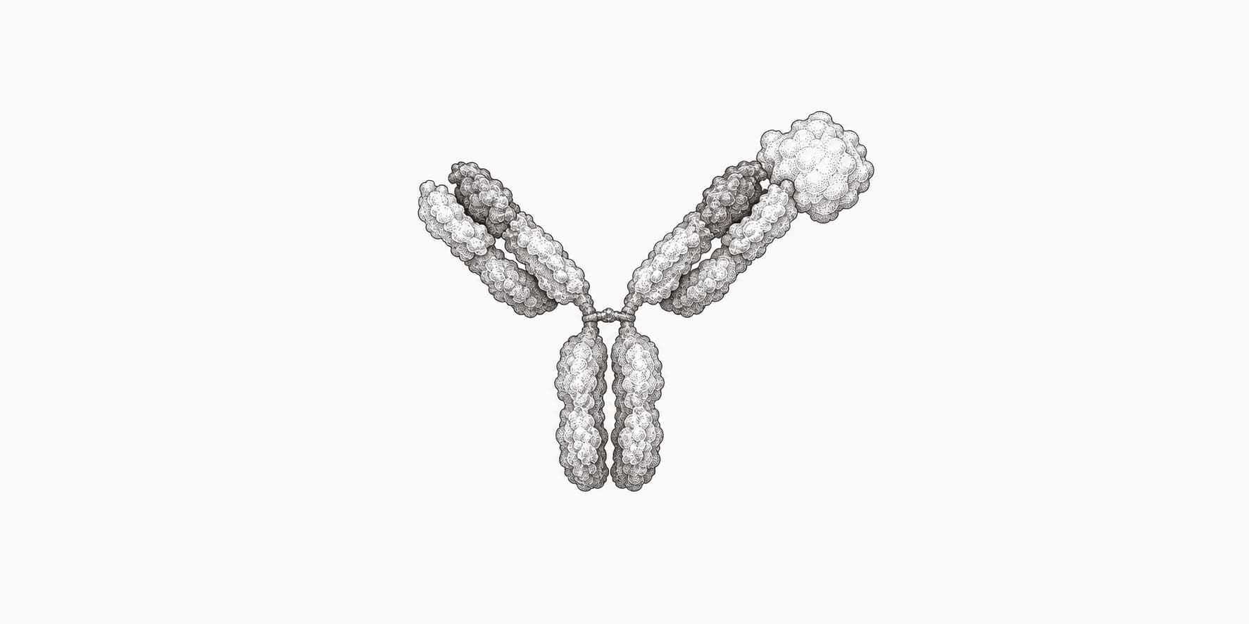
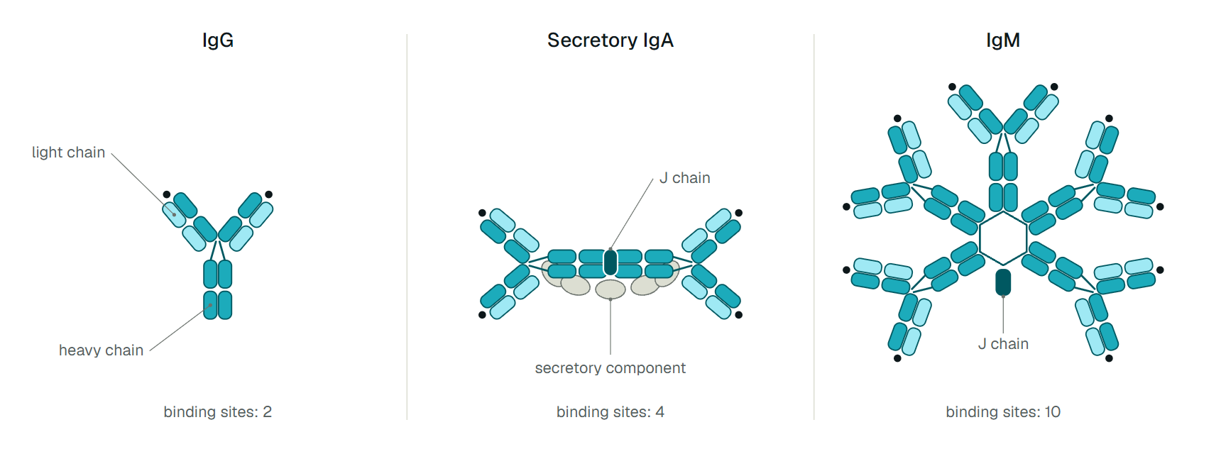
IgG, IgM and IgA share one Y-shaped antibody unit and differ in the heavy-chain constant region. That region decides how many units are joined together, where the antibody works and how long it lasts. IgG is a single Y that stays in blood for about three weeks. IgM is five Y units around a J chain, made early and cleared within days. IgA is a monomer in blood and a dimer in secretions.[1][2][5]
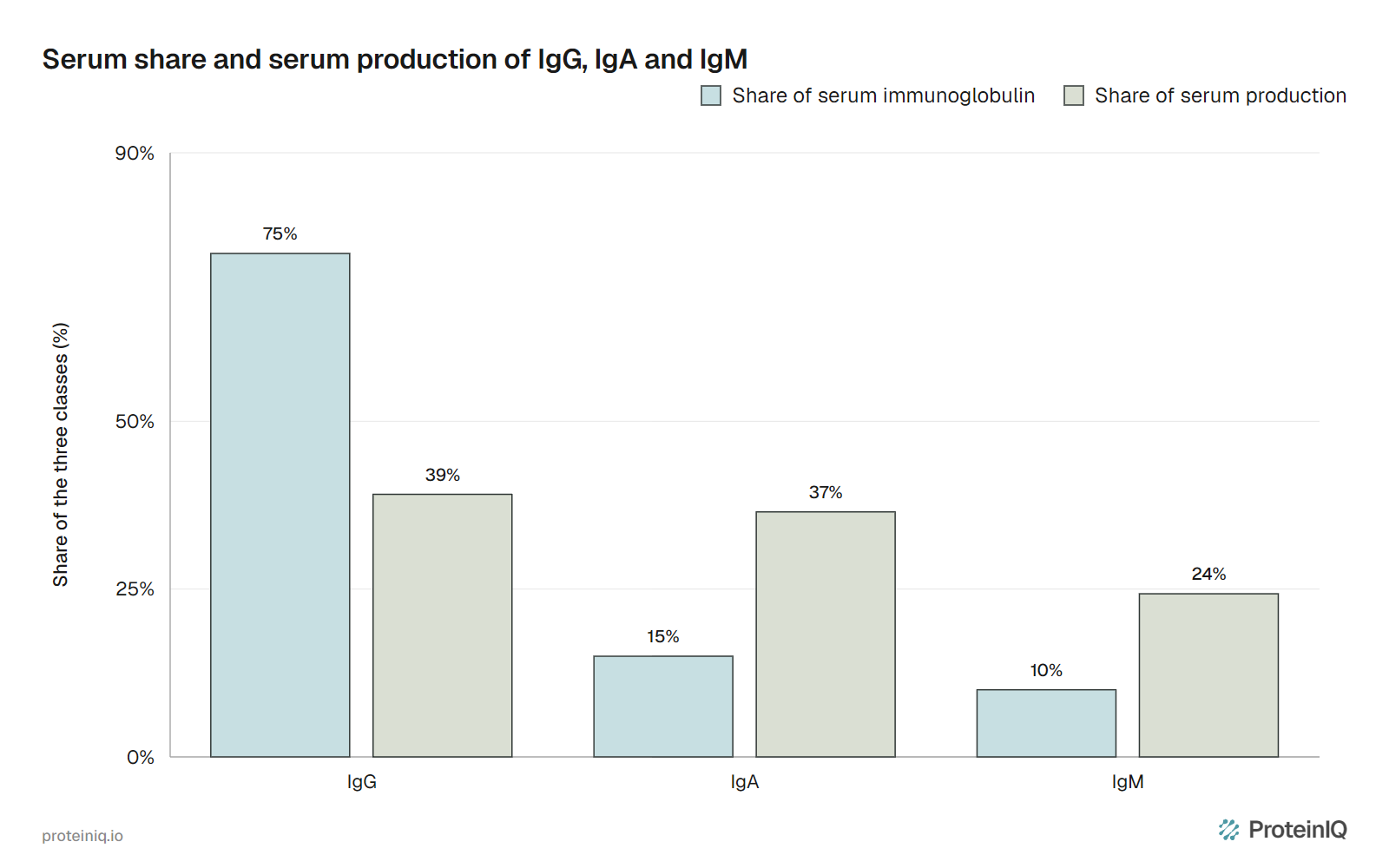
A blood test makes IgG look like the whole story. It is about 75% of serum immunoglobulin, against 15% for IgA and 10% for IgM.[2] But a serum level is a stock, not a rate. Dividing each share by the class's half-life (21 days for IgG, 4 to 5 days for IgA and IgM) gives its relative serum production at steady state, and IgA lands within three points of IgG (Figure 1).[1][5]
Kerr reached the same result from turnover: serum IgA is about a fifth of the IgG concentration but is broken down about five times faster.[4] And blood is only part of IgA's output. Conley and Delacroix estimated that about a third of daily IgA goes into the blood, and that more IgA is made each day than IgG and IgM combined.[3]

How IgG, IgM and IgA are built
Every class uses the same unit: two heavy chains and two light chains, with a Fab arm at each end of the Y and one Fc stem. What changes is the constant region of the heavy chain, which sets the class and, for IgM and IgA, whether units can be joined into larger molecules.[2]
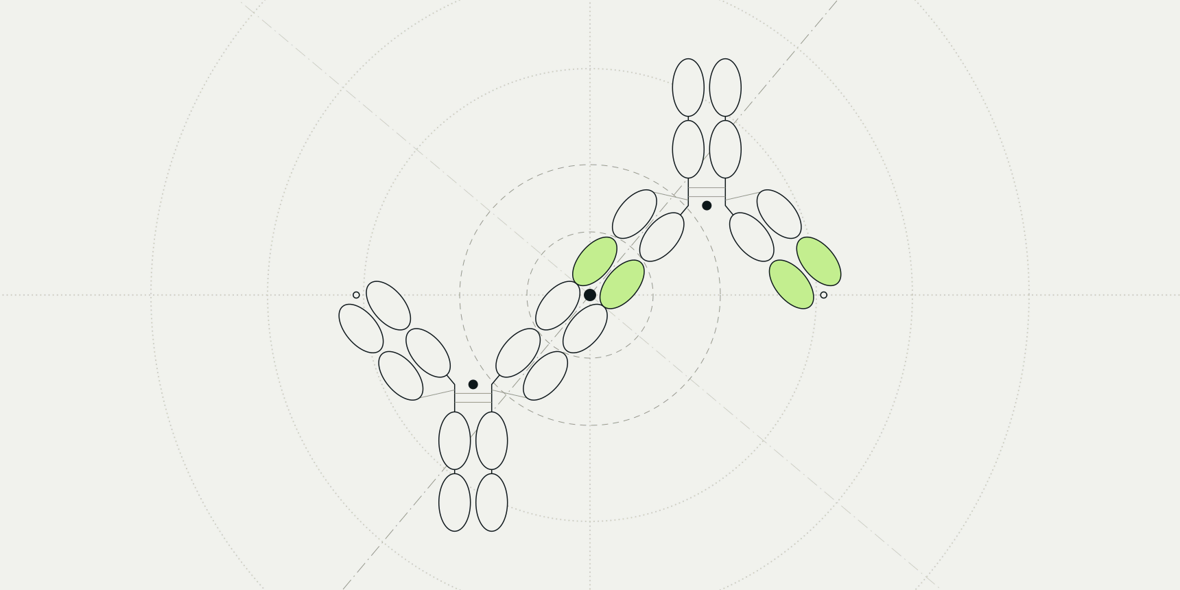
IgM and IgA heavy chains end in an 18-residue tailpiece whose cysteine bonds one unit to the next and to the J chain, a 15 to 16 kDa polypeptide the B cell adds before secretion. IgA usually forms dimers and IgM pentamers.[2] The cryo-EM structure shows the IgM pentamer is not a closed ring but a hexagon with a missing triangle, capped by the J chain.[6] Without a J chain, recombinant IgM mostly forms hexamers.[7]
Counting units gives the number of binding sites: two for IgG, ten for pentameric IgM and four for secretory IgA (Figure 2). The molecular weights scale the same way.
Table 1 sets the three classes side by side. Read the rows as trade-offs: the long-lived class has the fewest sites, and the class with the most sites lives for days.
| Property | IgG | IgM | IgA |
|---|---|---|---|
| Secreted form | Monomer | Pentamer with J chain | Monomer in serum, dimer with J chain and secretory component in secretions |
| Antigen-binding sites per molecule | 2 | 10 | 2 in serum, 4 in secretions |
| Share of serum immunoglobulin | 75% | 10% | 15% |
| Half-life in blood | About 21 days (IgG3 about 7 days in most allotypes) | 4 to 5 days | 4 to 5 days |
| Subclasses | 4 | None | 2 |
| Main role | Secondary response; neutralizes toxins and viruses | Primary response | Mucosal response |
IgG, the class that lasts
IgG makes up 10 to 20% of plasma protein.[1] Its long life comes from FcRn, a receptor that binds IgG in acidic endosomes and releases it at the neutral pH of blood, so IgG taken up by cells is recycled instead of destroyed. Recycling saves about four times as much IgG as is produced.[1]
FcRn also carries IgG across the placenta. The Fc stem binds Fcγ receptors on immune cells and C1q, which start cell killing, engulfment and the classical complement pathway. IgG1 and IgG3 trigger these strongly, IgG2 and IgG4 more subtly.[1]
IgM, ten sites and an early start
IgM is the first immunoglobulin a B cell expresses. Naive B cells carry it as a monomer on their surface and, after antigen stimulation, secrete it mostly as a pentamer.[2] Its half-life of 4 to 5 days keeps it at about 10% of serum immunoglobulin, and that fast turnover is why it is used to date an infection.[5]
Ten sites on one ring suit IgM to surfaces dense with antigen, and to complement. Oskam and colleagues found that recombinant IgM matched IgG1 at complement activation on dense antigen, and that hexameric IgM beat both pentameric IgM and IgG1 when antigen was sparse.[7] These were test surfaces, so they show what the molecule can do, not what serum IgM does in a patient.
IgA, the class made for secretions
Serum IgA and secretory IgA are different molecules made by different cells. Serum IgA is mostly monomeric IgA1 made in the bone marrow, while IgA in secretions is mostly polymeric, made locally and enriched in IgA2.[4] The mucosal surfaces are neither the source nor the destination of serum IgA.[3]
Polymeric IgA with its J chain binds the polymeric immunoglobulin receptor at the base of an epithelial cell, is carried across and is released at the surface with part of the receptor attached as the secretory component. IgA can make up to 50% of the protein in colostrum.[2]
IgA1 has a longer hinge than IgA2, with a duplicated stretch that makes it more sensitive to bacterial proteases. More than 90% of serum IgA is IgA1, while IgA2 predominates in several secretions such as the genital tract.[2]
How IgG1 and IgG4 differ
IgG1 and IgG4 are more than 90% identical in amino acid sequence, and the differences cluster in the hinge and the upper CH2 domain.[1] They are also the two ends of the IgG spectrum. IgG1 makes up about 60% of serum IgG and IgG4 about 4%, and IgG1 engages Fcγ receptors and complement far better (Table 2).[1]
| Property | IgG1 | IgG2 | IgG3 | IgG4 |
|---|---|---|---|---|
| Hinge residues | 15 | 12 | 62 | 12 |
| Inter-heavy chain disulfide bonds | 2 | 4 | 11 | 2 |
| Mean serum level (g/L) | 6.98 | 3.8 | 0.51 | 0.56 |
| Share of serum IgG | 60% | 32% | 4% | 4% |
| Half-life (days) | 21 | 21 | 7 | 21 |
| Effector triggering | Strong | Subtle | Strong | Subtle |
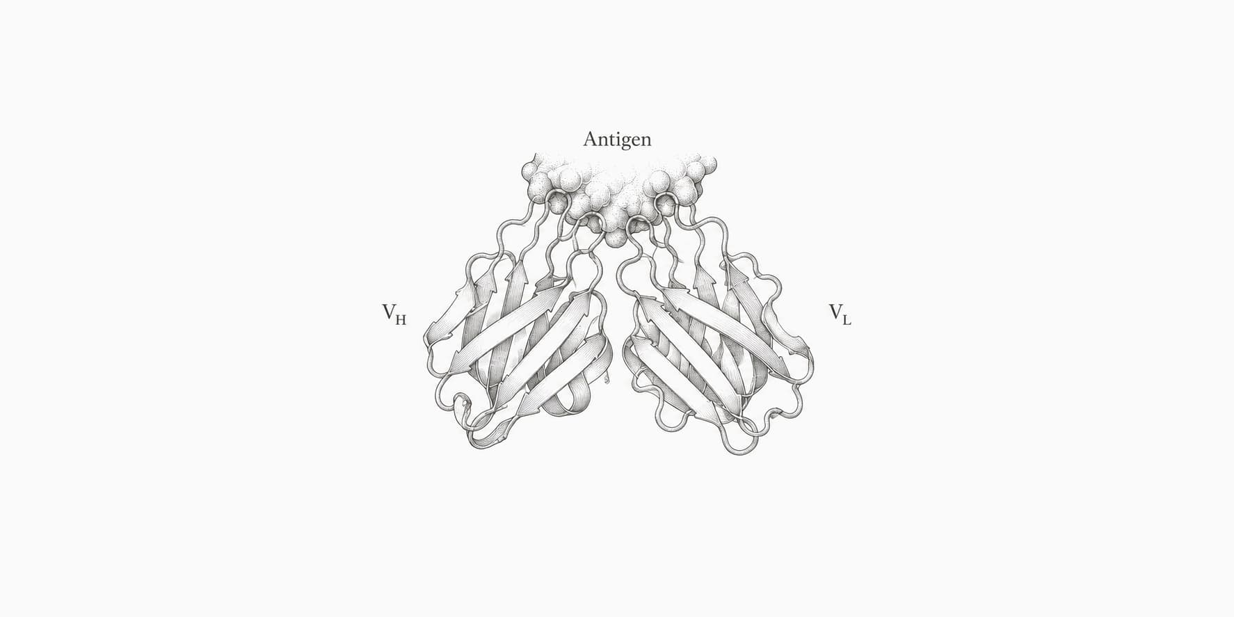
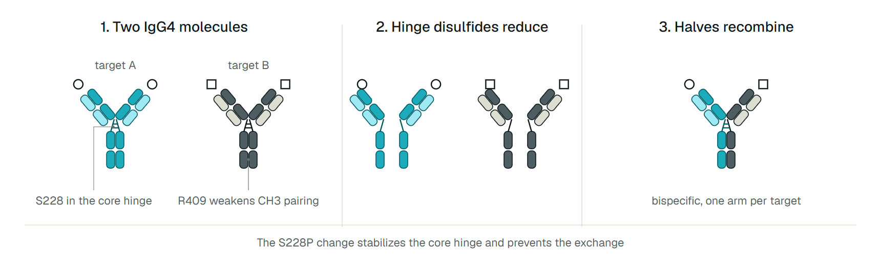
The surprising difference is not in the table. Serine 228 in the IgG4 core hinge lets its two hinge disulfides form within a chain instead of between chains, and arginine 409, lysine in IgG1, weakens CH3 pairing. IgG4 can therefore split into half-molecules that recombine at random with halves from other IgG4 clones (Figure 3), and most human IgG4 ends up as a hybrid with one arm for each of two targets.[1]
A hybrid cannot crosslink antigen. The Vidarsson review proposes that this, with weak binding to activating Fcγ receptors, suits IgG4 to repeated exposure to harmless antigens such as bee venom, where it acts as a blocking antibody by competing with IgE for allergen.[1]
For drugs the same property is a problem. A therapeutic IgG4 dosed into cynomolgus monkeys swapped arms with the animals' own IgG4, leaving drug molecules monovalent for the target, and the S228P substitution largely prevented it.[12] Silva and colleagues found that S228P brought exchange to undetectable levels in vitro and in vivo, depending on redox conditions.[11]
Heavy chains and light chains
The heavy chain decides the class: γ, α, μ, δ and ε heavy chains make IgG, IgA, IgM, IgD and IgE. In IgM it extends to a fourth constant domain, CH4, where the units bond to one another.[2] Heavy chains are about 50 kDa and light chains about 25 kDa, as the antibody molecular weight comparison shows chain by chain.
The light chain does not set the class. Kappa and lambda chains pair with heavy chains of any class, so a clone keeps its light chain and variable regions while its descendants switch from IgM to IgG or IgA. Humans use about 60% kappa and 40% lambda, yet most approved antibody drugs carry kappa.[13]
The light chain also tells you less. A variable region can be called heavy, kappa or lambda from sequence, but no variable-domain tool can tell you whether the full antibody was IgG, IgM or IgA.
Reading IgG, IgM and IgA in a blood test
Textbooks draw IgM first and IgG later, and SARS-CoV-2 shows how loosely that fits. In 285 patients, IgG and IgM seroconversion was simultaneous or sequential.[9] In 343 patients, the median time to seroconversion was nearly 12 days for IgG, IgA and IgM alike.[8]
The classes separated afterwards. Anti-RBD IgM and IgA seroreverted at medians of 49 and 71 days, while IgG decayed slowly and only three people lost it within 90 days.[8] IgM can also outlast its window: 7 of 21 chikungunya patients with chronic arthralgia still had IgM beyond 720 days.[10] It marks an early response on average and is a poor clock for any one patient.
| Pattern | Usual reading | What it does not show |
|---|---|---|
| IgM positive, IgG negative | Early or primary response | That the infection is recent: IgM was still detectable beyond 720 days in 7 of 21 chikungunya patients with chronic arthralgia |
| IgM and IgG positive | Response under way or recent | Which came first: in 285 COVID-19 patients, IgG and IgM seroconversion was simultaneous or sequential |
| IgM negative, IgG positive | Earlier infection or vaccination | How long ago: anti-RBD IgG decayed slowly, while IgM seroreverted at a median of 49 days |
| Serum IgA positive | An IgA response, with sensitivity close to IgG in COVID-19 (90% against 95% between days 15 and 28) | Mucosal immunity: serum IgA is mostly monomeric IgA1 from bone marrow, not secretory IgA |
State the assay, the antigen and the days since onset next to any call about timing.
IgG, IgM and IgA tools on ProteinIQ
Antibody class lives in the constant region, so these tools work on the variable domains and structures around it. Call the chain and number its residues, assign germline genes, then model the structure.
- ANARCI: call a chain as heavy, kappa or lambda and number its variable domain
- IgBLAST: assign V, D and J genes from nucleotide sequences
- ABodyBuilder3: predict an antibody Fv structure from heavy and light chain sequences
- ImmuneBuilder: predict antibody and nanobody structures from sequence
- BioPhi: propose humanizing mutations for a mouse antibody and score how human the sequence looks
- Molecular Weight Calculator: estimate the mass of each chain from its sequence
 ANARCIClassify each variable domain and number its residues in IMGT, Kabat, Chothia or other schemes.Open tool
Frequently asked questions
What is the main difference between IgG and IgM?
IgG is a single unit with two binding sites and a half-life of about 21 days. Secreted IgM is a pentamer with ten sites and a half-life of 4 to 5 days, and it leads the primary response while IgG carries the secondary one.[1][2][5]
Which is more abundant, IgG or IgA?
IgG in blood, at about 75% of serum immunoglobulin against 15% for IgA.[2] IgA overall, because more IgA is made each day than IgG and IgM combined, most of it for secretions.[3]
What is the difference between IgG1 and IgG4?
IgG1 is about 60% of serum IgG and triggers Fcγ receptors and complement strongly; IgG4 is about 4% and triggers them weakly.[1] IgG4 also swaps Fab arms into hybrid molecules, which the S228P substitution prevents.[11]
Does IgM always appear before IgG?
No. In COVID-19, IgG and IgM seroconversion was simultaneous or sequential, with a median of nearly 12 days for all three classes.[9][8]