PDB vs mmCIF vs PDBQT structure file formats
Three coordinate formats, and what quietly breaks between them

Matic Broz, PhD14 min
What is the difference between PDB, mmCIF and PDBQT?
PDB, PDBx/mmCIF (.cif) and PDBQT all store 3D atomic coordinates in ångströms, yet each serves a different job. PDB is the fixed-column archive format frozen in 2012, mmCIF is the Protein Data Bank's table format, and PDBQT is AutoDock's docking input format with partial charges, atom types and rotatable bonds.[3][10] The PDB viewer opens all three in the browser.
Many pipelines treat .pdb as universal and assume mmCIF matters only for giant ribosomes. In our query of the RCSB PDB Search API on November 19, 2026, 30.7% of structures released in 2026 (5,013 of 16,347) have no .pdb file, and only 506 pass 99,999 atoms or 62 chains.[8] Most are small X-ray complexes like WRN helicase (10AJ, 3,424 atoms) whose five-character ligand (A1C4J) cannot fit in three PDB columns.[15]
Even when a structure fits every format, converting between them silently drops data. PDB loses dual numbering and bond orders, PDBQT removes nonpolar hydrogens and all bond records, and small-molecule formats such as MOL2 and SDF keep ligand chemistry while leaving out polymer chains and experimental metadata.
 PDB to CIF ConverterConvert a legacy PDB file or fetched archive entry to mmCIF in the browser.Open tool
How the PDB file format works and where it breaks
The PDB file format defines every field by character column inside an 80-character line, a punch-card layout frozen at version 3.30 on November 21, 2012.[1][3] Because columns never move, anyone can read coordinates in a text editor, as opening a PDB file walks through column by column.
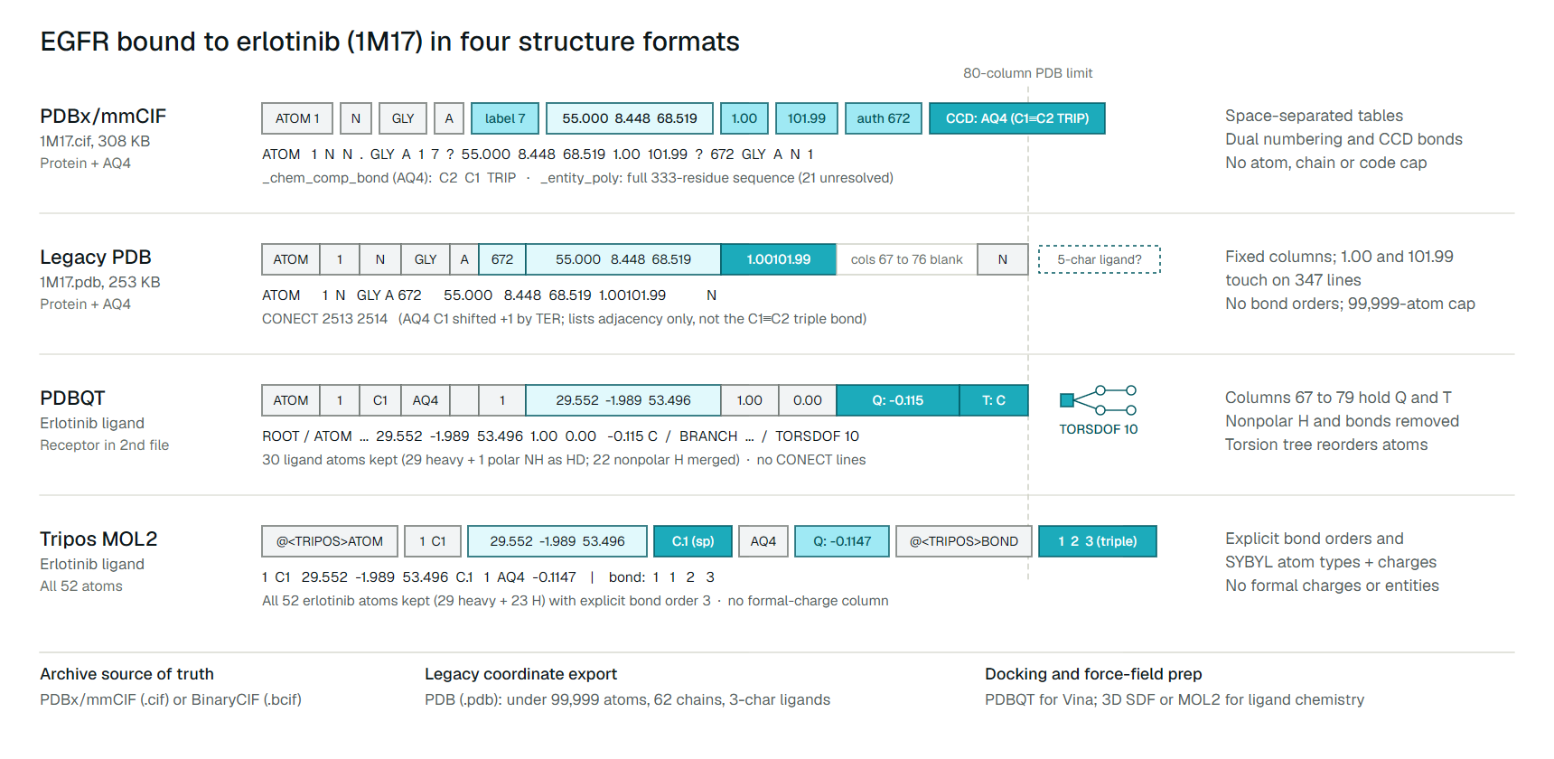
That fixed grid breaks quietly on ordinary proteins. Look at atom 1 of the EGFR kinase domain bound to erlotinib, PDB entry 1M17:[14]
ATOM 1 N GLY A 672 55.000 8.448 68.519 1.00101.99 NColumns 55 to 60 hold occupancy (1.00) and columns 61 to 66 hold the B-factor (101.99). Because 101.99 fills all six characters, the two fields touch as 1.00101.99 with no space between them. That happens on 347 lines of 1M17.pdb, so any script that splits PDB lines on whitespace breaks on the very first atom.
Those fixed widths also impose hard ceilings:[1][5]
- Atom serials span columns 7 to 11, capping a model at 99,999 atoms.
- Chain IDs occupy column 22 alone, capping a file at 62 single-character names (A to Z, a to z, 0 to 9).
- Residue and ligand codes span columns 18 to 20, capping names at three characters.
- Residue numbers span columns 23 to 26 (
-999to9,999), coordinates span eight characters (-999.999to9999.999Å), and B-factors stop at999.99.
Bond chemistry is missing too. CONECT lines list only bonded atom serials, with no column for single, double, triple or aromatic order.[2] In 1M17.pdb, erlotinib's terminal alkyne (C1 to C2, 1.202 Å apart) reads CONECT 2513 2514, identical to a single bond.
Metadata fares little better. Resolution, missing loops (REMARK 465) and biological assembly transforms live in free-text REMARK blocks, and structure predictors reuse the B-factor column for pLDDT confidence instead of atomic mobility.
Why mmCIF replaced PDB in the archive
PDBx/mmCIF replaces fixed columns with self-describing STAR tables: data_ blocks, category.item names and whitespace-separated loop_ rows.[4] Every column's meaning sits above the table, so fields never collide and no counter has a width cap. Its full syntax and how it differs from small-molecule crystal files are covered in small-molecule CIF and mmCIF.
Here is atom 1 of EGFR in 1M17.cif:[14]
ATOM 1 N N . GLY A 1 7 ? 55.000 8.448 68.519 1.00 101.99 ? 672 GLY A N 1Occupancy (1.00) and B-factor (101.99) sit cleanly apart, and the residue carries two numbers: label_seq_id is 7 along the 333-residue crystallized construct (residues 1 to 6 are disordered), while auth_seq_id is 672 in full-length human EGFR.
Categories above atom_site hold the complete sequence (_entity_poly), biological assembly matrices (_pdbx_struct_assembly) and Chemical Component Dictionary links (_chem_comp). For 1M17, that dictionary defines erlotinib (AQ4) with C1 to C2 explicitly marked TRIP (triple bond).
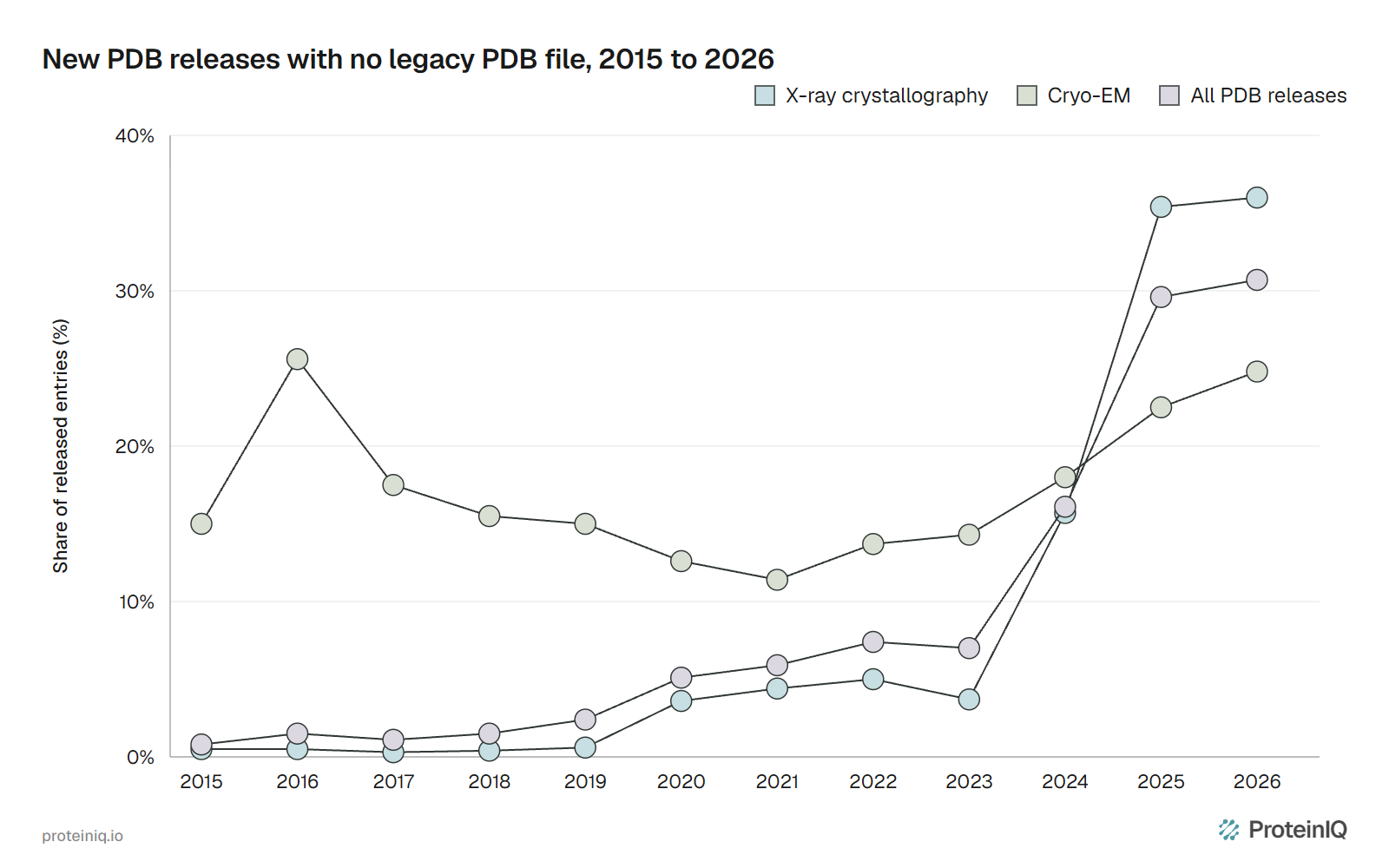
The wwPDB made PDBx/mmCIF the archive standard in 2014 and mandatory for crystallographic depositions in 2019.[3] Across the whole Protein Data Bank archive, 17,318 of 263,567 experimental entries (6.6%) lacked a .pdb file on November 19, 2026.[8] That overall figure hides how fast new releases shifted once five-character ligand codes began in December 2023 (Figure 1).[6]
From 2015 through 2023, cryo-EM led the mmCIF-only share (11.4% to 25.6%) while X-ray stayed at 0.3% to 5.0%. After three-character ligand codes ran out in late 2023, X-ray passed cryo-EM, climbing to 15.7% in 2024, 35.4% in 2025 and 36.0% in 2026 (3,244 of 9,003 X-ray entries).[6][8] Only 10 of those 3,244 X-ray entries exceed 99,999 atoms or 62 chains.[5][8]
Forcing one of those entries into .pdb breaks its identifiers. When we converted 10AJ.cif (WRN helicase with ligand A1C4J) to PDB with gemmi, A1C4J silently truncated to A1C, severing its dictionary link, and chain A fused with residue 1002 into A1002.[15][16] On July 21, 2027, extended 12-character IDs such as pdb_1000xxxx end legacy .pdb files for all new depositions.[7]
Co-folding predictors such as Boltz-2, Protenix and OpenFold3 also write mmCIF natively. For huge assemblies, RCSB serves BinaryCIF (.bcif), a binary encoding of the same data model.[9] On the 237,685-atom human 80S ribosome (4V6X), 4V6X.bcif.gz is 3.02 MB versus 5.69 MB for 4V6X.cif.gz; on small 1M17, gzipped sizes are close (55.8 KB BinaryCIF, 59.7 KB PDB, 79.9 KB mmCIF).
What PDBQT changes for molecular docking
A PDBQT file keeps PDB columns 1 to 66, then overwrites columns 67 to 76 with a partial charge (Q) and columns 78 to 79 with an AutoDock atom type (T), such as A for aromatic carbon, OA for acceptor oxygen and HD for donor hydrogen.[10] The AutoDock Vina FAQ notes plainly that PDBQT is not a standard structure format.[12]
Three alterations turn PDBQT into a one-way docking input rather than an archive format:[10][11]
- Nonpolar hydrogens are merged into their parent carbons, leaving only polar hydrogens (
HDon N, O or S). For erlotinib (C22H23N3O4), 22 of 23 hydrogens vanish, leaving 29 heavy atoms plus 1 aniline NH hydrogen. - Bond records disappear completely. AutoDock neither reads nor writes
CONECTlines, and receptor and ligand must sit in separate files (plus aBEGIN_RES/END_RESfile for flexible side chains). - Ligand atoms are reordered into a
ROOT/ENDROOTand nestedBRANCH/ENDBRANCHtorsion tree, ending withTORSDOFfor rotatable bonds.
Those fields affect docking differently than they appear. Vina ignores user-supplied partial charges in its default scoring function (AutoDock4 uses them), yet missing polar hydrogens or altered torsion trees shift scores materially.[12] Because PDB has no bond orders, Vina's docs warn against preparing ligands from .pdb: start from a protonated 3D SDF or MOL2, and keep in mind that a docking score is not a binding affinity.[11]
Where MOL2 and chemical formats fit
Tripos MOL2 sits between coordinate-only PDB files and cheminformatics connection tables. Built for SYBYL, it divides each molecule into tagged ASCII blocks (@<TRIPOS>MOLECULE, @<TRIPOS>ATOM, @<TRIPOS>BOND).[13] Each atom stores 3D coordinates, a SYBYL atom type (C.1 alkyne, C.ar aromatic, N.am amide, O.co2 carboxylate) and a partial charge in column 9, with the charge method named in the header.[13]
Unlike PDB and PDBQT, MOL2 writes explicit bond types (1, 2, 3, ar, am), so erlotinib's alkyne survives as C.1 atoms joined by bond 3.[13] That suits force-field setup (AMBER Antechamber) and docking suites such as DOCK 6 and GOLD, and PDB to MOL2 writes SYBYL types from coordinate ligands.
The catch is that standard MOL2 has no formal-charge column and no polymer entity metadata.[13] Parsers infer formal charges from atom types such as N.4 or O.co2 and bond valences, and toolkits often disagree on protonated heterocycles. SDF is safer for storing 3D small molecules because it records explicit bond orders and formal charges; GNINA and DiffDock read SDF directly without PDBQT conversion.
Two neighbouring format families handle tasks outside 3D coordinates. Line notations such as SMILES (including canonical SMILES and SELFIES) and InChI or InChIKey drop coordinates for tables and database lookup, as compared across chemical file formats. On the visualization side, .pse and .pml files (PyMOL file types) save viewer scenes and scripts rather than portable coordinates.
One complex across four structure formats
Entry 1M17 is the 2.6 Å crystal structure of the human EGFR kinase domain bound to erlotinib (AQ4, C22H23N3O4, 29 heavy atoms, 23 hydrogens and 10 rotatable bonds).[14] Figure 2 lines up protein atom 1 (Gly 672 N) and erlotinib's terminal alkyne (C1 to C2) across mmCIF, PDB, PDBQT and MOL2.
Even atom serial numbers diverge on the same complex: 1M17.pdb gives serial 2512 to the chain A TER line, so erlotinib's C1 is atom 2513 in PDB and _atom_site.id 2512 in mmCIF. Moving down the figure, each downstream export preserves coordinates while trading away a different layer of chemistry or metadata. Table 1 puts those trade-offs side by side.
| Property | PDB (.pdb) | PDBx/mmCIF (.cif) | PDBQT (.pdbqt) | Tripos MOL2 (.mol2) |
|---|---|---|---|---|
| Size and ID limits | 99,999 atoms, 62 chains, 3-char ligands, 4-char PDB IDs | No atom, chain, ligand-code or ID length limit | Inherits PDB column caps; separate receptor and ligand files | No fixed atom limit; designed for small molecules |
| Field layout | Fixed 80-character columns; values can touch | Whitespace-separated named loop_ tables | Fixed PDB columns 1 to 66, plus Q (67 to 76) and T (78 to 79) | Tagged @<TRIPOS> sections, whitespace-separated |
| Bond orders | None; CONECT lists bonded pairs only | Explicit for ligands via CCD (_chem_comp_bond) | None; CONECT ignored and omitted | Explicit (1, 2, 3, ar, am) |
| Hydrogens and charges | Usually omitted in X-ray; 2-char formal charge column | Explicit when modelled; _atom_site.pdbx_formal_charge | Polar H (HD) only; per-atom partial charge Q | Usually all H; partial charge column, no formal-charge column |
| Macromolecular metadata | Free-text REMARK; author numbering only | Structured entities, assemblies, label_ and auth_ numbering | None; ROOT/BRANCH/TORSDOF torsion tree instead | Substructure IDs only; no entity or assembly tables |
| Typical software | Legacy MD, older scripts, all viewers | RCSB PDB, Mol*, PyMOL, ChimeraX, Boltz-2, gemmi | AutoDock4, AutoDock Vina, Meeko | Antechamber/AMBER, DOCK 6, GOLD, Open Babel |
Which structure format should you use?
Match the file to the stage of your pipeline rather than defaulting to .pdb. Table 2 lists the right starting format for each task, its blind spot and the check that catches problems before a run.
| Task | Start with | What it cannot carry | Check first |
|---|---|---|---|
| Download an archive structure, view in 3D or save a prediction | PDBx/mmCIF (or BinaryCIF for large assemblies) | Nothing from the archive; older scripts may lack an mmCIF parser | Whether your tool reads auth_seq_id or label_seq_id |
Feed a legacy MD setup or script that insists on .pdb | PDB exported from mmCIF | Entity tables, label_ numbering, >99,999 atoms, >62 chains, 5-char ligands | No truncated ligand codes, missing loops or glued 1.00101.99 columns |
| Dock a small molecule in AutoDock Vina | PDBQT (receptor from repaired mmCIF/PDB; ligand from 3D SDF) | Bond orders and nonpolar hydrogens | Polar hydrogens (HD), protonation state and TORSDOF count |
| Set up small-molecule force-field parameters or SYBYL docking | MOL2 alongside a 3D SDF | Explicit formal charges and polymer entity metadata | Inferred formal charges on amines, carboxylates and heterocycles |
| Archive docked poses or a full complex for reuse | mmCIF for the protein, 3D SDF for the ligand | Vina's torsion tree (regenerate from SDF when re-docking) | Bond orders and stereochemistry survived export from PDBQT |
Keep two source-of-truth files for every complex: the archive mmCIF for the macromolecule and a protonated 3D SDF for the ligand. Treat .pdb, .pdbqt and .mol2 as disposable exports generated right before the program that demands them, repairing missing loops with PDBFixer and receptor hydrogens with PDB2PQR before docking.
Structure file tools on ProteinIQ
Fetch and inspect the archive structure first, repair missing atoms or protonation second, and convert to docking or cheminformatics formats last.
- PDB viewer: inspect PDB, mmCIF, PDBQT, MOL2 and SDF files together in the browser
- PDB download: fetch archive mmCIF or legacy PDB files by accession
- PDB to CIF: convert legacy PDB files to mmCIF locally without uploading
- PDBFixer: rebuild missing residues and atoms before simulation or molecular docking
- SDF to PDBQT and PDBQT to SDF: prepare ligands for AutoDock Vina and export docked poses back to SDF
- Open Babel: convert between mmCIF, PDB, MOL2, SDF and PDBQT when a tool expects another format

Frequently asked questions
What is the difference between PDB and CIF files?
PDB is a frozen fixed-column format capped at 99,999 atoms, 62 chains and three-character ligand codes.[1][3] PDBx/mmCIF (.cif) stores the same 3D coordinates in whitespace-separated named tables with no size caps, dual label_ and auth_ numbering and Chemical Component Dictionary bond definitions.[4]
Why do some PDB entries have no legacy PDB file?
The wwPDB omits legacy .pdb files when an entry passes any fixed-column ceiling: more than 99,999 atoms, more than 62 chains, multi-character chain IDs, B-factors above 999.99 or five-character ligand codes.[5][6] In 2026 through November 19, 30.7% of new releases (5,013 of 16,347) lacked a .pdb file, and from July 21, 2027 all new entries will be mmCIF-only.[7][8]
Can I convert mmCIF to PDB without losing data?
Not without losing metadata. When a structure fits under 99,999 atoms, 62 single-character chains and three-character residue codes, gemmi or Open Babel keeps coordinates and author numbering while dropping entity tables, label_ numbering and assembly matrices.[16] Above those limits, conversion fails or truncates identifiers so they no longer match the archive.
What is the difference between PDB and PDBQT?
PDBQT keeps PDB columns 1 to 66, writes a partial charge (Q) and AutoDock atom type (T) into columns 67 to 79, merges away nonpolar hydrogens, drops CONECT records and reorders ligand atoms into a ROOT/BRANCH/TORSDOF torsion tree.[10] PDB is a general coordinate file; PDBQT is an input and output format built specifically for AutoDock and AutoDock Vina.[12]
When should I use a MOL2 file instead of PDB or SDF?
Use Tripos MOL2 when software expects hybridization-aware SYBYL atom types (such as C.1 or C.ar) and partial charges alongside explicit bond orders, as in AMBER Antechamber, DOCK 6 and GOLD.[13] Because standard MOL2 has no formal-charge column, keep a 3D SDF as your primary small-molecule file and export MOL2 when needed.