N-terminus vs C-terminus: how to tell them apart in a sequence and a structure
Why the first residue you see is often not the real end

Matic Broz, PhD10 min
What are the N-terminus and C-terminus?
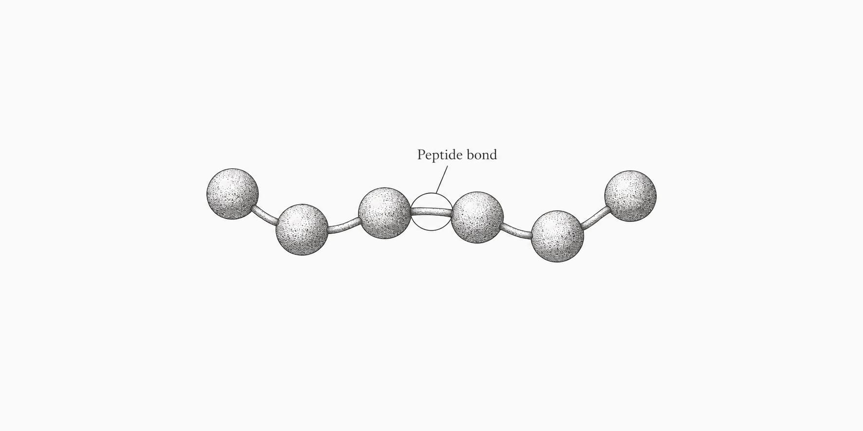
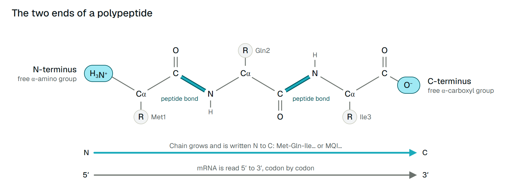
The N-terminus is the end of a protein chain whose first residue still has a free α-amino group; the C-terminus is the end whose last residue still has a free α-carboxyl group. Every other amino and carboxyl group in the backbone is used up in a peptide bond. Sequences are written from N on the left to C on the right, the order in which the ribosome builds the chain.[1][2]
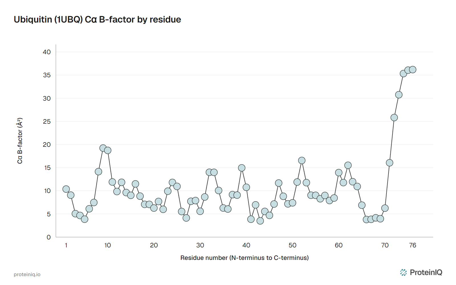
Finding the ends in real data is harder. In 1UBQ, the classic crystal structure of the 76-residue protein ubiquitin, Met1 sits inside the first β-strand, about as well ordered as the rest of the chain. The C-terminal tail sticks out into the solvent, and Vijay-Kumar and colleagues wrote that its last four residues "appear to have partial occupancy or large thermal motion".[3] Yet Gly76 is the residue ubiquitin uses to attach to other proteins. The end that matters most is the one the experiment saw least.
That is common. In a random sample of X-ray structures we drew from the PDB, 58% of protein chains were missing residues at the N-terminus and 45% at the C-terminus. So the ends have to be found three times: in the sequence, in the structure file and in 3D. Which answer is right depends on whether you mean the protein as encoded, as made in the cell or as resolved in an experiment.

How to tell them apart in a sequence
In a one-letter sequence, as in a FASTA file, the first letter is the N-terminal residue and the last is the C-terminal one, and numbering starts at 1 at the N-terminus. Nothing in the file says so; the IUPAC convention does, and every tool assumes it.[1]
The catch is that a database holds the encoded chain, not the mature protein. Hirel and colleagues showed in Escherichia coli that methionine aminopeptidase removes the initiator methionine when the second residue has a short side chain, and less often as that side chain gets longer.[6] Secreted proteins also lose an N-terminal signal peptide, and many hormones are cut from longer precursors. After any of these, residue 1 in the database is not the N-terminus in the cell.
Ubiquitin follows both rules. Gln2 is long, so Met1 stays. But ubiquitin is never translated alone: yeast UBI4 encodes five copies joined head to tail with no spacer, and other genes fuse it to unrelated proteins.[4] Deubiquitinases cut after each Gly76 to release the free C-terminus ubiquitin needs.[5] Every copy after the first gets its N-terminus from that same cut.
From DNA, the 5′ end of the coding strand gives the N-terminus, because mRNA is read 5′ to 3′. A gene on the minus strand read as written gives a wrong protein; DNA to Protein can translate all six frames, which catches that early.
How to tell them apart in a structure file
A PDB or mmCIF file lists each chain's residues from N to C, so a chain's first ATOM record is the first residue modelled, not necessarily the N-terminus. The full sequence is in SEQRES, and residues that were in the sample but not in the model are listed under REMARK 465 (pdbx_unobs_or_zero_occ_residues in mmCIF).[10] Reading a PDB file line by line shows where each record sits.
Two more clues help. The second oxygen of a terminal carboxyl group is named OXT, so a residue that has one is a modelled C-terminus; its absence proves nothing, since some refinement programs leave it out. And numbering can start anywhere: an expression tag often gets zero or negative numbers so the protein keeps its UniProt numbering.
How often are the ends missing? On 16 November 2026 we drew 500 of the 263,196 PDB entries at random and read unobserved residues from the RCSB Data API.[12] Keeping the first chain of each X-ray protein entity of at least 20 residues left 593 entities from 401 entries. The first residue of the deposited sequence was unmodelled in 58%, the last in 45%, and at least one end in 69%, with a median gap of five residues. Our per-chain results are available to download.
Expression tags, which count as part of the deposited sequence, explain some of this. The consequence is the same either way: in most crystal structures, the first residue on screen is not residue 1.
How to see the two ends in 3D
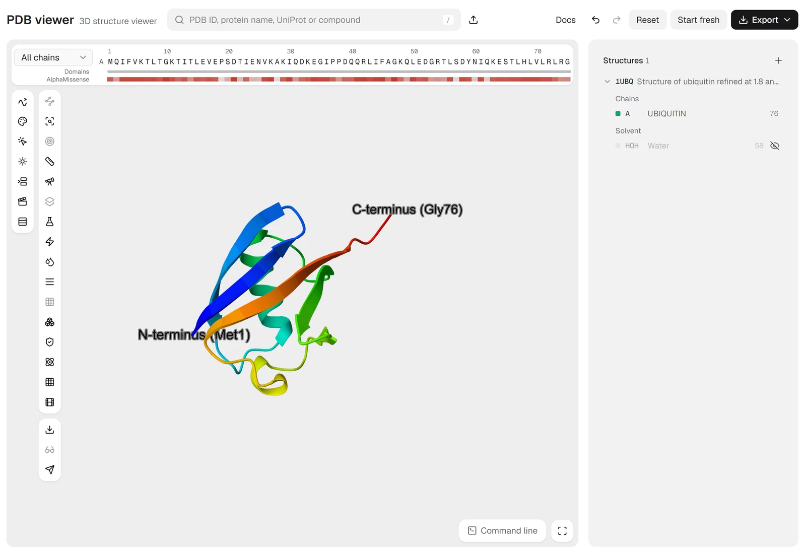
Two conventions give a chain's direction at a glance. In a cartoon, every β-strand is an arrow pointing toward the C-terminus, and rainbow colouring runs from blue at the N-terminus to red at the C-terminus. In the PDB viewer by ProteinIQ, type 1UBQ in the search box, open Command line and run spectrum count, rainbow, polymer. label polymer and resi 76 and name OXT, "C-terminus" puts a label on the last residue, and set seq_view, 1 adds a one-letter strip numbered from the N-terminus.
We measured 37 Å between the Cα atoms of Met1 and Gly76 in 1UBQ, more than the 29 Å across the folded core. Whether a protein's ends are free, buried or close together decides whether a tag or fusion partner can go there.
N-terminal vs C-terminal: why the two ends behave differently
The two ends carry opposite charges. Grimsley and colleagues collected measured pK values in folded proteins: 7.7 on average for the N-terminal amino group (16 values) and 3.3 for the C-terminal carboxyl group (22 values).[7] At pH 7.4 the N-terminus is positive only about two thirds of the time, while the C-terminus is almost always negative. Both count toward a protein's isoelectric point, and cells read different signals at each end, as Table 1 shows.
| Property | N-terminus | C-terminus |
|---|---|---|
| Free group | α-amino, NH₃⁺ | α-carboxyl, COO⁻ |
| Order of synthesis | Made first | Made last |
| Position in a written sequence | Left, residue 1 | Right, last residue |
| Targeting signals | Signal peptides, mitochondrial and chloroplast transit peptides | KDEL retention in the ER, GPI anchor signals |
| Common modifications | Methionine removal, acetylation, myristoylation | Amidation, lipid anchors |
The N-terminus leaves the ribosome first and can be recognized while the chain is still growing, which is why secretion signals and most co-translational modifications sit there. Its first residue can also mark a protein for destruction; Varshavsky notes that all 20 amino acids can do so in some context.[9] The C-terminus is read once the chain is finished. Munro and Pelham found that luminal ER proteins end in Lys-Asp-Glu-Leu, and that deleting it sent the protein out of the cell.[8]
So check which signals live at an end before you put a tag there. A C-terminal tag on an ER protein hides its KDEL, and an N-terminal tag on a secreted protein is cut off with the signal peptide.
Ubiquitin from sequence to structure
Human ubiquitin's sequence is 76 residues, MQIFVKTLTG…LVLRLRGG, and keeps Met1 as its long Gln2 predicts.[6] The 1UBQ file models all 76, with Met1 as its first ATOM record and an OXT atom on Gly76, so file and sequence agree.[11]
The structure adds what neither says. Figure 3 plots the B-factor of each Cα, a measure of how smeared out an atom's position is in the crystal. Met1 sits at 10.4 Ų, on the chain average of 10.6 Ų. From Leu71 the values climb to 36 Ų at Gly76, and occupancy falls to 0.45 for Leu73 and Arg74 and 0.25 for Gly75 and Gly76.
The sequence says the C-terminus is Gly76 and the file says Gly76 was modelled. Only the structure says its position is barely supported by the data. Another crystal form of the same protein, or an AlphaFold model with low pLDDT at the tail, can stop a few residues earlier or point the tail elsewhere. Before measuring anything at a terminus, check its B-factors or pLDDT and the missing-residue list.
N-terminus and C-terminus tools on ProteinIQ
Start from the sequence, then check what the structure file contains:
- DNA to Protein: translate a gene in all six frames and confirm where the N-terminus starts
- Protein Parameters: mass, isoelectric point and composition of a chain from its sequence
- PDB to FASTA: extract each chain's sequence from a structure to compare with the database entry
- PDBFixer: add missing atoms, including a terminal
OXT, and rebuild missing loops before docking or simulation
Frequently asked questions
Which end of a protein is made first?
The N-terminus. The ribosome reads mRNA from 5′ to 3′ and adds each new amino acid to the C-terminal end of the growing chain, as Dintzis showed by timing the labelling of hemoglobin chains in 1961.[2] The C-terminal residue is the last one added.
Is the N-terminus positive and the C-terminus negative?
Usually. The N-terminal amino group has an average measured pK of 7.7 in folded proteins and the C-terminal carboxyl group 3.3, so near pH 7 the C-terminus is almost always negative and the N-terminus is positive most of the time.[7] Modifications such as N-terminal acetylation or C-terminal amidation remove those charges.
Why are protein sequences written from N to C?
It matches the direction of synthesis, and IUPAC made it the standard: the residue with the free amino group is written on the left and numbered 1.[1] Every sequence database and tool assumes this order.
Is the first residue of a protein always methionine?
Only in the encoded sequence. Translation starts with methionine, but methionine aminopeptidase removes it when the next residue is small, and signal peptides and propeptides are cut off later.[6] The N-terminus of the mature protein is often a different residue.
Why does my PDB structure not start at residue 1?
Either the first residues were not resolved, or the numbering does not start at 1. In 593 random protein chains from X-ray structures, we found 58% missing residues at the N-terminus, a median of five. They are listed under REMARK 465.[10]