Post-translational modifications: types, examples and what they do
What cells change in a protein after the ribosome, and how to detect it

Matic Broz, PhD19 min
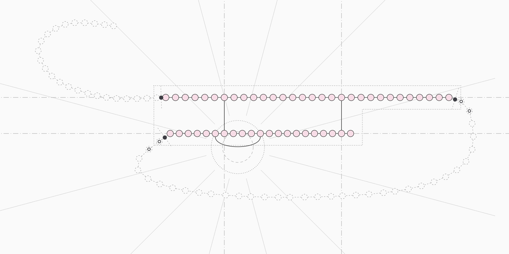
The human insulin gene encodes a chain of 110 amino acids. The hormone that leaves a pancreatic beta cell has 51, in two separate chains held together by three disulfide bonds.[18] Between the ribosome and the blood, the cell cuts off a 24-residue signal peptide, removes a 31-residue connecting peptide from the middle and links six cysteines in pairs. None of those steps changes the gene, and all of them decide whether the hormone works.
Erythropoietin shows the same gap from another side. Made in Chinese hamster ovary cells, the recombinant hormone weighs about 30,400 Da, and 39% of that mass is carbohydrate the cell attached after making the chain.[24] A molecular weight calculated from the sequence alone misses two fifths of the molecule.
Both are post-translational modifications (PTMs): covalent changes to a protein made during or after its synthesis. Some add a chemical group to a side chain or terminus, some cut the backbone, and some link two residues. In the current UniProtKB/Swiss-Prot release, 13,759 of the 20,431 reviewed human entries, or 67%, carry at least one annotated modified residue, glycosylation site, lipid attachment, cross-link or disulfide bond.[1]
Sequence tools such as the glycosylation site finder and the motif scanner mark where some of these can occur; whether they do occur has to be measured.
Co-translational and post-translational modification
The name suggests the protein is finished first, but many modifications start while the chain is still leaving the ribosome. N-terminal acetylation is one. In a proteomic survey of 742 human protein N-termini, 84% were acetylated, compared with 57% in yeast, and the acetyltransferases responsible bind the ribosome and act on the nascent chain.[5]
N-myristoylation is another: methionine aminopeptidase removes the initiator methionine so that a glycine becomes the first residue, then N-myristoyltransferase attaches the fatty acid, both during translation.[17] N-linked glycosylation of secreted and membrane proteins starts this early too, as stretches of the chain pass through the translocon into the endoplasmic reticulum (ER).[6]
In practice "post-translational modification" covers both kinds, and the more useful distinction is where the protein is when it is modified. Proteins bound for secretion or the membrane pass through the ER and then the Golgi, and they collect a different set of modifications from proteins that stay in the cytosol or nucleus. Table 1 sorts the main types by compartment.
| Compartment | Typical modifications | Typical substrates |
|---|---|---|
| Ribosome exit (co-translational) | Initiator methionine removal, N-terminal acetylation, N-myristoylation | Most cytosolic proteins |
| Endoplasmic reticulum | Signal peptide cleavage, N-glycan transfer, disulfide bonds, prolyl hydroxylation, GPI anchors | Secreted and membrane proteins, collagens, antibodies |
| Golgi apparatus | N-glycan trimming and extension, O-GalNAc (mucin-type) glycosylation | Secreted and cell-surface glycoproteins, mucins |
| Cytosol and nucleus | Phosphorylation, lysine acetylation and methylation, ubiquitination, SUMOylation, O-GlcNAc, S-palmitoylation at membranes | Signaling proteins, enzymes, histones, transcription factors |
Types of post-translational modifications
UniProt's controlled vocabulary lists 750 distinct modified residues, glycans, lipids and cross-links as of release 2026_03.[2] A handful of them account for most annotated sites, and each targets particular residues.
| Modification | What is added or changed | Main residues | Reversible in cells |
|---|---|---|---|
| Phosphorylation | Phosphate group | Ser, Thr, Tyr | Yes (phosphatases) |
| N-linked glycosylation | Branched glycan on the side-chain amide | Asn in Asn-X-Ser/Thr, X not Pro | No |
| O-linked glycosylation | GalNAc-initiated glycans, or a single GlcNAc | Ser, Thr | O-GlcNAc yes; mucin-type no |
| Acetylation | Acetyl group | Protein N-terminus, Lys | Lysine yes; N-terminal no |
| Methylation | One to three methyl groups | Lys, Arg | Yes |
| Ubiquitination, SUMOylation | Small protein joined by an isopeptide bond | Lys (ubiquitin also Met1 of another ubiquitin) | Yes (deubiquitinases, SUMO proteases) |
| Lipidation | Myristate, palmitate, prenyl group or GPI anchor | N-terminal Gly, Cys, C-terminus | Palmitoylation yes; others mostly no |
| Proteolytic processing | Peptide bonds cut | Signal peptides, propeptides, dibasic sites | No |
| Disulfide bond | Covalent S-S link between two cysteines | Cys | Yes, by reduction |
| Hydroxylation | Hydroxyl group | Pro, Lys (collagen) | No |
Phosphorylation
Phosphorylation adds a phosphate to the hydroxyl of serine, threonine or tyrosine. Manning and colleagues counted 518 protein kinase genes in the human genome, and phosphatases remove what kinases add.[10] That ready reversibility makes phosphorylation the standard switch of cell signaling.
The phosphate changes the residue more than its size suggests. The second pKa of a phosphate ester is about 5.8, so at pH 7 the group is dianionic, and a neutral serine becomes a residue carrying one full negative charge and most of a second.[11] Hunter points out that the result resembles no natural amino acid, and specific binding domains in other proteins recognize it, which is how a phosphorylation event can switch on a protein interaction.[11]
Serine dominates. Among human Swiss-Prot sites with experimental evidence, 25,239 are phosphoserine, 4,579 phosphothreonine and 1,307 phosphotyrosine, a split of 81%, 15% and 4%.[1] Kinases recognize short sequence patterns around the site, and the motif scanner searches the simplified consensus patterns for PKA, PKC and casein kinase 2.
Glycosylation
N-linked glycosylation attaches a glycan to the side-chain nitrogen of asparagine. The minimal acceptor is the sequon Asn-X-Ser/Thr, where X is any amino acid except proline, but not every sequon is used.[6] The oligosaccharyltransferase moves a 14-sugar precursor, Glc₃Man₉GlcNAc₂, onto the asparagine in one step, and enzymes in the ER and Golgi then trim and rebuild it into the three types found on mature proteins: oligomannose (high-mannose), hybrid and complex glycans.[6][9]
O-linked glycosylation has no such sequon. The common mucin-type form starts in the Golgi, where one of 20 human polypeptide GalNAc-transferases adds N-acetylgalactosamine to a serine or threonine, and further sugars extend it.[7] A different O-linked sugar, a single N-acetylglucosamine (O-GlcNAc), decorates nuclear and cytoplasmic proteins and is added and removed dynamically, closer in behavior to phosphorylation than to the glycans of the secretory pathway.[8]
Sequons are easy to find and hard to confirm. Human Swiss-Prot lists 16,263 N-linked sites, but only 3,234 of them carry experimental evidence. Another 12,737, or 78% of the total, are annotated from sequence analysis alone.[1] The glycosylation site finder lists every candidate sequon in a sequence, which is the same first step.
Acetylation
Acetylation adds an acetyl group, either to the free amine at the protein's N-terminus or to the ε-amine of a lysine. The N-terminal form is co-translational, permanent and very common, as the 84% figure above shows.[5] Lysine acetylation is reversible, and it neutralizes the positive charge of the lysine side chain.
Histones made it famous. In 1964 Allfrey, Faulkner and Mirsky reported that histones are acetylated and methylated after synthesis and proposed that this regulates RNA synthesis, an early link between histone modification and gene activity.[12] Human Swiss-Prot now records 4,381 acetyl-lysine sites, 2,493 of them with experimental evidence.[1]
Methylation
Methylation adds one, two or three methyl groups to lysine, or one or two to arginine. Unlike acetylation, it keeps the positive charge, so it changes what binds a residue without changing the residue's charge. Allfrey's 1964 paper described histone methylation alongside acetylation.[12] Arginine methylation is the more often annotated of the two in human Swiss-Prot, with 1,711 sites against 541 for lysine.[1]
Ubiquitination and SUMOylation
Ubiquitin is a 76-residue protein. Its C-terminal glycine forms an isopeptide bond with the side-chain amine of a lysine on the target, and ubiquitin's own seven lysines and N-terminal methionine let further ubiquitins attach to form chains.[13] Chains linked through lysine 48 are the most common, often more than half of all linkages, and they send proteins to the proteasome for degradation; chains through lysine 63 have non-degradative roles in signaling.[13] This is the main route by which cells set how long proteins last.
SUMO, the small ubiquitin-related modifier, attaches to lysines by the same isopeptide chemistry. Its conjugation sites often sit in the motif ψKxE, a large hydrophobic residue, the lysine, any residue and glutamate, and in Rodriguez's experiments modification in cells also required the protein to reach the nucleus.[14] Hundreds of proteins in chromatin organization, transcription and DNA repair are reversibly SUMOylated.[15]
Lipidation
Lipidation anchors proteins to membranes. N-myristoylation attaches the 14-carbon myristate to an N-terminal glycine through an amide bond, and almost all co-translationally myristoylated proteins begin Met-Gly-X-X-X-Ser/Thr.[17] The bond is about as stable as a peptide bond, so the protein keeps its myristate for life and regulates membrane binding in other ways, such as a myristoyl switch that tucks the chain into the protein or releases it to the membrane.[17]
S-palmitoylation attaches the 16-carbon palmitate to a cysteine through a thioester, which thioesterases can cleave, so proteins such as H-Ras cycle on and off membranes.[17] Prenylation adds a 15-carbon farnesyl or 20-carbon geranylgeranyl group to a cysteine.[3] GPI anchors work from the other side: a glycolipid is attached to the C-terminus of a protein in the ER, and the protein ends up on the outer leaflet of the plasma membrane.[29]
Proteolytic processing
Cutting the backbone is permanent and very common. Most secreted proteins lose an N-terminal signal peptide in the ER, which predictors such as SignalP 6 locate from the sequence. Many hormones and enzymes are made as inactive precursors and activated by later cuts.
Insulin is the textbook example. Steiner and colleagues showed in 1967 that islets first make a larger precursor, which they named proinsulin.[19] In UniProt's annotation of human preproinsulin, the 110-residue chain loses its signal peptide (residues 1 to 24), the dibasic pairs Arg-Arg and Lys-Arg are removed, and the C-peptide (residues 57 to 87) is excised.
What remains is the 30-residue B chain and the 21-residue A chain, which is why insulin has 51 amino acids.[18] The Peptide Cutter maps cleavage sites for 41 proteases and chemical reagents in any sequence.
Disulfide bonds
A disulfide bond forms when the thiols of two cysteines are oxidized to a covalent S-S link, usually in the ER of secreted proteins. Insulin keeps three: two between the A and B chains and one within the A chain.[18] Human Swiss-Prot annotates 18,825 disulfide bonds on 3,801 entries.[1] Because they form, break and rearrange in their own ways, disulfide bonds have a guide of their own.
Hydroxylation in collagen
Collagen strands repeat the triplet Gly-Xaa-Yaa. Proline fills 28% of Xaa positions and 4-hydroxyproline 38% of Yaa positions, and Pro-Hyp-Gly is the most common triplet at 10.5%.[20] Hydroxyproline is not encoded: prolyl 4-hydroxylases in the ER convert proline after it is built into the chain.[21] These enzymes depend on ascorbate to keep their iron reduced, which is the link between vitamin C and collagen synthesis.[22]
The hydroxyl matters for structure. Shoulders and Raines describe how hydroxyproline in the Yaa position stabilizes the collagen triple helix through stereoelectronic effects that preorganize each strand into the right shape before it folds.[20] Collagen lysines are hydroxylated too, and the resulting hydroxylysines can carry glycans of their own.[20]
How common each modification is
Counting annotations in UniProtKB/Swiss-Prot gives a rough ranking. We tallied every modified-residue, glycosylation, lipidation, cross-link and disulfide feature in the 20,431 reviewed human entries of release 2026_03, and separately counted sites whose evidence code marks an experimental source (ECO:0000269 or ECO:0007744) rather than similarity or sequence analysis.[1]
| Modification | All sites | Experimental sites | Proteins (experimental) |
|---|---|---|---|
| Phosphoserine | 33,295 | 25,239 | 6,070 |
| Disulfide bond | 18,825 | 4,779 | 1,255 |
| N-linked glycosylation | 16,263 | 3,234 | 1,196 |
| Phosphothreonine | 6,065 | 4,579 | 2,466 |
| SUMOylation (isopeptide) | 5,923 | 5,668 | 1,439 |
| Lysine acetylation | 4,381 | 2,493 | 1,273 |
| N-terminal acetylation | 2,390 | 2,237 | 2,133 |
| Phosphotyrosine | 2,153 | 1,307 | 706 |
| O-linked glycosylation | 1,889 | 852 | 241 |
| Arginine methylation | 1,711 | 913 | 366 |
| Ubiquitination (isopeptide) | 980 | 722 | 297 |
| Hydroxyproline | 579 | 366 | 31 |
| S-palmitoylation | 576 | 222 | 124 |
| Lysine methylation | 541 | 348 | 144 |
| N-myristoylation | 209 | 133 | 125 |
Phosphorylation leads by a wide margin: 6,569 human entries, 32% of the reviewed proteome, have at least one experimentally supported phosphosite.[1] Glycosylation is the other modification people expect to be everywhere, and the numbers temper that. Only 6.6% of entries carry an experimentally supported glycan, and 22.6% carry any glycosylation annotation. Khoury and colleagues reached a similar conclusion from Swiss-Prot in 2011, that at best fewer than one fifth of proteins are glycosylated, against an older belief that more than half are.[4]
These are counts of curated annotations, not of biology. Ubiquitination shows the gap most clearly. Swiss-Prot records 980 human ubiquitination sites, yet a single 2011 proteomics study identified about 19,000 ubiquitin-modified lysines in about 5,000 human proteins.[16] Olsen and Mann noted that for phosphorylation, ubiquitination, glycosylation and acetylation alike, mass spectrometry now identifies tens of thousands of sites, but no complete inventory exists for any PTM.[23]
What PTMs do to a protein
A modification changes the chemistry of one residue, and the protein feels it in five ways.
Charge
Phosphorylation adds close to two negative charges at pH 7, lysine acetylation removes one positive charge, and methylation leaves the charge as it was.[11] Sialic acids at the ends of complex glycans are acidic, and adding them makes a protein more negative overall.[25] Each change moves the isoelectric point. Sequence-based tools such as the IPC 2.0 isoelectric point calculator predict the pI of the unmodified chain, so treat their value as the starting point before modifications shift it.
Mass
Every PTM has a mass, and the larger ones dominate. Erythropoietin's glycans make up 39% of its 30,400 Da.[24] Protein Parameters and the molecular weight calculator return the mass of the polypeptide; the shifts in Table 4 below have to be added for anything they do not model.
Localization
Lipids put proteins on membranes, myristate and palmitate on the cytoplasmic face and GPI anchors on the outside.[17][29] Signal peptides and N-glycans belong to the secretory route, and SUMO conjugation in cells depends on the protein reaching the nucleus.[14]
Stability
Lysine 48-linked ubiquitin chains target a protein for destruction by the proteasome.[13] Glycosylation can do the opposite: darbepoetin alfa, erythropoietin engineered with five N-glycans instead of three, carries more sialic acid and lasts about three times longer in serum.[25] Hydroxyproline stabilizes the collagen triple helix.[20]
Activity and binding
Phosphorylation switches enzymes on or off and creates docking sites for phosphoserine-, phosphothreonine- and phosphotyrosine-binding domains.[11] Proteolytic cleavage turns an inactive proinsulin into active insulin.[19] Glycans on an antibody change which immune receptors it binds, covered in the last section below.
How PTMs are detected
Most PTMs are found by mass spectrometry. Proteins are digested, usually with trypsin, and each modification appears as a fixed mass shift on the peptide that carries it, and on the fragment ions that contain the modified residue, which is how the site is pinned to one residue. For a peptide of mass carrying modifications with mass shifts , an ion of charge appears at:
where is the proton mass, 1.00728 Da. A phosphate on a doubly charged peptide therefore moves the signal by m/z units. Unimod is the reference database for these shifts, and Table 4 lists the common ones.[3]
| Modification | Residues | Composition change | Monoisotopic shift (Da) |
|---|---|---|---|
| Phosphorylation | S, T, Y | +HPO₃ | +79.9663 |
| Sulfation | Y | +SO₃ | +79.9568 |
| Acetylation | K, N-terminus | +C₂H₂O | +42.0106 |
| Methylation | K, R | +CH₂ | +14.0157 |
| Dimethylation | K, R | +C₂H₄ | +28.0313 |
| Trimethylation | K | +C₃H₆ | +42.0470 |
| Hydroxylation | P, K | +O | +15.9949 |
| Ubiquitin remnant (Gly-Gly) after trypsin | K | +C₄H₆N₂O₂ | +114.0429 |
| HexNAc (GlcNAc, GalNAc) | N, S, T | +C₈H₁₃NO₅ | +203.0794 |
| Hexose | N, S, T, K | +C₆H₁₀O₅ | +162.0528 |
| Sialic acid (NeuAc) | On glycans | +C₁₁H₁₇NO₈ | +291.0954 |
| Myristoylation | N-terminal G | +C₁₄H₂₆O | +210.1984 |
| Palmitoylation | C | +C₁₆H₃₀O | +238.2297 |
| Farnesylation | C | +C₁₅H₂₄ | +204.1878 |
| Geranylgeranylation | C | +C₂₀H₃₂ | +272.2504 |
| Deamidation, citrullination | N, Q; R | −NH, +O | +0.9840 |
| C-terminal amidation | C-terminus | +NH, −O | −0.9840 |
| Pyroglutamate | N-terminal Q | −NH₃ | −17.0265 |
| Disulfide bond | C + C | −2H | −2.0157 |
Some pairs are close enough to fool a low-resolution instrument. Trimethylation and acetylation differ by 0.0364 Da, and phosphorylation and sulfation by 0.0095 Da. Hydroxylation has exactly the mass of methionine oxidation, a common artifact of sample handling, so the two can only be told apart by which residue carries the extra oxygen.[3]
Modified peptides are usually a small fraction of a digest, so most PTM workflows enrich them first. Phosphopeptides are captured by metal affinity, for instance with titanium dioxide, and phosphotyrosine with antibodies.[23] Trypsin cuts ubiquitin so that two glycines stay attached to the modified lysine, and antibodies against that Gly-Gly remnant made it possible to map ubiquitination sites in the tens of thousands.[16][23]
For planning such an experiment, Peptide Mass predicts the unmodified peptides of a digest, with fixed cysteine alkylation and optional methionine oxidation, and the Table 4 shifts can be added to the peptides that carry a candidate site.
PTMs in recombinant and therapeutic proteins
For a protein drug, the modifications are part of the product. The same sequence made in a different cell line, or in the same cells under different conditions, can carry different glycans, and the difference can change how the drug works. Erythropoietin and many therapeutic antibodies are produced in Chinese hamster ovary (CHO) cells, and the product carries whatever glycans those cells attach.[24][26]
Antibodies are the clearest case. IgG1 carries an N-linked glycan at Asn297 in its Fc region, the part that engages immune cells.[26] When Shields and colleagues produced IgG1 lacking the core fucose of that glycan, its binding to the receptor FcγRIIIa improved up to 50-fold and its antibody-dependent cellular cytotoxicity increased, while binding to FcγRI, C1q and the neonatal Fc receptor did not change.[26]
Two approved monoclonal antibodies are built on that finding. Obinutuzumab is a glycoengineered anti-CD20 antibody that depletes B cells mostly through antibody-dependent cellular cytotoxicity and phagocytosis rather than complement.[27] Mogamulizumab is a defucosylated anti-CCR4 antibody approved for relapsed or refractory mycosis fungoides and Sézary syndrome, two forms of cutaneous T-cell lymphoma.[28] Glycan variation also means one antibody preparation holds several glycoforms of slightly different mass, which is why antibody molecular weight is an approximate figure.
Erythropoietin went the other way, toward more glycosylation. Amgen added two N-glycosylation sites to make darbepoetin alfa, which carries more sialic acid, has a higher molecular weight and greater negative charge, and lasts about three times as long in serum, so patients can be dosed less often.[25] Adding a sequon was enough to change how the body handles the protein.
PTM tools on ProteinIQ
The glycosylation site finder lists every Asn-X-Ser/Thr sequon in a sequence, with its position and site density, as candidates rather than confirmed glycans. The motif scanner searches 13 short consensus patterns, including N-glycosylation, kinase consensus sites for PKA, PKC and CK2, N-myristoylation, C-terminal amidation and SUMOylation, which is a quick way to see which modifications a sequence could plausibly carry.
For masses, Peptide Mass predicts digest peptides and their m/z with fixed cysteine modifications and methionine oxidation, and the molecular weight calculator weighs an intact chain with N-terminal acetylation, C-terminal amidation and disulfide bonds. Protein Parameters reports mass, pI and composition of the unmodified chain in one profile, and the Peptide Cutter maps protease cleavage sites for processing and tag removal.
Frequently asked questions
What are the most common post-translational modifications?
By curated annotations in human UniProtKB/Swiss-Prot, phosphorylation is the most common, followed by disulfide bonds, N-linked glycosylation, SUMOylation and acetylation. About 32% of reviewed human proteins have at least one experimentally supported phosphosite. Large proteomics studies find many more sites than databases record, especially for ubiquitination.
What is the difference between co-translational and post-translational modification?
Co-translational modifications are made while the chain is still being synthesized, such as N-terminal acetylation, N-myristoylation and the transfer of N-glycans in the ER. Post-translational ones follow after the protein is released, such as most phosphorylation and ubiquitination. Both are usually grouped together as PTMs.
How many types of post-translational modifications are there?
UniProt's controlled vocabulary lists 750 distinct modified residues, glycans, lipids and cross-links in release 2026_03, with disulfide bonds and proteolytic processing annotated separately. Unimod, which also includes chemical artifacts and labeling reagents, lists far more. A dozen or so types cover most of the sites found in practice.
Can PTMs be predicted from the sequence?
Partly. Sequence rules such as the N-glycosylation sequon, the ψKxE SUMO motif or the Met-Gly start of myristoylated proteins tell you where a modification can occur, not whether it does. In human Swiss-Prot, only about one in five annotated N-glycosylation sites is backed by experimental evidence. Confirming a site needs mass spectrometry or another direct measurement.
Which PTMs are reversible?
Phosphorylation, lysine acetylation and methylation, ubiquitination, SUMOylation, O-GlcNAc and S-palmitoylation are all removed by dedicated enzymes, which is why they work as signals. Proteolytic cleavage, N-terminal acetylation, N-myristoylation and mucin-type O-glycosylation are effectively permanent for the life of the protein.
How does phosphorylation change a protein's isoelectric point?
Each phosphate adds close to two negative charges at neutral pH, so phosphorylation lowers the pI. The size of the shift depends on how many other charged groups titrate near the pI, so the same modification moves different proteins by different amounts.