Disulfide bonds in proteins: how they form and what they do
The covalent cross-link that holds secreted proteins together

Matic Broz, PhD19 min
Bovine ribonuclease A has eight cysteines, and there are 105 ways to pair them into four disulfide bonds. Only one gives an active enzyme. When Christian Anfinsen's group reduced the bonds and let them re-form in 8 M urea, where the chain cannot fold, the cysteines paired more or less at random and the product kept on the order of 1% of the native activity.[1]
Then they removed the urea and added a trace of mercaptoethanol, a small thiol that can break and remake disulfides. The scrambled mixture eventually rearranged itself into a single product indistinguishable from native ribonuclease.[1] The fold had chosen the pairs. That experiment holds most of what matters about disulfide bonds: they are covalent and strong, yet they can still be swapped, and the folded structure decides which cysteines end up joined.
A disulfide bond is a covalent link between the sulfur atoms of two cysteine side chains, and the bonded pair is called cystine. Most proteins that have one leave the cytosol: in UniProt release 2026_03, 70% of reviewed human proteins annotated as secreted carry at least one disulfide, against 1% of those annotated only in the cytoplasm or nucleus.[2]
Across all 20,431 reviewed human entries, 3,809 carry 18,945 annotated disulfides.[2] Each bond in a solved structure is recorded in the file, and DSSP marks every bonded pair when it assigns secondary structure.
The chemistry: two thiols become one disulfide
A cysteine side chain ends in a thiol, . Two thiols form a disulfide by oxidation: each loses its hydrogen, the two sulfur atoms bond, and two electrons go to an oxidant.
Here stands for the rest of each cysteine. The electrons have to go somewhere, so the reaction needs an oxidant. In practice, cells move disulfides from one protein to another by thiol-disulfide exchange, in which a deprotonated thiol (a thiolate) attacks an existing disulfide and takes one of its partners:
Exchange is how enzymes donate disulfides to folding proteins, how wrong pairs get corrected, and how mercaptoethanol and dithiothreitol (DTT) break disulfides in the lab. Because the thiolate is the reactive form, exchange speeds up at alkaline pH, which is also why disulfides can scramble during sample preparation in basic buffers.[19]
Two small physical consequences matter in daily lab work. Each disulfide removes two hydrogen atoms, so a protein with disulfides is Da lighter than the same chain with free cysteines, a correction the molecular weight calculator applies. And each cystine absorbs weakly at 280 nm, about 125 , which is why the extinction coefficient of a protein has reduced and oxidized versions.[23]
Where disulfide bonds form
Disulfides need an oxidizing environment and a catalyst. Cells provide both in only a few places, so a protein's location is a strong hint about whether it has any.
The endoplasmic reticulum: PDI and Ero1
In eukaryotes, proteins bound for secretion or the cell surface fold in the endoplasmic reticulum (ER). Hwang, Sinskey and Lodish measured the redox state there with a small cysteine-containing peptide that diffused into cells and became trapped in the secretory pathway. The ratio of reduced to oxidized glutathione was between 1:1 and 3:1 in the secretory pathway, against 30:1 to 100:1 for the cell as a whole.[3]
The disulfides themselves come from a relay of two enzymes. Ero1, a flavin-dependent oxidase, passes electrons to molecular oxygen and in turn oxidizes protein disulfide isomerase (PDI), which then forms disulfides directly in folding proteins.[4]
Oxygen is reduced to hydrogen peroxide in the process: with purified yeast Ero1p under aerobic conditions, the peroxide forms in stoichiometric amounts.[5] PDI also does what its name says: it rearranges wrong pairs by exchange until the native set forms, the cellular version of Anfinsen's mercaptoethanol.
Insulin folds this way. Its three disulfides form in the ER while it is still a single chain, proinsulin, before the connecting peptide is cut out and the hormone becomes two chains held together only by those bonds.
The bacterial periplasm: DsbA and DsbB
Gram-negative bacteria form disulfides in the periplasm, the space between their two membranes. James Bardwell, Jon Beckwith and colleagues found the key enzyme in 1991: in Escherichia coli with a mutated dsbA gene, secreted proteins such as beta-lactamase, alkaline phosphatase and OmpA largely lacked their disulfides. DsbA is a 21 kDa periplasmic protein with a Cys-Pro-His-Cys active site.[6]
DsbA hands its own disulfide to a substrate and is left reduced. The membrane protein DsbB reoxidizes it and passes the electrons to quinones, which links disulfide formation directly to the respiratory chain; under anaerobic conditions menaquinone carries them to other acceptors such as fumarate.[7] A second pathway, DsbC and DsbD, corrects cysteines that DsbA joined to the wrong partner.[8]
Why cytosolic proteins rarely have them
The cytosol is kept reducing. Glutathione is mostly in its reduced form there, and the thioredoxin and glutaredoxin systems actively reduce any disulfide that forms by accident. In E. coli the cytoplasm is reduced enough to strongly disfavor stable disulfides; only mutants lacking both thioredoxin reductase and glutathione reductase (trxB gor) accumulate oxidized, active alkaline phosphatase there.[9]
Those mutants became a practical tool. Expression strains built on them, such as SHuffle, which also carries a cytoplasmic copy of the isomerase DsbC, fold multi-disulfide proteins in the cytoplasm.[10] For an ordinary recombinant protein with disulfides, the choice is between secreting it (to the periplasm, or from yeast, insect or mammalian cells) and using one of these oxidizing strains.
There are exceptions in nature too. Genome analysis by Mallick and colleagues suggested that intracellular proteins of some hyperthermophilic archaea, especially Pyrobaculum aerophilum and Aeropyrum pernix, are rich in disulfides.[11] And the enzymes that keep the cytosol reduced use a disulfide in their own active site, opened and closed in every catalytic cycle. Schmidt and Hogg call these catalytic disulfides, to distinguish them from the structural ones that hold a fold together.[13]
The geometry of a disulfide bond
To see what a real disulfide looks like, we measured every intrachain disulfide in a set of high-resolution crystal structures: X-ray entries at 1.2 Å or better, one protein per group of 30% sequence identity, 1,808 proteins in all. We counted two cysteines as bonded when their sulfur atoms were closer than 2.5 Å, and found 941 disulfides in 378 entries.
| Measure | Mean | Median | 5th to 95th percentile |
|---|---|---|---|
| Sγ to Sγ distance (the S-S bond) | 2.06 Å | 2.05 Å | 2.01 to 2.13 Å |
| Cβ to Cβ distance | 3.82 Å | 3.80 Å | 3.54 to 4.13 Å |
| Cα to Cα distance | 5.46 Å | 5.56 Å | 4.07 to 6.39 Å |
| Cβ-Sγ-Sγ bond angle | 104.0° | 103.8° | 100.8 to 107.7° |
| Residues between the two cysteines | 44 | 27 | 5 to 128 |
The sulfur-sulfur distance is the tightest number in the table: 90% of bonds fall within about 0.1 Å of 2.05 Å. In the 0.65 Å structure of hen lysozyme, the four bonds measure 2.026 to 2.038 Å.[24] The Cα atoms, by contrast, can sit anywhere from about 4 to 6.5 Å apart, because the two side chains can swing around the bond in several ways.
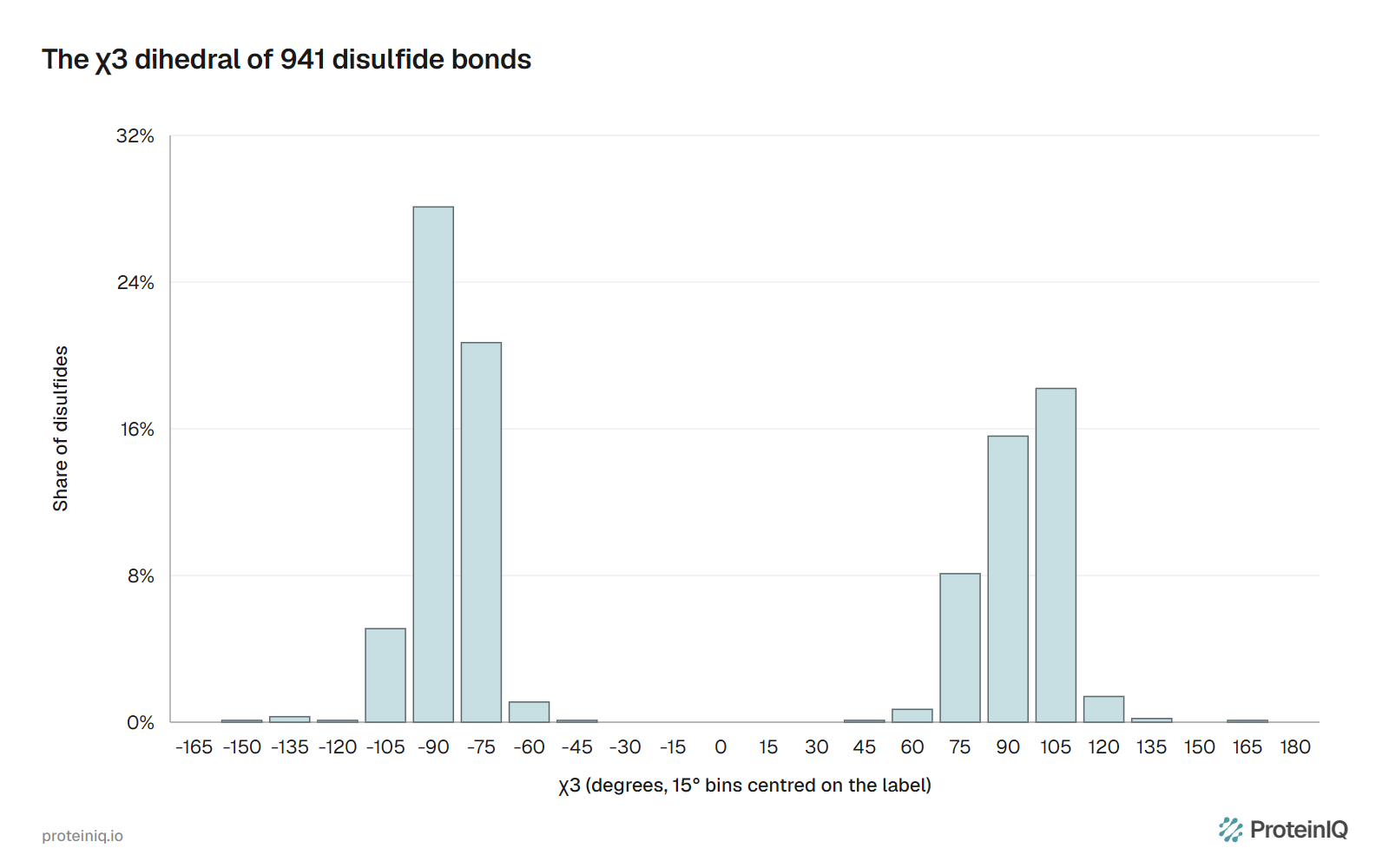
The angle that decides those shapes is , the dihedral angle about the S-S bond itself, defined by the atoms ---. It sits close to , where the lone pairs on the two sulfur atoms clash least. In our set, 929 of 941 values lie within 30° of or . Bonds with negative averaged and made up 56% of the set; those with positive averaged .
The sign of gives a disulfide its handedness, negative for left-handed and positive for right-handed. Together with the signs of the four other side-chain dihedrals, it sorts every disulfide into one of 20 configurations named after the shapes they resemble: spirals, hooks and staples.[12] The most common is the left-handed spiral with both angles negative, written -LHSpiral. It made up 32% of our 941 bonds and 29.7% of the 42,690 disulfides that Schmidt and Hogg classified in X-ray structures.[13]
Configuration says something about function. Catalytic disulfides in oxidoreductases are almost always right-handed hooks, while the allosteric disulfides that switch a protein's activity when they break are -RHStaples, with Cα atoms unusually close together.[13] Our -RHStaples averaged 4.23 Å between Cα atoms, against 5.46 Å for all bonds, close to the 4.18 Å Schmidt and Hogg reported.[13]
How disulfide bonds stabilize a protein
A disulfide changes the folded state little, because the two cysteines already sit next to each other there. The large effect is on the unfolded state. Without the cross-link, an unfolded chain can take an enormous number of conformations; with it, every conformation has to bring two distant residues within about 5 Å of each other. Removing all those conformations lowers the entropy of the unfolded chain, and because the folded state loses little, the balance shifts toward folding.
Nick Pace and colleagues measured this in ribonuclease T1, which has two disulfides, Cys2-Cys10 and Cys6-Cys103. The native enzyme melted at 59.3 °C with a conformational stability of 10.2 kcal/mol. Breaking the 2-10 bond cut the stability to 6.8 kcal/mol and the melting temperature to 53.3 °C. With both bonds reduced, the protein still folded and kept about half its activity, but it melted at 27.2 °C and its stability fell to 3.0 kcal/mol.[14]
From those data they proposed an equation for the entropy a cross-link removes from the unfolded chain, where is the number of residues between the two linked cysteines and is the gas constant:[14]
The longer the loop the bond closes, the larger the effect, but only logarithmically. With in , we calculated that a disulfide closing the median loop in our structure set, 27 residues, is worth about 3.6 kcal/mol at 25 °C. For ribonuclease T1 the equation gives about 2.5 kcal/mol for the 2-10 bond and 4.7 kcal/mol for the 6-103 bond, 7.2 together, matching the 7.2 to 9.3 kcal/mol loss Pace's group measured when both bonds were broken.
The equation assumes the bond fits the folded state without strain, and natural disulfides usually do. Schmidt and Hogg estimated a mean dihedral strain energy of 13.1 kJ/mol for disulfides in X-ray structures, and our high-resolution set averaged 10.4 kJ/mol.[13] A strained bond pays part of its entropy gain back in the folded state, which is one reason engineered disulfides often disappoint.
Famous examples
Four proteins come up in almost every discussion of disulfides, and their bonds are worth knowing exactly. We checked each pairing against a high-resolution structure.
| Protein | Chains and length | Disulfides | Pairs | Source |
|---|---|---|---|---|
| Human insulin | A chain 21, B chain 30 residues | 3 | A6-A11 (within A), A7-B7, A20-B19 | PDB 3I40 |
| Hen egg-white lysozyme | 1 chain, 129 residues | 4 | 6-127, 30-115, 64-80, 76-94 | PDB 2VB1 |
| Bovine ribonuclease A | 1 chain, 124 residues | 4 | 26-84, 40-95, 58-110, 65-72 | PDB 7RSA |
| Human IgG1 antibody | 2 heavy and 2 light chains | 16 | 12 intrachain, one per domain; 2 between the heavy chains in the hinge; 2 linking each light chain to a heavy chain | Liu and May (2012) |
Insulin shows how far a disulfide can hold separate chains together: reduce its two interchain bonds and the A and B chains come apart. Its third bond, A6-A11, closes a loop of only five residues. In the 1.85 Å structure 3I40, the Cα atoms of that pair sit 3.86 Å apart, near the bottom of the range in Table 1, and its estimated dihedral strain of 22.6 kJ/mol is the highest of the eleven bonds we measured in insulin, lysozyme and ribonuclease A.[24]
In antibodies the disulfides do two jobs. The 12 intrachain bonds each sit inside one immunoglobulin domain and hold its two sheets together, while the interchain bonds assemble the four chains into one molecule. The hinge count differs by subclass: 2 in IgG1 and IgG4, 4 in IgG2 and 11 in IgG3.[15]
In IgG1 the light chain's last cysteine bonds to the fifth cysteine of the heavy chain, and in the other three subclasses to the third.[15] The hinge bonds are also where papain and pepsin cut on opposite sides to produce the Fab and Fc fragments.
Real antibodies are not always this tidy. Liu and May describe free cysteines detected in all IgG subclasses, trisulfide bonds with an extra sulfur atom, and non-reducible thioether cross-links that form when a disulfide degrades.[15] These variants matter for therapeutic antibodies, which is why their disulfides are routinely mapped by mass spectrometry.
How to find disulfide bonds
In a structure file
A PDB-format file lists each disulfide in an SSBOND record, giving the two cysteines, the symmetry operators that place them and the bond length.[16] The wwPDB uses lysozyme as its example:
SSBOND   1 CYS A    6    CYS A  127                          1555   1555  2.03
SSBOND   2 CYS A   30    CYS A  115                          1555   1555  2.07
SSBOND   3 CYS A   64    CYS A   80                          1555   1555  2.06
SSBOND   4 CYS A   76    CYS A   94                          1555   1555  2.04
In the newer mmCIF format the same information sits in the struct_conn table as rows with conn_type_id set to disulf, with the distance in pdbx_dist_value. The CIF file is the archive's main format, and the records match: entry 2VB1 lists the four lysozyme bonds at 2.034, 2.038, 2.033 and 2.026 Å, the same values we computed from its coordinates.[24]
Predicted models have no such records. AlphaFold2 and ESMFold write coordinates only, so the only evidence for a disulfide is the distance between two sulfur atoms. In our crystal structures, 95% of bonded sulfur pairs sat within 2.13 Å of each other. Two cysteines whose sulfurs are 3 to 4 Å apart in a prediction are a candidate pair, not a modeled bond.
The PDB viewer shows the pair in 3D, DSSP labels bonded cysteines with matching lowercase letters, and MolProbity reports clashes and covalent-geometry outliers around them if the local model looks doubtful.
Ellman's assay
Ellman's reagent, 5,5'-dithiobis(2-nitrobenzoic acid) or DTNB, reacts with free thiols and releases a yellow anion, TNB, that absorbs at 412 nm.[17] Ellman reported a molar absorption coefficient of 13,600 ; Riddles and colleagues later measured 14,150, the value most protocols now use.[17][18]
The assay counts free cysteines, so it finds disulfides by difference. Measure the thiols in the native protein, then again after reduction (with the reductant removed, since it would react too), and halve the increase. Lysozyme gives no free thiols before reduction and eight per molecule after, which means four disulfides. Run both measurements in a denaturant so that buried cysteines react.
Reducing and non-reducing SDS-PAGE
Standard SDS-PAGE sample buffer contains DTT or mercaptoethanol, which breaks every disulfide. Leave the reductant out and interchain bonds survive the gel. The comparison shows how chains are linked: an IgG runs near 150 kDa without reductant and splits into heavy chains near 50 kDa and light chains near 25 kDa with it, as described in the guide to antibody molecular weight.
Mass spectrometry
Mass spectrometry gives both the number of bonds and the pairs. The intact mass shifts by 2.016 Da per disulfide on reduction, so lysozyme gains about 8 Da when its four bonds open. To find which cysteines are paired, the usual approach alkylates any free cysteines, digests the protein without reducing it, and compares the peptide map with that of a reduced digest; peptides that appear only in the non-reduced map are disulfide-linked pairs.[19]
Fragmentation method matters. Collision-induced dissociation mainly breaks the peptide backbone and leaves the disulfide intact, while in electron-transfer dissociation cleavage of the disulfide is the main pathway, which releases the two linked peptides for identification.[19] Slightly acidic conditions during digestion limit artifacts such as disulfide scrambling, which alkaline pH favors.[19]
Engineering disulfide bonds for stability
If a natural disulfide stabilizes a protein through entropy, an added one should too, and sometimes it does. Brian Matthews' group put three disulfides, linking residues 3-97, 9-164 and 21-142, into T4 lysozyme, which naturally has none. Each made the protein more stable, their effects on melting temperature were roughly additive, and the triple mutant melted 23.4 °C higher than the wild type.[20]
Many attempts do worse. Dombkowski and colleagues note that, contrary to expectation, many designed disulfides have decreased the stability of the protein they were put into.[21] A new bond can strain the folded state, remove a stabilizing side chain at each end, or link two regions that need to move. Design tools such as Disulfide by Design 2.0 therefore screen residue pairs for geometry that a disulfide can adopt and also look at the B-factors of the regions involved, favoring sites expected to raise thermal stability.[22]
The numbers in Table 1 are a fair first filter. Candidate pairs in a model should have Cβ atoms about 3.5 to 4.1 Å apart and Cα atoms about 4 to 6.5 Å apart, and a pair that would need far from is a poor bet. Single-mutation stability predictors such as ThermoMPNN score each substitution alone and handle combinations poorly, so they cannot judge a cysteine pair. The new bond has to be modeled in the structure and then tested, the same build-and-test loop used in protein design.
Disulfide bond tools on ProteinIQ
Protein Parameters reports the cysteine count of a sequence with both the reduced and oxidized extinction coefficients, and the extinction coefficient calculator lets you set the exact number of disulfides when converting an A280 reading into a concentration. The molecular weight calculator subtracts 2.016 Da for each disulfide you specify, which matters when matching an intact mass.
For structures, the PDB viewer displays bonded cysteines in context, DSSP lists every disulfide bridge alongside its secondary structure assignment, and MolProbity checks the geometry around them. For a sequence without a structure, AlphaFold2 or ESMFold give a model in which candidate pairs can be measured, with the caveat that the bond itself is never written into the file.
Our disulfide geometry dataset lists all 941 bonds behind Table 1 and Figure 1 with their distances, angles, dihedrals and configuration, and the summary and human UniProt counts hold the other figures on this page. They are free to reuse under CC BY 4.0 with credit to ProteinIQ.
Frequently asked questions
Which amino acid forms disulfide bonds?
Cysteine, the only one of the 20 standard amino acids with a thiol side chain. Two cysteines form one disulfide, and the bonded pair is called cystine. Cysteine is one of the least common amino acids, about 1.4% of residues in Swiss-Prot, so most proteins have few chances to make one.
Is a disulfide bond covalent?
Yes. It is a covalent bond between two sulfur atoms, about 2.05 Å long, unlike the hydrogen bonds, salt bridges and hydrophobic contacts that hold most of a folded protein together. It can still be broken by reduction or by exchange with another thiol, which is how reducing agents and enzymes such as PDI work.
Is a disulfide bond part of tertiary or quaternary structure?
Both, depending on what it links. A bond between two cysteines in the same chain, like the four in lysozyme, stabilizes tertiary structure. A bond between different chains, like the two that join insulin's A and B chains or the hinge bonds of an antibody, holds quaternary structure together. Disulfides also count as a post-translational modification, since they form after the chain is made.
What breaks disulfide bonds?
Reducing agents. Dithiothreitol (DTT) and 2-mercaptoethanol break them by thiol-disulfide exchange, and TCEP reduces them without being a thiol itself. Inside the cell, the thioredoxin and glutathione systems do the same job, which is why the cytosol has so few disulfides.
What is the difference between a disulfide bond and a disulfide bridge?
None. Disulfide bond, disulfide bridge, S-S bond and SS-bond all name the same link, and cystine names the pair of cysteines it joins. The PDB format calls the record SSBOND, and DSSP reports "SS-bridges".
Do disulfide bonds affect A280 readings?
Slightly. Each cystine absorbs about 125 at 280 nm, against 5,500 for a tryptophan, so lysozyme's four bonds add about 1% to its extinction coefficient and ribonuclease A's four, with no tryptophan to dominate, about 6%.[23] Use the oxidized coefficient for a protein with its native disulfides and the reduced one for a sample in DTT or TCEP.