What is protein denaturation?
Why a cooked egg stays cooked, and some proteins refold

Matic Broz, PhD17 min
Crack an egg into a hot pan and the white turns from clear to opaque within seconds, long before anything in the pan boils. Nothing burns off and no bond in the protein chains breaks. The proteins only lose their shape. Heated slowly in a calorimeter at pH 9, close to the pH of a stored egg, ovotransferrin in egg white unfolds at about 63 °C and ovalbumin, its main protein, at about 76 °C.[1][2]
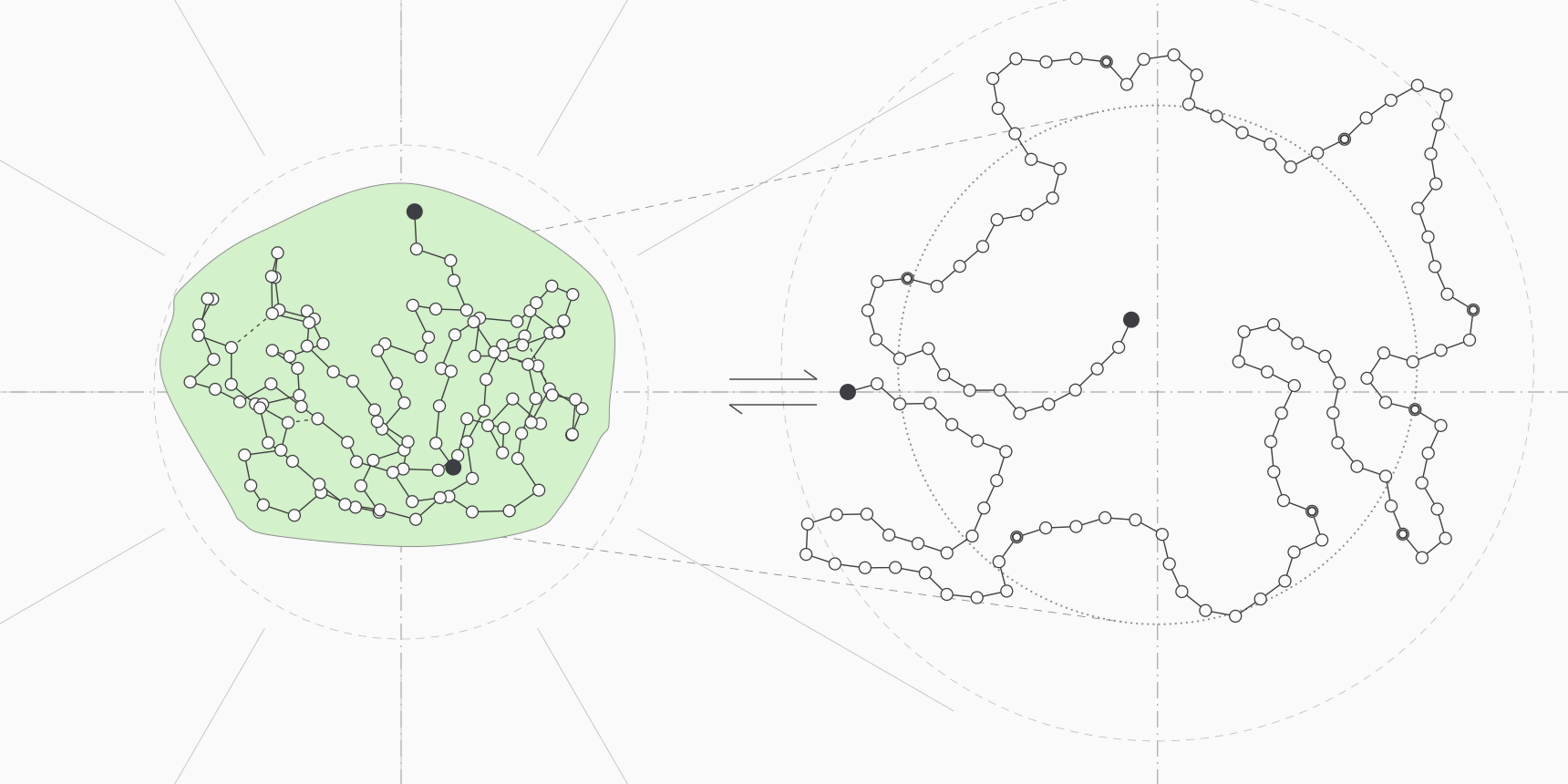
Christian Anfinsen did something much harsher to ribonuclease A. He unfolded it in 8 M urea, cut all four of its disulfide bonds with mercaptoethanol, then removed the urea and let the chain reoxidize in air. The enzyme folded back into its native form and recovered its activity.[3][4] Both are protein denaturation. The egg stays cooked and the enzyme comes back, and the difference lies in what happens to the chain after it unfolds.
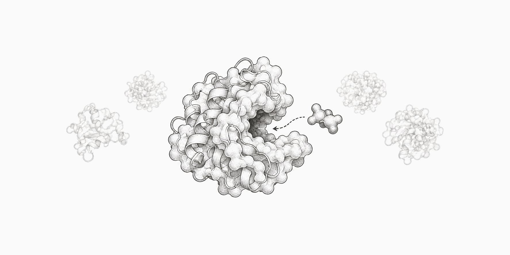
Denaturation is the loss of a protein's native three-dimensional structure: its secondary, tertiary and quaternary structure fall apart while the amino acid sequence stays intact. How easily that happens is set by the protein's stability, the free energy gap between the folded and unfolded states. ThermoMPNN predicts how each possible mutation shifts that gap from a protein structure.
What denaturation changes and what stays intact
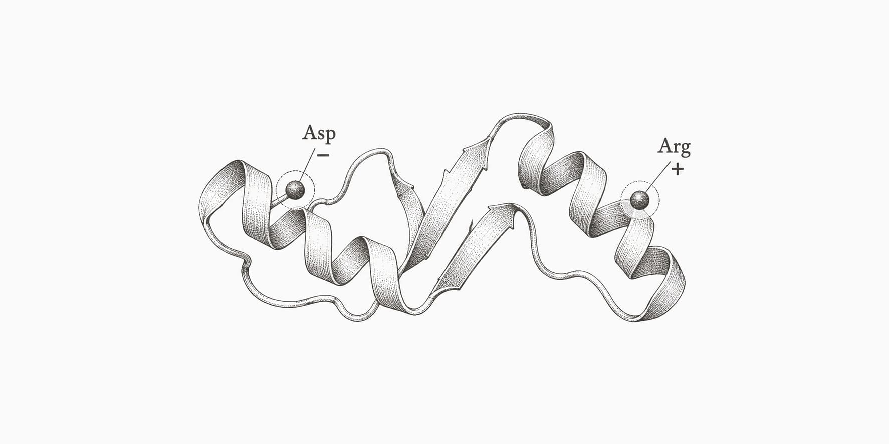
Protein structure is usually described at four levels, and denaturation removes the top three:
- Primary structure, the sequence of amino acids joined by peptide bonds, stays intact.
- Secondary structure, the alpha helices and beta sheets held together by backbone hydrogen bonds, is lost.
- Tertiary structure, the packing of helices and sheets into a fold around a hydrophobic core, is lost.
- Quaternary structure, the assembly of several chains into one complex, is lost as the chains separate or unfold.
The peptide bonds survive because heat, urea or a detergent only disrupt noncovalent interactions: hydrogen bonds, salt bridges and hydrophobic contacts. Cutting the backbone is hydrolysis, which needs a protease or hot concentrated acid. That is why a cooked egg still supplies the same amino acids as a raw one.
Disulfide bonds sit in between. They are covalent and survive heating, so a heat-denatured protein can keep its cysteine crosslinks. Reducing agents such as mercaptoethanol or dithiothreitol break them, which is why Anfinsen needed one to unfold ribonuclease completely and why SDS-PAGE sample buffers usually contain one.
The denatured state is not a single structure either. In 8 M urea a chain behaves close to a random coil, but milder conditions leave compact, partly structured states, so "denatured" really means "not native".
Acid shows the range. Goto and colleagues found that apomyoglobin, beta-lactamase and cytochrome c unfold almost fully near pH 2 at low salt, then fold back into a compact molten globule when more acid is added, because the extra anions screen the charge repulsion that unfolded them.[5]
Why proteins are only marginally stable
Many small proteins unfold in a single cooperative step, with no stable intermediates. That two-state view treats the folded (N) and unfolded (U) forms as an equilibrium, and the stability is the free energy of unfolding:
A positive means the folded state is favored. The range usually quoted for globular proteins is 5 to 15 kcal/mol, or about 21 to 63 kJ/mol.[6] Measured values bear that out, with small domains at the low end: Nick Pace's group measured 2.7 kcal/mol for the 36-residue villin headpiece subdomain and 6.4 kcal/mol for ribonuclease T1 at pH 7.[7]
Those numbers sound abstract until you convert them into the fraction of molecules that are unfolded at any moment, . We calculated it for 25 °C and 37 °C:
| Stability (kcal/mol) | Unfolded at 25 °C | Unfolded at 37 °C |
|---|---|---|
| 2.7 | 1 in 95 | 1 in 80 |
| 5 | 1 in 4,600 | 1 in 3,300 |
| 6.4 | 1 in 49,000 | 1 in 32,000 |
| 10 | 1 in 21 million | 1 in 11 million |
| 15 | 1 in 99 billion | 1 in 37 billion |
The margin is small because it is the difference between two large opposing terms. Pace's group estimated that hydrophobic interactions supply about 60% of the stabilizing free energy and hydrogen bonds about 40%, while lost conformational entropy costs about 2.4 kcal/mol per residue.[7] For a 76-residue protein like ubiquitin, the stabilizing side adds up to roughly 180 kcal/mol, and nearly all of it pays for ordering the chain.
The same study found that burying one extra CH₂ group adds 1.1 kcal/mol on average.[7] A single mutation that removes a buried methyl can therefore cost a sizeable share of the whole margin, and a few of them can unfold the protein at body temperature. Predicting that change, called ΔΔG, is what stability predictors do.
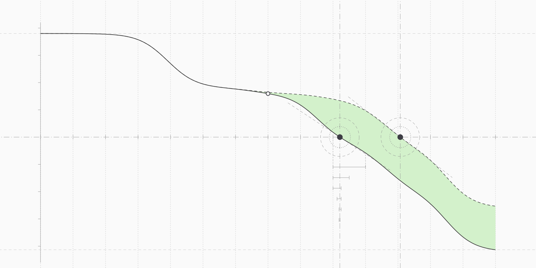
The stability curve
Stability changes with temperature in a way the Gibbs-Helmholtz equation captures. With the melting temperature, where , the enthalpy of unfolding at , and the heat capacity change on unfolding:
is positive because unfolding exposes nonpolar surface to water, and it scales with the surface exposed.[8] That curvature bends the stability curve down at both ends. peaks at one temperature and falls toward zero on heating and, for many proteins, on cooling too.
The curve also explains why a high does not mean a protein is especially stable at room temperature. Myers, Pace and Scholtz showed it with two hypothetical proteins: one melts at 90 °C but has 6.3 kcal/mol of stability at 25 °C, the other melts at 70 °C with 8.0 kcal/mol, because its larger makes its curve steeper.[8] Small proteins often melt high while being only averagely stable.
What causes protein denaturation
Anything that makes the unfolded state relatively more favorable can denature a protein: adding heat, removing the hydrophobic effect, piling up charge, giving the unfolded chain a better solvent or simply pulling it apart.
Heat
Heating favors the unfolded state because its higher entropy counts for more as temperature rises, the in . At , half the molecules are unfolded. For the major egg white proteins the melting points measured by calorimetry at pH 9 are about 63 °C for ovotransferrin, 69 °C for lysozyme, 76 °C for ovalbumin and 83 °C for S-ovalbumin, a more heat-stable form that builds up in stored eggs.[1][9]
The archaeon Pyrococcus furiosus grows best at 100 °C, and its rubredoxin is so stable that its melting temperature had to be extrapolated from hydrogen exchange data, to between 176 and 195 °C. At 100 °C it still has 15 kcal/mol of stability.[10] Across 13 species, from archaea to humans, the Meltome Atlas measured melting temperatures for 48,000 proteins between 30 and 90 °C.[11]
Human body temperature sits near 37 °C, and a fever adds a few degrees. Proteome-wide experiments suggest that cells killed by heat do not die because every protein melts: Leuenberger and colleagues traced heat-induced cell collapse to the unfolding of a subset of proteins with essential functions.[12] Human mitochondria in the Meltome study still respired close to normally at 46 °C.[11]
Cold
Because of the curved stability profile, some proteins unfold on cooling as well. Yeast frataxin is a rare protein that does it without any denaturant: in a salt-free buffer at pH 7 it unfolds below about 7 °C and above about 30 °C, and even at its most stable only about two thirds of the molecules are folded.[13]
Calorimetric data on cold denaturation are scarce, because most proteins would have to be cooled below the freezing point of water. One of the best examples is beta-lactoglobulin in 4 M urea, where the denaturant lowers the whole stability curve and the protein unfolds as it is cooled toward −14 °C.[6]
Acid and base
At extreme pH, many side chains carry the same charge. Near pH 2, for example, carboxyl groups are protonated and neutral while lysine, arginine and histidine are positive, so the folded protein carries a large net positive charge whose repulsion pushes the chain apart.[5] Precipitation is a different event: a protein near its isoelectric point has little net charge and can come out of solution without necessarily unfolding.
Urea and guanidinium chloride
Urea and guanidinium chloride (GdnHCl) are the standard chemical denaturants. Both are better solvents for the exposed backbone and side chains of the unfolded chain than water is, so they favor the state with more surface. In the linear extrapolation method, stability falls linearly with denaturant concentration , and the midpoint of the transition is where it reaches zero:
The slope grows with the amount of surface the protein exposes on unfolding. For proteins measured in both, the GdnHCl value is about 2.3 times the urea value, which makes guanidinium roughly twice as potent per mole.[8] That is why full unfolding is usually done in 8 M urea or 6 M GdnHCl, the conditions of Anfinsen's ribonuclease work and of the Edelhoch method for measuring a protein's extinction coefficient.
A midpoint alone does not rank stability. In the same hypothetical pair, the protein with 6.3 kcal/mol and a low value has its midpoint at 6.3 M, while the one with 8.0 kcal/mol and twice the value is half unfolded at 4.0 M.[8]
Detergents
Sodium dodecyl sulfate (SDS) unfolds proteins by coating them. Reynolds and Tanford found that once free SDS rises above about 0.5 mM, a wide range of proteins bind the same amount per gram, reaching a plateau of about 1.4 g of SDS per gram of protein through hydrophobic contacts.[14] Every chain ends up with a similar charge-to-mass ratio, so in SDS-PAGE it migrates by size alone. The same principle runs the gel step of a western blot.
Alcohols and organic solvents
Organic solvents weaken the hydrophobic effect that holds the core together, so the buried side chains no longer gain much by staying buried. Protein denaturation is also the most likely explanation for why alcohol kills microbes. The CDC gives the optimum bactericidal ethanol concentration as 60 to 90% in water and notes that absolute ethanol works less well, because water is needed for the proteins to denature quickly.[15]
Mechanical force and interfaces
A protein can also be pulled apart. Rief and colleagues stretched single molecules of titin, the giant muscle protein, with an atomic force microscope and saw its immunoglobulin domains unfold one at a time at 150 to 300 piconewtons, then refold once the tension was released.[16] Shear and air-water interfaces stress proteins in a similar way during manufacturing and administration, which is why surfactants are added to protein drugs.[17]
Is protein denaturation reversible?
It can be. Anfinsen's ribonuclease experiments made the case. Reduced in 8 M urea with mercaptoethanol, ribonuclease A became a random chain with eight free cysteines, and oxidation after the urea was removed restored the native disulfides.[3]
When the chain was instead reoxidized while still in 8 M urea, it formed a mixture of "scrambled" isomers drawn from 105 possible ways of pairing eight cysteines, with about 1% of the native activity. Removing the urea and adding a trace of mercaptoethanol let the disulfides reshuffle until the mixture became native enzyme again.[4]
From this Anfinsen drew his thermodynamic hypothesis: in its normal environment, the native structure is the one with the lowest Gibbs free energy, so the sequence alone determines the fold.[4] It is also the premise behind predicting structure from sequence alone, as ESMFold does.
Denaturation becomes irreversible when the unfolded chain does something it cannot undo before it refolds. Lumry and Eyring described the scheme in 1954, with a reversible unfolding step followed by an irreversible one:[18]
The irreversible step is usually protein aggregation, in which exposed hydrophobic surfaces of different molecules stick together, or a covalent change such as disulfide exchange between chains. Aggregation involves two or more molecules while refolding needs only one, so high protein concentration pushes the balance toward aggregates.
Egg white has every ingredient for that. Ovalbumin, 54% of egg white protein, is a 385-residue chain with four free thiols and one disulfide bond.[1] When it unfolds, a hydrophobic C-terminal segment carrying the free cysteine Cys368 is exposed and drives the molecules into aggregates.[2] Egg white starts to set at about 60 °C, where ovotransferrin denatures, and firms further around 70 to 75 °C as ovalbumin follows.[2][1]
Ovomucoid, another egg white protein, behaves differently. Its three domains are each held by internal disulfide bonds and do not link to each other, and it denatures irreversibly only after prolonged heating above 90 °C, such as boiling for more than 30 minutes.[2] Differences like these in how egg proteins respond to heat are part of why most children allergic to egg can still eat hard-boiled or baked egg.[2]
How denaturation is measured
Every method follows some property that differs between the folded and unfolded protein while temperature or denaturant concentration rises. The midpoint gives or , and a reversible two-state transition also gives .
| Method | What it detects | Typical use |
|---|---|---|
| Far-UV circular dichroism (CD) | Loss of helix and sheet signal from the backbone | Melting curves at one wavelength; Tm, van't Hoff enthalpy and ΔG if reversible |
| Intrinsic tryptophan fluorescence | Emission maximum shifting toward longer wavelengths as buried tryptophans meet water | Chemical denaturation curves for ΔG and m values |
| Differential scanning calorimetry (DSC) | Heat absorbed on unfolding, measured directly as heat capacity | Tm and calorimetric enthalpy without an optical probe; formulation screening |
| Differential scanning fluorimetry (DSF, thermal shift) | Fluorescence of a dye that binds hydrophobic surface exposed on unfolding | Fast Tm screens in a real-time PCR instrument; ligand binding from the Tm shift |
Circular dichroism reads the backbone, so it reports on secondary structure, and Greenfield's protocol uses it to extract the van't Hoff enthalpy, the midpoint and the free energy of unfolding when the melt is reversible.[19]
Tryptophan fluorescence reads the environment of individual side chains instead. The emission maximum ranges from about 308 nm for the buried tryptophan of azurin to about 355 nm for a fully exposed one and roughly tracks solvent exposure, so unfolding shifts it toward the red.[20]
Calorimetry needs no probe at all. DSC measures the heat capacity of the sample as it is heated, and the area under the unfolding peak is the enthalpy of unfolding.[21] DSF trades that rigor for speed: a dye fluoresces when it binds the hydrophobic surface that unfolding exposes, and a ligand that binds the native state raises the .[22]
For a two-state transition, the measured signal is a weighted average of the folded and unfolded baselines, and the fraction unfolded follows from :
Where denaturation matters
In the kitchen, heat denaturation is the point of cooking. It also changes digestion: native ovalbumin resists gastric digestion, while heat-aggregated ovalbumin is broken down more easily.[2]
Hospitals use the same chemistry on purpose. Steam sterilization of wrapped instruments runs at 121 °C for at least 30 minutes in a gravity displacement sterilizer, or 132 °C for 4 minutes in a prevacuum one, and kills microbes by irreversibly coagulating and denaturing their enzymes and structural proteins.[23]
In the lab, denaturation is a tool. SDS-PAGE depends on complete unfolding, urea and GdnHCl unfolding curves measure stability, and thermal shift assays screen for ligands. How long a protein survives in a cell is a separate question, governed by degradation rather than denaturation, with protein half-lives ranging from minutes to a lifetime.
For protein drugs, unfolding and the aggregation that follows it are what formulation scientists guard against for the whole shelf life. Polysorbate 20 and 80 are the most common surfactants, added to stop unfolding and aggregation driven by surface tension and shear, and histidine, sucrose, arginine and methionine are common excipients in approved antibody products.[17]
Protein engineers attack the problem at the sequence level. Trevino and colleagues replaced residues in beta turns of ribonuclease Sa with the proline or glycine those positions prefer, and five single mutations each raised its stability by 0.7 to 1.3 kcal/mol.[24] Against a margin of 5 to 15 kcal/mol, a few gains of that size make a large difference.
Protein stability tools on ProteinIQ
ThermoMPNN predicts the ΔΔG in kcal/mol of every single amino acid substitution in one chain from a PDB structure, so you can rank stabilizing mutations before making any. If you have no experimental structure, ESMFold predicts one from the sequence and returns a PDB file that ThermoMPNN accepts directly; check its pLDDT first, because a mutation scan on a low-confidence region inherits that uncertainty.
ESM-C Mutation Scoring works from sequence alone and ranks substitutions by how strongly a protein language model prefers them, which is a useful second opinion but not a free energy. Protein stability prediction reports sequence descriptors such as the instability index, aliphatic index and hydropathy, which flag likely problems without predicting .
For complexes, where denaturation includes losing quaternary structure, StaB-ddG predicts how mutations change the binding free energy between chains. It describes binding, not the folding stability of each chain.
Frequently asked questions
Is protein denaturation reversible?
Sometimes. Small proteins unfolded by urea or mild heat in dilute solution often refold when the conditions return to normal, as ribonuclease A does. Denaturation becomes irreversible when unfolded chains aggregate or undergo covalent changes before they can refold, which is what happens in a cooked egg.
At what temperature do proteins denature?
There is no single temperature. Proteome-wide measurements across 13 species found melting temperatures from 30 to 90 °C, egg white proteins between about 63 and 83 °C, and the rubredoxin of Pyrococcus furiosus is estimated to stay folded up to nearly 200 °C. Melting temperature also depends on pH, salt, concentration and, for irreversible unfolding, heating rate.
Does denaturation break peptide bonds?
No. Denaturation disrupts the noncovalent interactions that hold the fold together and leaves the peptide bonds and the amino acid sequence intact. Breaking peptide bonds is hydrolysis, done by digestive proteases or by strong acid. Disulfide bonds also survive heat, but reducing agents break them.
Does cooking destroy protein?
Cooking denatures protein but does not remove it. The amino acids are all still there, and denatured proteins are often easier to digest: heat-aggregated ovalbumin, for example, is broken down more readily in the stomach than native ovalbumin.
What is the difference between denaturation and aggregation?
Denaturation is the loss of a single molecule's native structure. Aggregation is what unfolded or partly unfolded molecules often do next, sticking to each other through exposed hydrophobic surfaces. Aggregation is the usual reason denaturation cannot be reversed.