The isoelectric point (pI) is the pH where a protein or peptide carries zero net charge. It determines how a molecule behaves during isoelectric focusing, ion exchange chromatography, and 2D gel electrophoresis. Predicting pI from sequence requires knowing the pKa of every ionizable group, which varies depending on the pKa scale used.
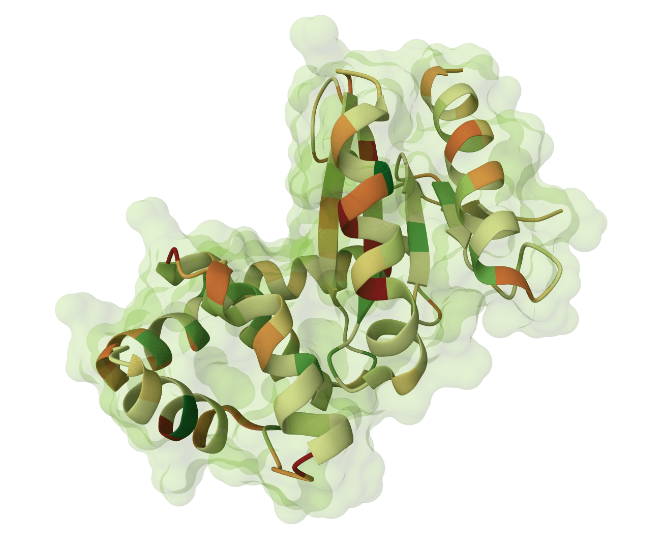
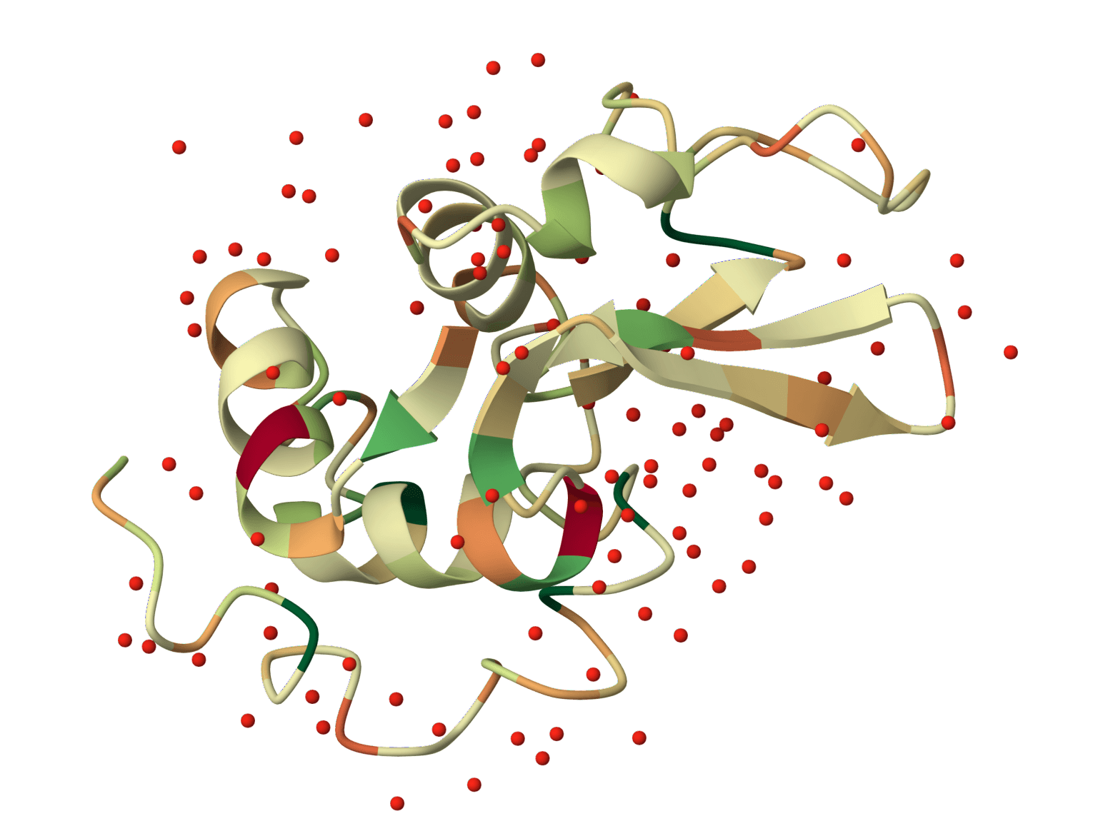
IPC 2.0 (Isoelectric Point Calculator 2.0) by Kozlowski combines 19 pKa scales with machine learning to predict pI more accurately than any single Henderson-Hasselbalch calculation. The SVR models trained on 2,324 proteins and 119,092 peptides achieve RMSD values of 0.85 and 0.23 respectively. A SepConv2D deep learning model pushes peptide accuracy further (RMSD 0.22). Version 2.0 also introduced per-residue pKa prediction, estimating the dissociation constant of each ionizable residue in context using an MLP-SVR ensemble.
For a quick single-sequence estimate, the pI Calculator or Protein Parameters tool runs instantly in the browser using the Bjellqvist scale. IPC 2.0 is the better choice when:
IPC 2.0 on ProteinIQ predicts the isoelectric point of proteins and peptides from amino acid sequence. Paste sequences in FASTA format (or fetch them from UniProt), choose a prediction method, and get pI values across up to 19 pKa scales plus machine learning predictions, along with per-residue pKa for every ionizable site.
| Input | Description |
|---|---|
Protein/Peptide Sequences | One or more amino acid sequences in FASTA format. Also accepts .fasta, .fa, .fas, or .txt files. UniProt IDs can be fetched directly using the batch fetcher. |
| Setting | Description |
|---|---|
Predictor | Prediction method. Default: IPC1_ALL. See predictor options below. |
| Predictor | Best for | What it does |
|---|---|---|
IPC1_ALL | General use | Henderson-Hasselbalch with all 19 pKa scales (18 literature scales + ProMoST). Fastest option. |
IPC2_SVR_protein | Proteins | SVR trained on 2,324 proteins from SWISS-2DPAGE and PIP-DB. Reports all 19 scales plus the SVR prediction. |
IPC2_SVR_peptide | Peptides | SVR trained on 119,092 peptides from HiRIEF experiments. Reports all 19 scales plus the SVR prediction. |
IPC2_DL_peptide | Peptides ≤60 aa | SepConv2D deep learning model. Highest accuracy for short peptides (RMSD 0.22). |
ALL | Method comparison | Runs every predictor and all pKa scales. Useful for assessing prediction confidence. |
The Results tab shows a spreadsheet with one row per sequence:
| Column | Description |
|---|---|
ID | Sequence identifier from the FASTA header. |
Length | Number of amino acid residues. |
MW | Molecular weight in Daltons. |
pI_[scale] | Predicted pI for each pKa scale (e.g., pI_IPC2_protein, pI_Bjellqvist, pI_ProMoST). |
pI_SVR_protein | SVR prediction optimized for proteins (when SVR or ALL method selected). |
pI_SVR_peptide | SVR prediction optimized for peptides (when SVR or ALL method selected). |
pI_DL_peptide | Deep learning prediction (when DL or ALL method selected). |
Avg_pI | Average pI across all calculated scales (excluding Patrickios). |
The Per-Residue pKa tab shows predicted pKa values for every ionizable site:
| Column | Description |
|---|---|
Sequence | Which input sequence this residue belongs to. |
Position | Residue position (1-indexed), or N-term/C-term for terminal groups. |
Residue | Amino acid type (D, E, H, K, Y, or the terminal residue). |
pKa | Predicted dissociation constant from the MLP-SVR ensemble. |
Most proteins have pI between 4 and 10. Knowing where a protein falls relative to pH 7 determines its behavior in common buffers:
| pI range | Character | At pH 7.4 (physiological) |
|---|---|---|
| <5 | Strongly acidic | Net negative charge |
| 5 to 7 | Weakly acidic | Slight negative charge |
| 7 to 9 | Weakly basic | Slight positive charge |
| >9 | Strongly basic | Net positive charge |
When multiple pKa scales agree within 0.3 pH units, the prediction is likely reliable. Disagreement of more than 1 pH unit suggests the protein has unusual charge properties (e.g., many histidines) where scale choice matters. In those cases, the SVR prediction is more trustworthy because it was trained on experimental data and can capture non-additive effects.
Per-residue pKa values show how sequence context shifts each ionizable group's dissociation constant away from its "standard" textbook value. For example, an aspartate surrounded by other negative residues will have a higher pKa (harder to deprotonate), while one near positive charges will have a lower pKa.
These values are useful for identifying residues with unusual protonation behavior, which matters for enzyme active sites, pH-dependent conformational changes, and designing mutations that shift pI.
The classical approach calculates net protein charge as a function of pH using:
Starting at pH 6.51, a bisection algorithm adjusts pH up or down until the sum of positive charges (Lys, Arg, His, N-terminus) equals negative charges (Asp, Glu, Cys, Tyr, C-terminus) within a precision of 0.01 pH units.
Each pKa scale assigns different dissociation constants to these groups, producing different pI estimates. The Bjellqvist scale additionally uses position-specific pKa values for N-terminal and C-terminal residues (e.g., N-terminal Ala has pKa 7.59 instead of the generic 7.5), which matters for proteins starting with A, M, S, P, T, V, or E.
ProMoST uses a separate model with three pKa values per residue (N-terminal, middle, C-terminal positions), giving it slightly different behavior from the other 18 scales.
IPC 2.0 includes 19 pKa scales:
| Scale | Origin |
|---|---|
IPC2_protein | Optimized for proteins (Kozlowski 2021) |
IPC2_peptide | Optimized for peptides (Kozlowski 2021) |
IPC_protein | Original IPC scale (Kozlowski 2016) |
IPC_peptide | Original IPC scale (Kozlowski 2016) |
ProMoST | Position-specific model (Halligan 2004) |
Bjellqvist | 2D electrophoresis standard with terminal corrections |
EMBOSS | EMBOSS software suite |
DTASelect | Proteomics analysis |
Grimsley | Experimental NMR measurements |
Lehninger | Biochemistry textbook |
Solomon | Protein chemistry |
Sillero | Theoretical calculations |
Rodwell | Biochemistry reference |
Thurlkill | NMR measurements in pentapeptides |
Toseland | Statistical analysis of PDB structures |
Nozaki | Model compound measurements |
Dawson | Data compilation |
Wikipedia | General reference values |
Patrickios | Simplified (only D/E/K/R, ignores C/H/Y) |
The Avg_pI column averages all scales except Patrickios, whose simplified model (ignoring cysteine, histidine, and tyrosine) makes it an outlier.
The SVR models take a 19-dimensional feature vector (pI predictions from all 18 H-H scales + ProMoST) and feed it into a support vector machine with RBF kernel. The protein SVR was trained on 2,324 proteins from SWISS-2DPAGE and PIP-DB. The peptide SVR was trained on 119,092 peptides from high-resolution isoelectric focusing (HiRIEF) experiments.
Because the SVR sees all 19 scale predictions simultaneously, it learns which scales are most informative for different sequence compositions. It consistently outperforms any individual scale.
The deep learning model uses a separable convolutional architecture with four input channels:
Sequences longer than 60 residues undergo truncation that preferentially removes non-charged residues (Ala, Gly, Leu, etc.) while preserving ionizable residues that determine pI. This truncation uses random selection among non-charged positions, so predictions for long sequences may vary slightly between runs.
The per-residue pKa predictor estimates the dissociation constant of each ionizable residue (D, E, H, K, Y) plus the N- and C-terminal groups. It uses a stacking ensemble of 9 multilayer perceptrons (MLPs) feeding into a final SVR:
Each MLP encodes the local sequence context around an ionizable residue using one-hot encoding (and optionally AAindex physicochemical features). The 9 MLP predictions are stacked as features for the final SVR, which outputs a single pKa value. Charged N- and C-terminal residues are flanked with alanine padding before prediction to ensure they are treated as terminal groups.
Match experimental peptide masses against theoretical digest fragments of a protein sequence. Identify peptides from mass spectrometry data by peptide mass fingerprinting.
Predict protease and chemical cleavage sites across a protein sequence for up to 39 enzymes simultaneously. Identify where each enzyme cuts, the cleavage residue, and context window around each site.
Faithful static-mode Aggrescan3D tool for per-residue aggregation propensity analysis from a single protein structure.
Plot net charge vs pH for protein sequences. Visualize how protein charge changes across pH 0-14 and identify the isoelectric point (pI) where the net charge crosses zero.
Generate Kyte-Doolittle hydropathy plots to visualize hydrophobic and hydrophilic regions along protein sequences. Identify transmembrane domains and surface-exposed regions.
Generate hydrophobicity plots using 24 different amino acid scales. Visualize hydrophobic and hydrophilic regions for protein analysis, epitope prediction, and membrane protein studies.
Cleave a protein sequence with a chosen protease and compute the masses of the resulting peptides. Supports multiple enzymes, missed cleavages, chemical modifications, and different ion types for mass spectrometry experiment planning.
Predict pKa values of ionizable groups in proteins and protein-ligand complexes from 3D structure. PROPKA calculates environment-driven pKa shifts for standard ionizable residues, terminal groups, and supported ligand atom types.

Calculate protein parameters, including molecular weight, theoretical pI, extinction coefficients, aromaticity, secondary structure fractions, atomic composition, estimated half-life, and several indices, including instability, aliphatic index, and GRAVY.
Generate amino acid property profiles using 42 different scales spanning hydrophobicity, secondary structure propensity, flexibility, polarity, surface accessibility, antigenicity, and more.
The isoelectric point (pI) is the pH where a protein or peptide carries zero net charge. It determines how a molecule behaves during isoelectric focusing, ion exchange chromatography, and 2D gel electrophoresis. Predicting pI from sequence requires knowing the pKa of every ionizable group, which varies depending on the pKa scale used.
IPC 2.0 (Isoelectric Point Calculator 2.0) by Kozlowski combines 19 pKa scales with machine learning to predict pI more accurately than any single Henderson-Hasselbalch calculation. The SVR models trained on 2,324 proteins and 119,092 peptides achieve RMSD values of 0.85 and 0.23 respectively. A SepConv2D deep learning model pushes peptide accuracy further (RMSD 0.22). Version 2.0 also introduced per-residue pKa prediction, estimating the dissociation constant of each ionizable residue in context using an MLP-SVR ensemble.
For a quick single-sequence estimate, the pI Calculator or Protein Parameters tool runs instantly in the browser using the Bjellqvist scale. IPC 2.0 is the better choice when:
IPC 2.0 on ProteinIQ predicts the isoelectric point of proteins and peptides from amino acid sequence. Paste sequences in FASTA format (or fetch them from UniProt), choose a prediction method, and get pI values across up to 19 pKa scales plus machine learning predictions, along with per-residue pKa for every ionizable site.
| Input | Description |
|---|---|
Protein/Peptide Sequences | One or more amino acid sequences in FASTA format. Also accepts .fasta, .fa, .fas, or .txt files. UniProt IDs can be fetched directly using the batch fetcher. |
| Setting | Description |
|---|---|
Predictor | Prediction method. Default: IPC1_ALL. See predictor options below. |
| Predictor | Best for | What it does |
|---|---|---|
IPC1_ALL | General use | Henderson-Hasselbalch with all 19 pKa scales (18 literature scales + ProMoST). Fastest option. |
IPC2_SVR_protein | Proteins | SVR trained on 2,324 proteins from SWISS-2DPAGE and PIP-DB. Reports all 19 scales plus the SVR prediction. |
IPC2_SVR_peptide | Peptides | SVR trained on 119,092 peptides from HiRIEF experiments. Reports all 19 scales plus the SVR prediction. |
IPC2_DL_peptide | Peptides ≤60 aa | SepConv2D deep learning model. Highest accuracy for short peptides (RMSD 0.22). |
ALL | Method comparison | Runs every predictor and all pKa scales. Useful for assessing prediction confidence. |
The Results tab shows a spreadsheet with one row per sequence:
| Column | Description |
|---|---|
ID | Sequence identifier from the FASTA header. |
Length | Number of amino acid residues. |
MW | Molecular weight in Daltons. |
pI_[scale] | Predicted pI for each pKa scale (e.g., pI_IPC2_protein, pI_Bjellqvist, pI_ProMoST). |
pI_SVR_protein | SVR prediction optimized for proteins (when SVR or ALL method selected). |
pI_SVR_peptide | SVR prediction optimized for peptides (when SVR or ALL method selected). |
pI_DL_peptide | Deep learning prediction (when DL or ALL method selected). |
Avg_pI | Average pI across all calculated scales (excluding Patrickios). |
The Per-Residue pKa tab shows predicted pKa values for every ionizable site:
| Column | Description |
|---|---|
Sequence | Which input sequence this residue belongs to. |
Position | Residue position (1-indexed), or N-term/C-term for terminal groups. |
Residue | Amino acid type (D, E, H, K, Y, or the terminal residue). |
pKa | Predicted dissociation constant from the MLP-SVR ensemble. |
Most proteins have pI between 4 and 10. Knowing where a protein falls relative to pH 7 determines its behavior in common buffers:
| pI range | Character | At pH 7.4 (physiological) |
|---|---|---|
| <5 | Strongly acidic | Net negative charge |
| 5 to 7 | Weakly acidic | Slight negative charge |
| 7 to 9 | Weakly basic | Slight positive charge |
| >9 | Strongly basic | Net positive charge |
When multiple pKa scales agree within 0.3 pH units, the prediction is likely reliable. Disagreement of more than 1 pH unit suggests the protein has unusual charge properties (e.g., many histidines) where scale choice matters. In those cases, the SVR prediction is more trustworthy because it was trained on experimental data and can capture non-additive effects.
Per-residue pKa values show how sequence context shifts each ionizable group's dissociation constant away from its "standard" textbook value. For example, an aspartate surrounded by other negative residues will have a higher pKa (harder to deprotonate), while one near positive charges will have a lower pKa.
These values are useful for identifying residues with unusual protonation behavior, which matters for enzyme active sites, pH-dependent conformational changes, and designing mutations that shift pI.
The classical approach calculates net protein charge as a function of pH using:
Starting at pH 6.51, a bisection algorithm adjusts pH up or down until the sum of positive charges (Lys, Arg, His, N-terminus) equals negative charges (Asp, Glu, Cys, Tyr, C-terminus) within a precision of 0.01 pH units.
Each pKa scale assigns different dissociation constants to these groups, producing different pI estimates. The Bjellqvist scale additionally uses position-specific pKa values for N-terminal and C-terminal residues (e.g., N-terminal Ala has pKa 7.59 instead of the generic 7.5), which matters for proteins starting with A, M, S, P, T, V, or E.
ProMoST uses a separate model with three pKa values per residue (N-terminal, middle, C-terminal positions), giving it slightly different behavior from the other 18 scales.
IPC 2.0 includes 19 pKa scales:
| Scale | Origin |
|---|---|
IPC2_protein | Optimized for proteins (Kozlowski 2021) |
IPC2_peptide | Optimized for peptides (Kozlowski 2021) |
IPC_protein | Original IPC scale (Kozlowski 2016) |
IPC_peptide | Original IPC scale (Kozlowski 2016) |
ProMoST | Position-specific model (Halligan 2004) |
Bjellqvist | 2D electrophoresis standard with terminal corrections |
EMBOSS | EMBOSS software suite |
DTASelect | Proteomics analysis |
Grimsley | Experimental NMR measurements |
Lehninger | Biochemistry textbook |
Solomon | Protein chemistry |
Sillero | Theoretical calculations |
Rodwell | Biochemistry reference |
Thurlkill | NMR measurements in pentapeptides |
Toseland | Statistical analysis of PDB structures |
Nozaki | Model compound measurements |
Dawson | Data compilation |
Wikipedia | General reference values |
Patrickios | Simplified (only D/E/K/R, ignores C/H/Y) |
The Avg_pI column averages all scales except Patrickios, whose simplified model (ignoring cysteine, histidine, and tyrosine) makes it an outlier.
The SVR models take a 19-dimensional feature vector (pI predictions from all 18 H-H scales + ProMoST) and feed it into a support vector machine with RBF kernel. The protein SVR was trained on 2,324 proteins from SWISS-2DPAGE and PIP-DB. The peptide SVR was trained on 119,092 peptides from high-resolution isoelectric focusing (HiRIEF) experiments.
Because the SVR sees all 19 scale predictions simultaneously, it learns which scales are most informative for different sequence compositions. It consistently outperforms any individual scale.
The deep learning model uses a separable convolutional architecture with four input channels:
Sequences longer than 60 residues undergo truncation that preferentially removes non-charged residues (Ala, Gly, Leu, etc.) while preserving ionizable residues that determine pI. This truncation uses random selection among non-charged positions, so predictions for long sequences may vary slightly between runs.
The per-residue pKa predictor estimates the dissociation constant of each ionizable residue (D, E, H, K, Y) plus the N- and C-terminal groups. It uses a stacking ensemble of 9 multilayer perceptrons (MLPs) feeding into a final SVR:
Each MLP encodes the local sequence context around an ionizable residue using one-hot encoding (and optionally AAindex physicochemical features). The 9 MLP predictions are stacked as features for the final SVR, which outputs a single pKa value. Charged N- and C-terminal residues are flanked with alanine padding before prediction to ensure they are treated as terminal groups.
Match experimental peptide masses against theoretical digest fragments of a protein sequence. Identify peptides from mass spectrometry data by peptide mass fingerprinting.
Predict protease and chemical cleavage sites across a protein sequence for up to 39 enzymes simultaneously. Identify where each enzyme cuts, the cleavage residue, and context window around each site.
Faithful static-mode Aggrescan3D tool for per-residue aggregation propensity analysis from a single protein structure.
Plot net charge vs pH for protein sequences. Visualize how protein charge changes across pH 0-14 and identify the isoelectric point (pI) where the net charge crosses zero.
Generate Kyte-Doolittle hydropathy plots to visualize hydrophobic and hydrophilic regions along protein sequences. Identify transmembrane domains and surface-exposed regions.
Generate hydrophobicity plots using 24 different amino acid scales. Visualize hydrophobic and hydrophilic regions for protein analysis, epitope prediction, and membrane protein studies.
Cleave a protein sequence with a chosen protease and compute the masses of the resulting peptides. Supports multiple enzymes, missed cleavages, chemical modifications, and different ion types for mass spectrometry experiment planning.
Predict pKa values of ionizable groups in proteins and protein-ligand complexes from 3D structure. PROPKA calculates environment-driven pKa shifts for standard ionizable residues, terminal groups, and supported ligand atom types.
Calculate protein parameters, including molecular weight, theoretical pI, extinction coefficients, aromaticity, secondary structure fractions, atomic composition, estimated half-life, and several indices, including instability, aliphatic index, and GRAVY.
Generate amino acid property profiles using 42 different scales spanning hydrophobicity, secondary structure propensity, flexibility, polarity, surface accessibility, antigenicity, and more.