Computer-aided drug design: structure-based vs ligand-based
What each approach needs, where it fails and how to combine them

Matic Broz, PhD13 min
What is computer-aided drug design?
Computer-aided drug design (CADD) is the use of calculation to find, rank and improve molecules that might become drugs. It has two branches. Structure-based methods use the three-dimensional structure of the target protein; ligand-based methods use only molecules already known to act on it.[1]
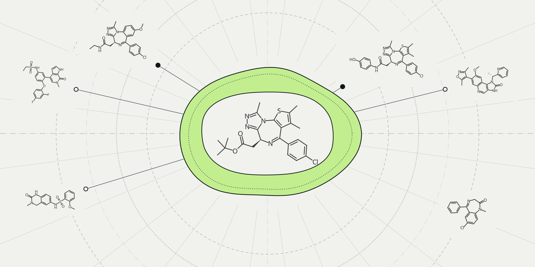
Each branch fails where the other works. Take JQ1, the best-known inhibitor of the bromodomain protein BRD4, and use it as a similarity query against five other BET bromodomain inhibitors mixed with five unrelated drugs. Only two of the five inhibitors come out near the top. The other three, each from a different chemical family, rank below diazepam, and one ranks below caffeine (Table 3).
Docking fails the other way. It does not need a resemblance to anything, but it needs a score to find the right pose, and the GNINA example for JQ1 in BRD4 put every one of its eight poses 4.7 to 5.3 Å from the crystal ligand while printing a respectable −8.957 kcal/mol for the top one. Similarity search cannot see past the query's chemistry; docking cannot see past its scoring function.
What you already know decides which failure you can afford. With a good structure and few ligands, start from the structure. With many measured ligands and no structure, start from the ligands. With both, use both, because the evidence below says combining them beats either alone.
| Starting knowledge | Branch | Typical methods | What it needs | What it cannot see |
|---|---|---|---|---|
| A target structure, few or no known ligands | Structure-based | Pocket detection, docking, structure-based de novo design, free energy calculations | A reliable structure with a defined pocket | Anything the scoring function does not model: solvent, entropy, flexibility |
| Many measured ligands, no usable structure | Ligand-based | Similarity search, pharmacophores, shape matching, QSAR and machine-learning models | Actives, and ideally inactives, measured in a comparable assay | Chemistry unlike the references, and the reason the references bind |
| Both | Combined | Sequential filters, parallel selection, score fusion | Both inputs, and a protocol to merge them | Compounds that neither method ranks highly |
| Neither | Start by getting one | Structure prediction, or a screen for first actives | A sequence, or a few assay results | Not applicable until one input exists |
Structure-based design
Structure-based design starts from the target. The usual first step is to find or define the binding pocket, with a pocket detector such as fpocket or from a crystal ligand. The next is molecular docking: placing each candidate molecule in the pocket and scoring the pose. Free energy calculations and structure-guided design of new molecules come later, on the few compounds that survive.
Its best argument is what it finds. Lyu and colleagues docked 138 million make-on-demand molecules against the dopamine D4 receptor and tested 549 of the best-ranked. At 10 μM, 122 of them (22%) displaced the radioligand, and the screen yielded 81 new chemotypes, 30 of them submicromolar.[8] A virtual screen of that size does not depend on any known active D4 ligand as a starting point.
The survey of 419 prospective docking studies by Zhu and colleagues reads the same way from the other side. About one quarter of the hits were better than 1 μM, and the authors conclude that the main advantage of structure-based screening is new chemotypes, not high potency.[9] Docking is a way to widen the search, not to finish it.
The limits are in the score. Docking usually ranks poses better than it ranks affinities, as the guide to docking scoring functions shows. On a DUD-E subset of 280 actives and 280 decoys across 56 targets, AutoDock Vina reached a ROC AUC of only 0.68; Guterres and Im raised it to 0.83 by adding short molecular dynamics runs on each docked pose.[12]
A structure is also a snapshot: a pocket from one crystal may reject ligands that bind another conformation, which is why ensemble docking uses several.
Ligand-based design
Ligand-based design starts from molecules. If compound A is active, compounds like A may be active too. Similarity search turns that into a number, usually a Tanimoto score between fingerprints. Pharmacophore and shape methods compare 3D features instead of bonds, and QSAR or machine-learning models such as Chemprop learn activity from a larger set of labelled molecules.
It is cheaper than docking and, on retrospective benchmarks, often as good. Hu and colleagues compared 14 two-dimensional fingerprints and four 3D shape methods on a tailored version of the DUD benchmark, and found that ligand-based methods gave higher early enrichment than Glide docking with MM-GBSA rescoring.[4] Tan and colleagues saw the same on nine enzymes: without target-specific tuning of the docking, fingerprint searching was clearly preferred.[11]
The catch is what those benchmarks contain. Von Korff and colleagues compared fingerprints, the Flexophore pharmacophore descriptor and a docking tool on the 2,950 actives of DUD, and fingerprints won on enrichment. After they removed molecules chemically similar to the query, the ranking changed and the Flexophore came out ahead.[5] A fingerprint search is strongest where it recognises analogues, and benchmark actives are full of analogues.
That is the weak point of the whole branch: it finds more of what you already have. For a new chemotype, the query has nothing to match. Deep learning models can predict activity without a receptor structure,[18] though how far they generalise depends on the chemistry they were trained on.
What the benchmarks hide
The retrospective numbers in the CADD literature deserve some suspicion, for both branches. DUD-E, the most used set, has 102 targets and 22,886 ligands, each with 50 property-matched decoys.[2] That is one active for every 51 compounds, about 2%. Tran-Nguyen and colleagues report that DUD, DUD-E and MUV carry obvious and hidden chemical biases that overestimate the accuracy of virtual screening, and built LIT-PCBA from 149 dose-response assays to remove them.[6]
Their selection rule is telling. From 21 target sets, they kept the 15 where at least one of fingerprint similarity, 3D shape similarity or docking enriched the top 1% of the ranking in actives by a factor of at least 2.[6] A factor of 2 was the bar for a usable target, far below the 46-fold early enrichment reported on DUD-E+ below.
Stein and colleagues add a warning about optimising on matched decoys: it can give false confidence, because a real library contains molecules the decoys do not represent.[7] A method tuned on DUD-E has shown that it can separate DUD-E actives from DUD-E decoys, not that it will pick hits from your library.
Combining the two branches
The evidence for combining is stronger than the evidence for either branch alone. Cleves and colleagues compared docking and 3D ligand similarity (their eSim method) on DUD-E+ (Table 2). With one pocket, docking reached a mean ROC AUC of 0.81; with the one cognate ligand as a query, similarity reached 0.77. Five pockets lifted docking to 0.84, and five aligned ligands lifted similarity to 0.83. Combining ensemble docking with similarity gave 0.89 and a 1% early enrichment of 46-fold.[3]
Their advice is blunt: it is unwise to dock into one arbitrary structure or search with one arbitrary ligand.[3] A single query is a single chemotype, which is the lesson of the JQ1 example below. An ensemble of references covers more of the target's chemistry.
| Method | Mean ROC AUC | Compounds ranked above the average active |
|---|---|---|
| Similarity, one cognate ligand | 0.77 | 230,000 |
| Similarity, five ligands, automatic alignment | 0.80 | 200,000 |
| Docking, one pocket | 0.81 | 190,000 |
| Similarity, five crystallographically aligned ligands | 0.83 | 170,000 |
| Docking, five pockets | 0.84 | 160,000 |
| Ensemble docking plus similarity | 0.89 | 110,000 |
Table 2 shows why enrichment at the top matters more than AUC. An AUC of 0.89 still leaves the average active 110,000 places down a million-compound list, far beyond anything you would buy or dock again. What matters is the fraction of actives packed into the first few hundred, which is what an early enrichment factor measures.
Other groups found the same pattern with different tools. Sastry and colleagues fused 2D fingerprint, shape and docking scores with an augmented Z-score and improved top-1% enrichment by 9% on DUD and 25% on MDDR over the best single method.[10] Tan and colleagues found that taking candidates in parallel from each method's own ranking generally beat fusing the ranks.[11]
Drwal and Griffith's review sorts the strategies into sequential, parallel and hybrid, and notes that most published studies use the sequential one, with one method filtering and the other ranking.[17] Sequential is also the cheapest: similarity or a property filter cuts the library, and docking is spent on what remains.
Worked example with JQ1 and the BET inhibitors
JQ1 is a thienotriazolodiazepine that blocks the acetyl-lysine pocket of the BET bromodomains, including BRD4.[13] Other groups have found other chemical families that bind the same pocket. I-BET762 and OTX015 share JQ1's triazolodiazepine core, while I-BET151, PFI-1 and mivebresib do not.[14][15][16][19]
We built a ten-compound test library from these six inhibitors (JQ1 as the query, five others as the actives) plus five unrelated drugs: diazepam, celecoxib, caffeine, ibuprofen and aspirin. This is a constructed control, not a benchmark, and the unrelated drugs are only there to give the ranking something to fall below. We ranked them by Tanimoto similarity to JQ1 using Morgan fingerprints with radius 2 and 2048 bits, calculated with RDKit 2025.03.5.
| Rank | Compound | Binds BET bromodomains | Tanimoto to JQ1 |
|---|---|---|---|
| 1 | OTX015 (birabresib) | Yes | 0.671 |
| 2 | I-BET762 (molibresib) | Yes | 0.432 |
| 3 | Diazepam | No | 0.173 |
| 4 | I-BET151 | Yes | 0.135 |
| 5 | Celecoxib | No | 0.128 |
| 6 | Ibuprofen | No | 0.123 |
| 7 | Aspirin | No | 0.110 |
| 8 | PFI-1 | Yes | 0.096 |
| 9 | Caffeine | No | 0.093 |
| 10 | Mivebresib (ABBV-075) | Yes | 0.086 |
The two inhibitors with a triazolodiazepine core come first, as they should. The three with different cores sit among the unrelated drugs, and mivebresib comes last. Diazepam, a diazepine drug with a chlorinated aromatic ring, outranks I-BET151 on shared fragments alone, with no link to the bromodomain. With the radius of 3 that the ProteinIQ molecular similarity search uses by default, the picture is the same: I-BET151, PFI-1 and mivebresib still rank below diazepam.
The two branches fail in opposite ways on this one protein. Similarity finds the analogues and misses the new chemotypes. Docking does not care about chemistry, but in the GNINA run its score pointed at a pose about 5 Å from the crystal ligand. The shortcut is to use all six inhibitors as references, which is what Cleves and colleagues found worked best, and then to check the docked poses with PoseBusters and PLIP before reading any score.
Choosing an approach
Start from what you hold, not from the method you prefer (Table 4).
| Your situation | Start with | Add | Then check |
|---|---|---|---|
| One known active, no structure | Similarity search with that active | A second and third chemotype as queries as they appear | Whether hits are only analogues of the query |
| Dozens of measured actives and inactives | A QSAR or machine-learning model, plus similarity | Docking if a structure exists | Performance on compounds unlike the training set |
| Good structure, no ligands | Docking into the pocket | Several pocket conformations, a learned rescoring | Redocking of any known ligand, then pose geometry |
| Structure and ligands | Filter by similarity or properties, then dock the remainder | Parallel selection from both rankings | Known actives and decoys put through the same protocol |
| Only a predicted structure | Check its pocket confidence first | Co-folding with the ligand | Whether docking recovers any known ligand |
Three habits cover most of the table. Use several references, not one. Set the cutoff from your own target, using known actives docked or searched with the same protocol as the library, which is the idea behind redocking validation. And clean the shortlist before ordering anything: PAINS and drug-likeness filters remove compounds that look like hits for the wrong reasons.
Computer-aided drug design tools on ProteinIQ
The usual order is to collect known actives, rank or filter the library, dock what remains, check the poses, then spend the expensive calculations on a handful. Table 5 lists tools for each step in small-molecule discovery, and the virtual screening use case shows how they fit together.
| Task | Tool |
|---|---|
| Collect known actives for a target | ChEMBL download |
| Rank a library by similarity to a query molecule | Molecular similarity search |
| Calculate fingerprints and descriptors | Molecular descriptors |
| Train a property model on your own measurements | Chemprop |
| Find and rank binding pockets | fpocket |
| Dock compounds into a pocket | AutoDock Vina |
| Dock and rescore poses with a learned model | GNINA |
| Check that a pose is physically plausible | PoseBusters |
| List the contacts in a docked complex | PLIP |
| Remove assay-interference compounds | PAINS filter |
| Predict ADMET properties of the shortlist | ADMET-AI |
| Compare close analogues by relative binding free energy | OpenFE |
Frequently asked questions
What is computer-aided drug design?
It is the use of computational methods to find and improve drug candidates. The methods are grouped into structure-based approaches, which use the target's 3D structure, and ligand-based approaches, which use molecules already known to be active.[1]
What is the difference between structure-based and ligand-based drug design?
Structure-based design needs a target structure and asks whether a molecule fits the pocket. Ligand-based design needs known actives and asks whether a molecule resembles them. The first can propose new chemistry; the second is cheaper and tends to rank analogues of its references best.[4][5]
Which should I use first?
Use whichever input you have. If you have both, the evidence favors using both: in DUD-E+, combining ensemble docking with ligand similarity gave a mean ROC AUC of 0.89, against 0.81 for single-structure docking and 0.77 for a single ligand query.[3]
Is docking better than similarity searching?
Not reliably. On a tailored DUD set, ligand-based methods gave higher early enrichment than Glide docking with MM-GBSA rescoring, and on nine enzymes fingerprint searching beat untuned docking.[4][11] Docking is the better route to new chemotypes, as in the D4 screen where 81 were found.[8]
How reliable are virtual screening benchmarks?
Less than their headline numbers suggest. DUD, DUD-E and MUV carry chemical biases that inflate accuracy, and LIT-PCBA was built to avoid them.[6] Matched decoys can also mislead when a method is tuned against them.[7]
Do I need a crystal structure for structure-based design?
You need a 3D structure, and its quality sets the ceiling. When a crystal structure is not available, a predicted model can stand in, provided its pocket confidence is checked and docking can recover a known ligand first.