How to visualize AutoDock Vina results
From a docked .pdbqt file to poses, contacts and a redocking check

Matic Broz, PhD14 min
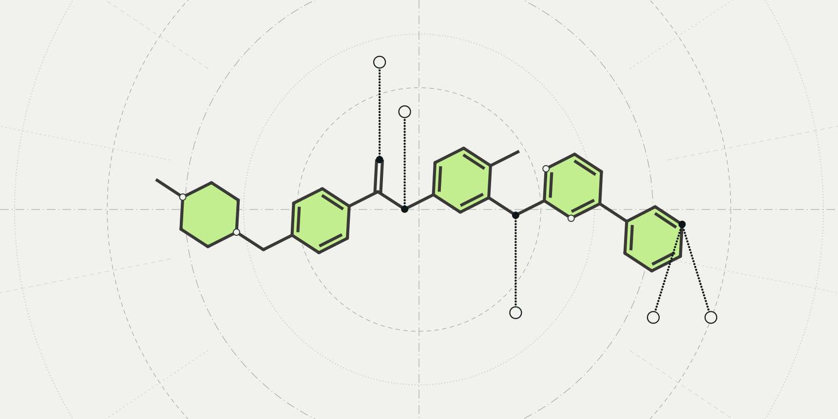
A Vina job ends with a table. For imatinib docked into c-Abl kinase it lists five poses, the best at −12.512 kcal/mol, and nothing in it says whether that pose sits in the ATP site, which residues it touches or whether it resembles the way the drug really binds.
To see that, open the receptor and the output PDBQT file together in a molecular viewer. PyMOL and ChimeraX both do it, and so does the PDB viewer by ProteinIQ, which lists every pose with its score and measures the pocket and the contacts for you.

The examples below use one public run, the c-Abl and imatinib example from AutoDock Vina on ProteinIQ. Its receptor is the kinase domain from PDB entry 1IEP, which already holds imatinib, so the docked poses can be compared with the real one.[9] How the job was set up is covered in the AutoDock Vina tutorial, and what the score means in the Vina score guide.
 AutoDock VinaDock ligands into protein structures and estimate binding modes, poses, and affinity scores.Open example
What AutoDock Vina writes, and what it leaves out
Vina writes the ligand poses and nothing else. The receptor is a separate file, so a pose opened alone floats in empty space. This job returned twelve files, of which four matter for viewing (Table 1). The poses use the same coordinates as the receptor you docked into, which is why any file of that same structure lines up with them.
| File | Holds | Use it for |
|---|---|---|
Imatinib_output.pdbqt | All five poses, one per MODEL, best first | Browsing every pose in one go |
pose_mode01.pdbqt to pose_mode05.pdbqt | One pose each | Opening or sharing a single pose |
protein.pdb | The receptor as submitted, chains A and B | The protein to show the poses in |
prepared_receptor.pdbqt | The receptor after preparation, with charges and atom types | Seeing exactly what Vina scored against |
Each pose starts with a REMARK VINA RESULT line holding the affinity and the two RMSD columns, followed by the atoms. This is the top of the combined file:
MODEL 1
REMARK VINA RESULT:   -12.512      0.000      0.000
REMARK INTER + INTRA:         -18.265
REMARK INTER:                 -17.633
REMARK INTRA:                  -0.632
REMARK UNBOUND:                -0.632
REMARK SMILES Cc1ccc(NC(=O)c2ccc(CN3CCN(C)CC3)cc2)cc1Nc1nccc(-c2cccnc2)n1
...
ATOM      1  N   UNL     1      16.660  51.746  14.707  1.00  0.00    -0.322 N
ATOM      2  C   UNL     1      15.779  51.683  15.772  1.00  0.00     0.255 C
Each pose has 39 atoms: the 37 heavy atoms of imatinib and the two polar hydrogens that Vina keeps. Every atom belongs to a residue called UNL, the generic ligand name, so there is no drug name to select by.
Vina also returned five poses where it found nine; the four others scored more than 3 kcal/mol worse than the best and were dropped. The PDBQT file guide explains the other columns, and the SMILES remark is what lets Meeko rebuild bond orders later.
How to view AutoDock Vina results in PyMOL
PyMOL's Load page lists .pdb, .mol, .mmod, .xplor, .ccp4, .r3d, .pse and .pkl and does not mention PDBQT.[5] The dependable route is therefore to convert the poses to SDF first, which also restores the bond orders that PDBQT drops. Meeko's mk_export.py, used in the AutoDock Vina tutorial, writes the poses of a Vina output file to SDF, and the PDBQT to SDF converter does it in the browser.
- Run
mk_export.py Imatinib_output.pdbqt -s Imatinib_output.sdf. - In PyMOL, run
load protein.pdb, thenload Imatinib_output.sdf, poses. - Run
show sticks, posesto draw the ligand. - Run
select pocket, byres (poses around 5), thenshow sticks, pocket, to draw every residue with an atom within 5 Å of any pose. - Run
distance contacts, poses, pocket, 3.5, mode=2to draw the polar contacts as dashed lines.
A file with several models loads as the states of one object; multiplex=1 on load loads them as separate objects instead, and split_states poses splits an object that is already loaded.[5][6] The selection byres (poses around 5) follows the selection algebra on the PyMOL wiki, and mode=2 is the distance option that shows only polar contacts.[7][8]
How to view AutoDock Vina results in ChimeraX
ChimeraX reads PDBQT directly. Run open protein.pdb, then open Imatinib_output.pdbqt. When a file holds several docked poses, ChimeraX starts the ViewDock tool automatically, with a table that has one row per pose.[1][2] The first two columns, ID and Rating, are made by ChimeraX; the others come from what the file contains. The tool's HBonds and Clashes buttons then draw the contacts of the poses you tick.[2]
From the command line, hbonds #2 restrict #1 reveal true finds hydrogen bonds between the poses (model 2) and the protein (model 1), where restrict #1 limits the search to bonds with the protein.[4]
The rmsd command measures the deviation between two atom sets without moving either, but it pairs atoms in order, so both sets must hold the same atoms in the same order.[3] A docked pose and a crystal ligand rarely do, which makes it a poor fit for the redocking check below.
How to visualize AutoDock Vina results in the PDB viewer
The PDB viewer opens PDBQT files directly and runs in the browser, and files you open are read there and not uploaded. Public example jobs need no account to open or download, so the c-Abl files used here can be fetched without signing in.
- On the job's Files tab, download
protein.pdbandImatinib_output.pdbqt. - Open the PDB viewer, click the upload button at the right of the search box, and select both files together. They open in one scene.
- Click Binding pocket in the left toolbar and check that Ligand reads
UNL 1 · Imatinib_output.pdbqt. - Click Ligand interactions to draw the contacts of the pose on screen.
Select both files in one go, because opening files replaces what was in the scene. If you opened the poses alone, the panel tells you a pose without a protein has nothing to contact.
Browse the poses by score
When a pose file holds several poses, the side panel lists each with its name and score, read from the REMARK VINA RESULT lines, under a header such as Pose 3 of 5 · Affinity (kcal/mol). The arrows beside it, the arrow keys or a click on a row move between poses, and the pocket follows the pose you pick. Overlay all poses draws the other poses as faded grey sticks.
That is the quickest way to see whether the top poses agree or sit in different places.
See the pocket and its contacts
Binding pocket shows which residues line the ligand's site, within a distance you set (5 Å by default). The panel lists each residue with its closest distance, and Copy residue IDs copies them in the A:25,B:50 form that Vina takes for flexible residues. Ligand interactions adds dashed 3D lines for each contact and a table by type: hydrogen bonds, salt bridges, hydrophobic contacts, π-stacking and the rest. Each kind can be switched on or off, and 2D diagram draws the ligand flat with its contacting residues around it, to save as SVG or PNG for a slide.
For this pose the geometry is easy to check by hand.
Counting residues with any heavy atom within 5 Å, pose 1 touches 25 residues of chain A, all of them also within 5 Å of the crystal imatinib, which has 28.
Pose 1 has five pairs of ligand and protein nitrogen or oxygen atoms within 3.5 Å, and the crystal ligand has the same five: Glu286, Thr315, Met318 (two atoms) and Asp381. The crystal also has two backbone carbonyl contacts, from Ile360 and His361, near the methylpiperazine end that this pose does not reproduce. That count uses distance alone, without the angle test that a hydrogen bond search adds, so treat it as a cross-check of what the viewer reports.
Compare a pose with the crystal ligand
Search 1IEP in the same scene, for example with Shift+Enter, to add the entry beside the poses. Its imatinib then sits in the site as a reference to look at, and Superpose with the Ligand method fits one ligand onto another.
That method fits first and then reports the RMSD, so the number it gives measures how well the two shapes match once the placement error is gone. A redocking test asks for the opposite, the distance without any fit. We calculated both for the c-Abl poses with a short script, and pose 1 gives 0.70 Å fitted against 0.94 Å in place.
Checking the top pose by redocking
Because 1IEP holds imatinib, the poses can be scored against the answer. We measured each pose against the crystal ligand in chain A of 1IEP (residue STI 201), on all 37 heavy atoms, in the receptor frame with no fitting, trying every symmetry-equivalent numbering of the atoms and keeping the lowest (Table 2).
| Pose | Affinity (kcal/mol) | Vina RMSD l.b. (Å) | Vina RMSD u.b. (Å) | RMSD to crystal (Å) |
|---|---|---|---|---|
| 1 | −12.512 | 0.000 | 0.000 | 0.94 |
| 2 | −11.290 | 1.405 | 2.102 | 1.43 |
| 3 | −10.489 | 2.879 | 12.419 | 12.24 |
| 4 | −10.287 | 1.790 | 13.534 | 13.39 |
| 5 | −9.775 | 2.279 | 12.652 | 12.54 |
Poses 1 and 2 both pass the usual 2 Å test, and pose 1 is within 1 Å. Poses 3 to 5 sit 12 to 13 Å from the crystal ligand, even though Vina's lower bound puts them only 1.8 to 2.9 Å from pose 1. The lower bound pairs each atom with the nearest atom of the same element, so a ligand that has moved can still read as close. The upper bound, 12 to 14 Å, is the column that shows it, and a 3D view shows it at once.
The wrong placements are also the poorer scores here, but a score gap is not a guarantee. The redocking and validation guide shows a cross-docking case where the best-scoring pose is 10 Å off. The RMSD guide explains why the atom pairing matters, and PoseBusters reports a symmetry-corrected RMSD against a reference ligand along with chemistry and geometry checks.
Common mistakes when viewing Vina results
| What you see | Cause | Fix |
|---|---|---|
| The ligand floats in empty space | Only the output file is open; Vina does not write the receptor | Open protein.pdb or prepared_receptor.pdbqt with it |
| The ligand sits outside the protein | A receptor from a different structure or frame | Use the receptor file from the same job |
| Poses appear in one chain only | The receptor has two chains and the box covers one site | Look at the chain the box was centred on |
| Bonds or hydrogens look wrong | PDBQT keeps only polar hydrogens and no bond orders | Convert to SDF with Meeko and judge the heavy atoms |
| Vina's RMSD looks small but the poses differ | The lower bound ignores which atom is which | Overlay the poses and check the upper bound |
After the poses
Once a pose looks right, check it with the PLIP interaction profiler, which lists contacts by type from a structure file, and read the protein-ligand interactions guide for what each contact type means. To fit the receptor of another structure onto this one, the superimpose protein structures guide shows the options.
Rescoring the same poses with GNINA gives a second opinion, and the viewer's Send to menu passes the receptor and a chosen pocket to Vina, with the box centred on it, to dock another ligand.
Frequently asked questions
How do I open a Vina output file?
Open the receptor and the output .pdbqt together in a viewer. ChimeraX and the PDB viewer read PDBQT directly. For PyMOL, convert the file to SDF first, because its Load page does not list PDBQT.
How do I see all the poses Vina returned?
All poses are in the one output file, one MODEL each, best score first. A viewer that understands them lists the poses and lets you step through or overlay them; in PyMOL they load as states of one object.
Why does Vina return fewer poses than I asked for?
The number of poses is a maximum. Vina drops poses that sit too close to a better one or lie more than the energy range, 3 kcal/mol by default, below the best. The c-Abl example returned five of nine.
Does a low RMSD in Vina's table mean the pose is correct?
No. Vina's RMSD columns compare each pose with pose 1 of the same run, not with an experimental structure, and the lower bound can read small for poses that are far apart. Accuracy needs the crystal ligand and a separate calculation.
Can I view Vina results without installing anything?
Yes. The PDB viewer opens the receptor and the poses in the browser, with no account for public example jobs. AutoDock Vina on ProteinIQ also shows the poses in its own Structure tab straight after the run.