Amyloid fibrils and protein misfolding
One architecture, many shapes, and what that means for disease

Matic Broz, PhD14 min
What is an amyloid fibril?
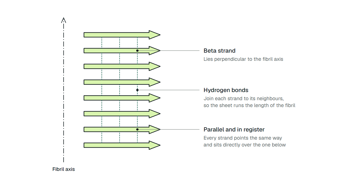
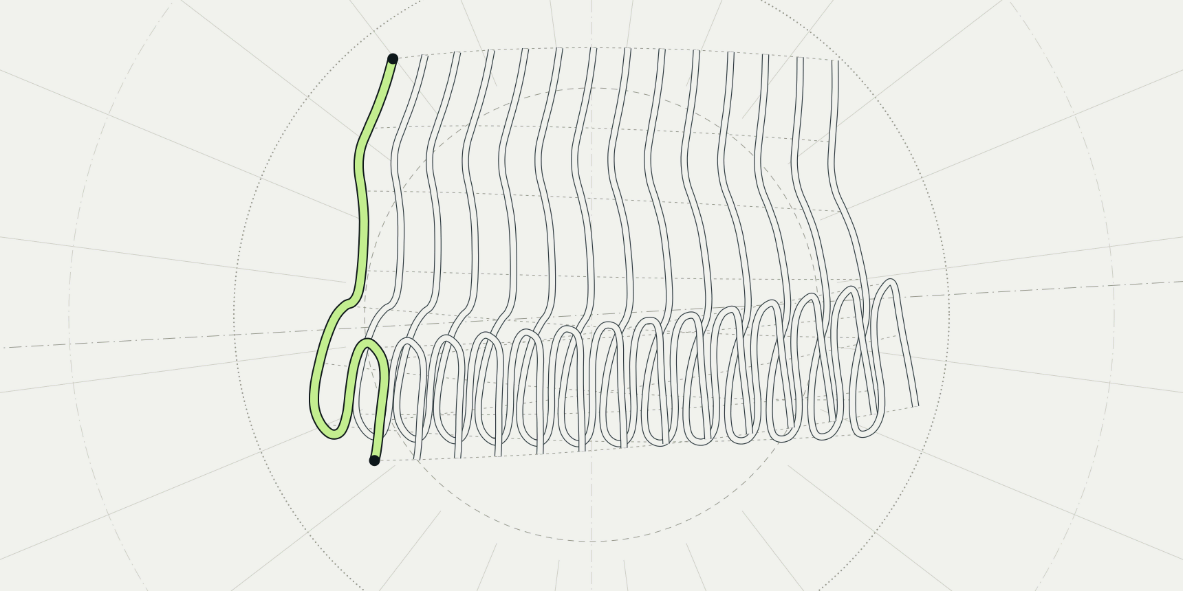
An amyloid fibril is a long, unbranched protein filament built from beta strands that stack across the fibril axis like rungs on a ladder, so that hydrogen-bonded sheets run its whole length. Proteins with unrelated sequences and native folds build this same cross-beta architecture. Sunde and colleagues found that six fibrils taken from patient tissue and two synthetic fibrils all gave similar X-ray fibre diffraction patterns.[1]
The shared architecture hides how much the details differ. Synthetic amyloid beta 42 (Aβ42), the Alzheimer peptide, forms two intertwined protofilaments with each subunit folded into an "LS" shape (PDB 5OQV, 4.0 Å).[2] Aβ42 filaments extracted from human brains are S-shaped instead, in two related types (PDB 7Q4B, 2.5 Å), and their authors report that they differ from filaments assembled in vitro.[3]
Same peptide, same architecture, different fold. What a fibril means for a cell depends on that fold, on how fast fibrils multiply once the first ones appear and on whether the cell controls where they assemble. Aggregation predictors such as Aggrescan3D speak only to the first step, which residues are prone to aggregate.

What is protein misfolding?
Protein misfolding means a chain fails to reach, or fails to stay in, the compact native fold that carries out its function. It is a broader idea than amyloid. A misfolded chain can be degraded, can clump into disordered amorphous aggregates or can assemble into ordered fibrils. Protein folding is the route to the native state, and denaturation is what heat, pH or chemicals do to take a protein off it.
Fibrils are not a quirk of a few disease proteins. Chiti and Dobson describe the amyloid state as an inherent tendency of peptides and proteins, and Knowles, Vendruscolo and Dobson call the ability to form it more general than previously imagined.[4][5] If almost any chain can form amyloid, the useful questions become what fold it takes and why cells usually stop it.
What the cross-beta structure looks like
The clearest atomic picture comes from a seven-residue segment. Nelson and colleagues solved GNNQQNY from the yeast prion protein Sup35 at 1.8 Å, from microcrystals that formed alongside amyloid-like fibrils (PDB 1YJP). It is a double beta sheet, each sheet built from parallel segments stacked in register. Side chains from the two sheets interlock in a dry interface called a steric zipper.[6]
Two properties of fibrils follow from that picture. Nelson and colleagues argue that the structure explains self-seeding, in which an existing fibril templates new growth, and the tendency to form polymorphic structures, so that one sequence can give several.[6] Eisenberg and Sawaya add that a polymorph can reproduce itself by seeding, and suggest that polymorphs may be the molecular basis of prion strains.[7]
Full-length fibrils were harder to solve. In 2017 Eisenberg and Sawaya counted only a few dozen amyloid structures, mostly short segments, against roughly 100,000 globular proteins.[7]
Cryo-electron microscopy and solid-state NMR changed that. Iadanza and colleagues report that the first near-atomic structures of fibrils formed in vitro, seeded from plaque material or taken directly from tissue were far more elaborate than anticipated.[8] Those structures are where the difference between test-tube and patient fibrils became visible.
How amyloid fibrils form
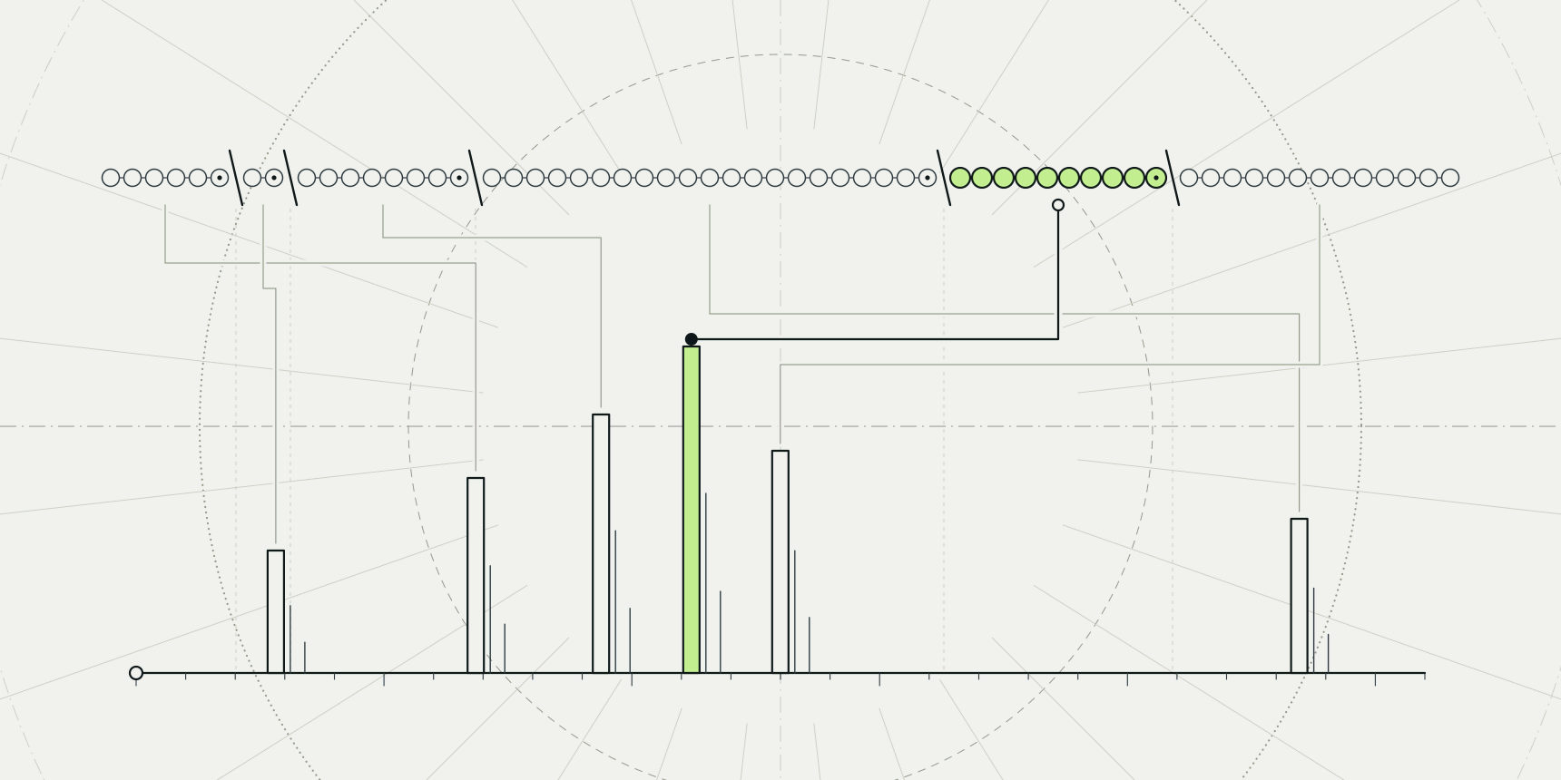
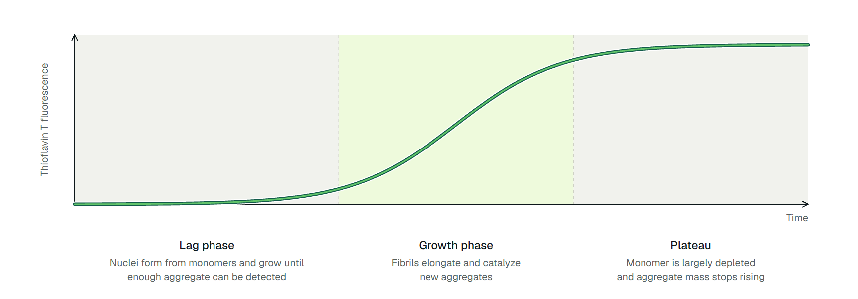
In a test tube, fibril growth is usually followed with thioflavin T, a dye whose fluorescence rises sharply when it binds fibrils.[9] Plotted against time, the signal is sigmoidal: a lag phase with little signal, a rapid growth phase and a plateau once the free protein is used up (Figure 2).[10]
The lag phase is not a wait for one rare nucleus. Arosio and colleagues argue that several processes run in parallel and that typically millions of primary nuclei form during the lag phase. The lag time is the time those early nuclei need to grow and multiply to a detectable concentration.[10]
The growth phase is steep because fibrils make more fibrils. Knowles and colleagues showed analytically that growth is often dominated by secondary nucleation, in which existing filaments generate new aggregates.[11]
Cohen and colleagues measured the consequence for Aβ42. Once enough fibrils had accumulated, toxic oligomers formed mainly through secondary nucleation catalysed by the fibrils, so a growing fibril load raises the oligomer load with it.[12]
Diseases that involve amyloid fibrils
Alzheimer disease is the most common cause of dementia. WHO reports 57 million people living with dementia in 2021 and says Alzheimer disease may contribute to 60 to 70% of cases.[13] For Parkinson disease, WHO gives global estimates of more than 8.5 million people in 2019.[14] Neither figure counts only amyloid, since dementia has other causes, but both diseases are marked by fibrils whose structures have now been solved from patient tissue.
Table 1 lists the main examples with a PDB entry to open in a viewer. Where the fibril material came from matters, because only some entries are filaments extracted from patients.
| Disease | Protein | Fibril core in the cited structure | Origin and PDB example |
|---|---|---|---|
| Alzheimer disease (plaques) | Amyloid beta 42 | S-shaped protofilament folds, type I mostly in sporadic disease | Human brain, 7Q4B |
| Alzheimer disease (tangles) | Tau | Two identical protofilaments of residues 306 to 378 in a combined cross-beta and beta-helix fold | Human brain, 5O3L |
| Parkinson disease, Lewy body dementia | Alpha-synuclein | Single protofilament, the Lewy fold | Human brain, 8A9L |
| Multiple system atrophy | Alpha-synuclein | Two filament types, each of two different protofilaments | Human brain, 6XYO |
| Prion disease | Prion protein | Parallel in-register beta sheets, one rung per monomer, no paired protofilaments | Brain-derived infectious scrapie fibril (263K strain), 7LNA |
| Type 2 diabetes (islet deposits) | Islet amyloid polypeptide | Two intertwined S-shaped protofilaments | Synthetic peptide fibrils formed in vitro, 6Y1A |
| Hereditary transthyretin amyloidosis | Transthyretin | Single protofilament from an N-terminal and a C-terminal fragment | Patient tissue, Val30Met, 6SDZ |
The fold changes with the disease. Fitzpatrick and colleagues found that tau filaments of other shapes characterize other neurodegenerative diseases.[15] The alpha-synuclein rows show the same split inside one protein: the Lewy fold of Parkinson disease is a single protofilament, unlike those of multiple system atrophy.[16] Schweighauser and colleagues suggest that distinct conformers, or strains, characterize specific synucleinopathies.[17]
Two rows need a caution. The islet amyloid polypeptide structure comes from synthetic peptide, and Röder and colleagues describe the fibrils as associated with beta-cell loss and implicated in type 2 diabetes, short of a proven cause.[18] The transthyretin fibril, from one of the most abundant forms of systemic amyloidosis, is built from fragments of the protein rather than whole chains, as Schmidt and colleagues found.[19]
The mature fibril is not necessarily the toxic species. Bucciantini and colleagues found that species formed early in the aggregation of two proteins unrelated to any disease were inherently highly cytotoxic.[21] That leaves open which species a treatment should go after.
The clinic shows the same ambiguity. In the 18-month Clarity AD trial of 1,795 people with early Alzheimer disease, lecanemab, an antibody that binds soluble Aβ protofibrils, slowed decline on the 0 to 18 CDR-SB scale by 0.45 points (1.21 against 1.66 on placebo, about 27% less decline). The authors call this moderately less decline.[22] Clearing the amyloid target helped, but not much, and not without adverse events.
Not all amyloids are harmful
Some amyloid is made on purpose. Fowler and colleagues reported that the mammalian protein Pmel17 forms amyloid that templates melanin synthesis in the melanosome, and concluded that amyloid is a fundamental nonpathological fold used from bacteria to humans.[23] Chapman and colleagues showed that the curli fibers of Escherichia coli are amyloid, assembled by a dedicated machinery.[24]
The contrast with disease is control. Functional amyloid assembly is directed by dedicated nucleator and chaperone-like proteins in bacteria.[24] Fowler and colleagues describe Pmel17 amyloidogenesis as carefully orchestrated by the secretory pathway, with membrane sequestration and proteolytic steps that protect the cell.[23] The fibril fold is not the problem. Uncontrolled assembly in the wrong place is.
How cells defend against misfolding
Cells keep their proteins folded with a network of chaperones that prevent aggregation and promote folding. Newly made chains need it most: Hartl and colleagues note that newly synthesized proteins are at great risk of aberrant folding and aggregation.[25]
Protein molecules are highly dynamic, so Hartl and colleagues argue that this surveillance has to be constant, and that an age-related decline in proteostasis capacity allows aggregation diseases such as Alzheimer and Parkinson disease to manifest.[25] Balch and colleagues call the wider set of activities the proteostasis network and link its deficiencies to metabolic, cancer, neurodegenerative and cardiovascular disorders.[26]
Chiti and Dobson reach the same point from the disease side: an efficient network actively combats aggregation and can still fail.[4] Disease therefore needs two things, a protein that can form amyloid, which is almost any protein, and a defence that stops keeping up.
What aggregation predictors can say about amyloid
Predictors answer a narrower question than fibril structure. They estimate which segments or residues of a protein are likely to drive aggregation, not what fold a fibril will take. TANGO models intermolecular beta-sheet formation with fully buried aggregate cores, and its authors report that it predicted the aggregation of 179 literature peptides and of a new set of 71 peptides from disease-related proteins.[27]
AGGRESCAN uses an aggregation-propensity scale derived from in vivo experiments and assumes that short, specific stretches modulate aggregation.[28] CamSol answers a neighbouring question: it screens tens of thousands of mutations for solubility gains, so it ranks variants rather than locating fibril cores.[29]
Structure changes what can be asked. The Aggrescan3D authors point out that sequence-only methods struggle with folded globular proteins, where aggregation-prone sites are often not contiguous in sequence or are buried in the native structure.[30] Aggrescan3D therefore applies the AGGRESCAN scale to the three-dimensional structure.
Version 2.0 extends its dynamic mode to larger and multimeric proteins and adds predicted solubility and stability changes on mutation.[31] ProteinIQ runs the static workflow, so the dynamic mode plays no part in its results.
Two limits follow for amyloid work. A flagged segment does not say which polymorph forms, because the predictor papers report propensity, not fibril structure. And intrinsically disordered peptides such as Aβ and alpha-synuclein have no single folded structure to score, so a structure-based tool suits folded precursors such as transthyretin better.
One peptide, three structures
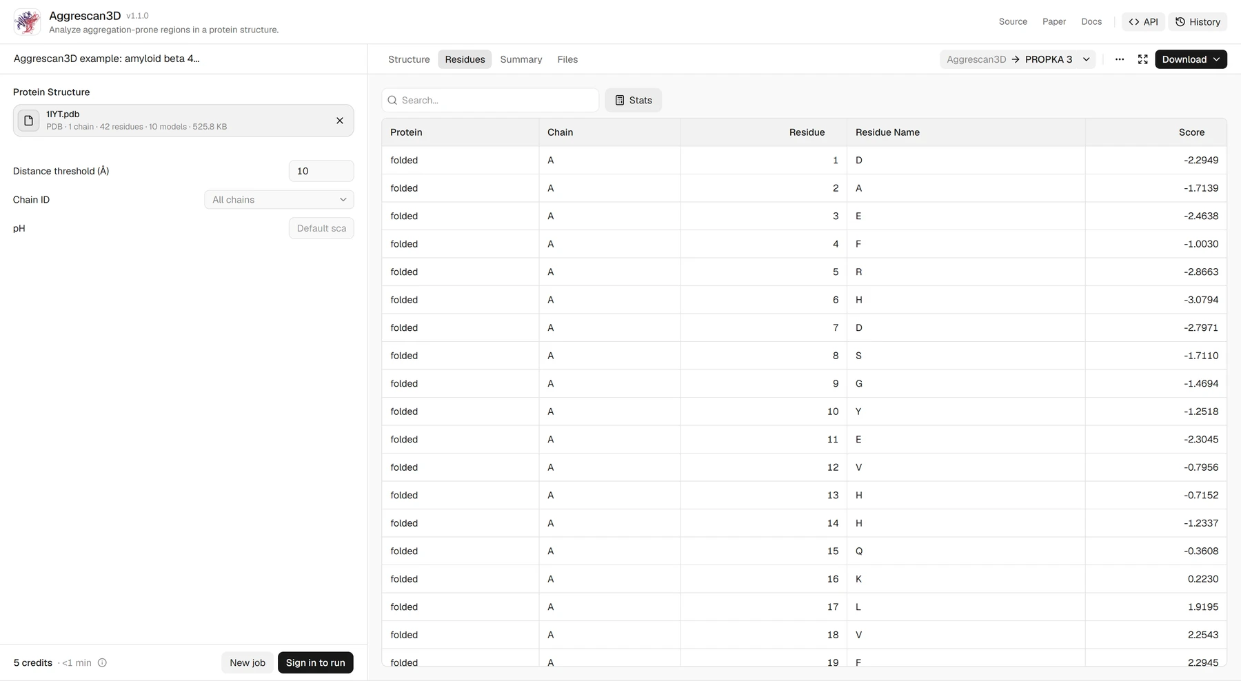
Aβ42 shows how far apart these answers sit. The two fibril structures from the start of this page already disagree with each other: LS-shaped subunits in synthetic fibrils (5OQV), S-shaped ones in brain filaments (7Q4B).[2][3] A predictor sees neither. The public Aggrescan3D example for Aβ42 scores PDB entry 1IYT, a solution NMR structure of the peptide in an apolar microenvironment.
The run uses the default 10 Å distance threshold, all chains and no pH, and costs 5 credits. Its Residues tab gives one score per residue, positive for aggregation-prone structural environments and negative for environments that favour solubility. The Summary tab reports an average A3D score of 0.16 and a maximum of 4.18.
Those scores describe the peptide in the conformation 1IYT gives it, which is neither fibril fold. An A3D score is a residue score, not a probability, and two runs are comparable only with the same pH, distance threshold and chain scope.
Read the disagreement as a difference in questions. The run says which residues of one conformation sit in sticky environments. The cryo-EM structures say what fold the chain actually takes in a fibril, and that answer changed between a test tube and a brain. Only the second kind of evidence settles the fold.
Matching the question to the method
Table 2 pairs each question with the method that can answer it and the experiment that checks it.
| Question | Start with | Check with |
|---|---|---|
| Which stretch of a peptide could form fibrils? | A sequence predictor such as TANGO or AGGRESCAN | Thioflavin T assay and electron microscopy |
| Where are the sticky patches on a folded protein? | Aggrescan3D on its structure | Mutate the top residues and measure aggregation |
| Is a deposit amyloid or amorphous? | Thioflavin T and fibre diffraction | Cross-beta signature by diffraction or cryo-EM |
| What fold does a fibril take? | Cryo-EM or solid-state NMR | Compare patient-derived and in vitro fibrils |
Run a predictor on the kind of input it was built for and treat its output as a shortlist for experiments. A high score for a segment says nothing about the fold of any fibril it might form; for that, the structure has to be determined, ideally from patient tissue.
Misfolding and aggregation tools on ProteinIQ
Get a structure first, score it for aggregation-prone regions, then compare against sequence-based solubility models.
- PDB Download: download an experimental structure by PDB ID
- ESMFold: predict a structure from a sequence when none exists
- Aggrescan3D: score residues for aggregation propensity in a folded structure
- Protein-Sol and NetSolP: estimate solubility from sequence
Frequently asked questions
What are amyloid fibrils made of?
Amyloid fibrils are built from a single protein or peptide, such as Aβ, tau, alpha-synuclein or transthyretin fragments, stacked as beta strands perpendicular to the fibril axis. Unrelated precursors share this cross-beta core.[1]
Is protein misfolding the same as aggregation?
No. Misfolding means the chain has not reached or has left its native fold. Aggregation is one possible result; chaperones can also prevent a chain from aggregating and promote its folding.[4][25]
Are amyloid fibrils always toxic?
No. Functional amyloids include bacterial curli and the mammalian protein Pmel17, which assemble under tight cellular control.[24][23] In disease, species formed early in aggregation can be more toxic than the mature fibrils.[21]
Do fibrils made in the lab match those in patients?
Not necessarily. For Aβ42 and for alpha-synuclein, filaments from human brain differ from those assembled in vitro.[3][17]
Can Aggrescan3D predict whether my protein forms amyloid?
It scores residues in a folded structure for aggregation-prone environments, which can point to candidate sticky regions. It does not report a probability or predict the fibril structure, and the ProteinIQ version runs the static workflow only.[30]
How many people live with amyloid-related diseases?
No single count exists. WHO reports 57 million people with dementia in 2021, of whom Alzheimer disease may account for 60 to 70%, and more than 8.5 million with Parkinson disease in 2019.[13][14]