Jalview alternatives and multiple sequence alignment viewers
What to open an alignment in, and how to see conservation on a structure

Matic Broz, PhD12 min
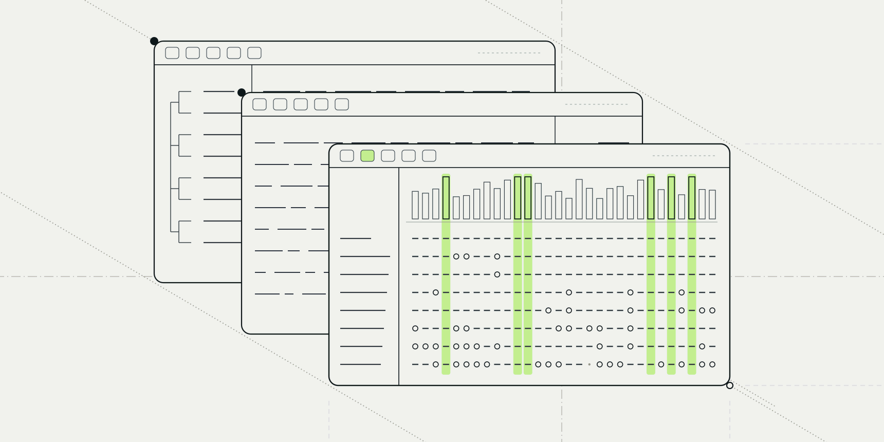
Sixteen cytochrome c sequences, from horse to yeast to a ciliate, went through Clustal Omega and came back as a text file of 115 columns. Nothing in that file says which positions matter. To see that, you need a viewer that colours residues, shows a conservation bar, and ideally tells you where the conserved columns sit in the protein.
Jalview is the name most people reach for, and it is a good answer. It is also a Java program you install, and its current release series has stopped getting new features. If you only want to look at an alignment, or put its conservation on a structure, lighter options exist.
For a browser-only look at an alignment, the MSA viewer by ProteinIQ opens it with no install or account.

What Jalview does, and what it costs
Jalview is free. Its site describes it as an open source project released under the GPL, and as a free cross-platform program; the pages we read do not say which GPL version.[3] It is an alignment editor and analysis workbench: you edit alignments, annotate them, draw trees and PCA plots, and link the columns to a 3D structure.[4] Jmol is built in, and Jalview can be set up to use ChimeraX, Chimera or PyMOL instead.[3]
The download page lists a macOS installer for Intel and Apple Silicon, a Windows x64 installer, Linux installers for x64 and aarch64, and a generic Unix archive. Its latest installer is 2.11.5.1, released on December 10, 2025, which updates itself to 2.11.5.3 (October 27, 2026) on launch.[1] The 2.11.5 release is described as the last of the 2.11 series, which is now in maintenance mode, and new work goes to 2.12.[2]
The cost is therefore not money. It is the install, a Java runtime behind it (the installer file names carry java_8), and a desktop interface with a lot of menus.[1] There is also JalviewJS, which compiles the same Java program to JavaScript so it runs in a browser.[3]
If you work with alignments every day, none of that is a reason to leave. It is a reason to look elsewhere when the job is one alignment, one figure or one question.
Jalview alternatives at a glance
| Program | Runs on | Price and licence | Linked to a 3D structure | Best for |
|---|---|---|---|---|
| Jalview | Windows, macOS, Linux; JalviewJS in a browser | Free, GPL | Yes (Jmol, ChimeraX, Chimera, PyMOL) | Editing, annotating and analysing alignments |
| AliView | Windows, macOS, Linux | Free, GPLv3 | No | Large alignments and sequence editing |
| MView | Anywhere Perl runs | Free, GPL | No | HTML alignments from the command line |
| UGENE | Windows, macOS, Linux | Free, GPLv2 | Structure viewer for PDB and MMDB files, not linked to columns in what we read | One program for alignment, trees and sequences |
| Geneious Prime | Windows, macOS, Linux | $200 student, $660 academic, $2,350 corporate per year | Not on its pricing page | A paid suite for a whole lab |
| MSAViewer and NCBI MSA Viewer | Web browser | Free | No | Embedding or sharing an alignment |
| MSA viewer by ProteinIQ | Web browser | Free, no account | Through the PDB viewer | Looking at an alignment without an install |
| PDB viewer by ProteinIQ | Web browser | Free | Yes, colours the chain by conservation | Seeing where the conserved residues are |
AliView
AliView is a free alignment viewer and editor, licensed under GPLv3, for Windows, macOS and Linux. It opens FASTA, NEXUS, PHYLIP, Clustal and MSF files with no stated size limit, and it can call MUSCLE or MAFFT to align sequences, translate nucleotides and search for degenerate primers. Its page lists version 1.33 from September 28, 2026.[8]
The pages we read describe no link to a 3D structure. AliView is the closest replacement for Jalview's editing side, and it is also a Java application.
MView
MView, by Nigel Brown, turns alignments and BLAST or FASTA search results into coloured HTML from the command line. It is free (GPL, version 2 or later), written in Perl, and reads Clustal, HSSP, MSF, FASTA, PIR and MAF alignments. It writes HTML, FASTA, Clustal, MSF, PIR and tab-separated tables.[9]
It has no interactive window, which is its point: a script can make the same figure from a hundred alignments, and the HTML pastes into a lab web page.
UGENE
UGENE is free software under GPLv2 for Windows, macOS and Linux. Its feature list covers alignment with ClustalW, Clustal Omega, MUSCLE, Kalign, MAFFT and T-Coffee, phylogeny, and a 3D viewer for PDB and MMDB files. The latest release it lists is 53.1 from March 2026.[10]
It suits someone who wants sequences, alignments and trees in one desktop program. We did not find a documented link between alignment columns and the structure view.
Geneious Prime
Geneious is the paid option. Its pricing page lists personal subscriptions per year at $200 for degree-enrolled students, $660 for academic and government users, and $2,350 for corporate users, with team plans above that.[11] It runs on Windows, macOS and Linux and covers far more than alignment. Its pricing page does not describe the alignment viewer or any structure link, and we did not review them.
It makes sense when a group wants one supported suite for cloning, assembly and alignment. It is not worth paying for to view an alignment.
Browser viewers
MSAViewer is an open source JavaScript component, published by Yachdav and colleagues in 2016, that draws large alignments in a web page with no backend. It reads FASTA and Clustal files, and it is built to be embedded.[12] The NCBI Multiple Sequence Alignment Viewer, at version 1.26.0 when we looked, displays nucleotide and protein alignments with an anchor sequence, a consensus, colouring and sorting, and NCBI's own tools link to it.[13]
Both are good for sharing or embedding an alignment. Neither connects to a structure.
MSA viewer by ProteinIQ
The MSA viewer reads aligned FASTA, A2M and A3M, Clustal, Stockholm, PHYLIP, MSF and SELEX or Vienna files entirely in the browser. Nothing is uploaded and no account is needed. It does not align sequences; for unaligned FASTA you run Clustal Omega, MAFFT or MUSCLE5 first, and open the result.
It lays the sequences out as a residue grid with a consensus row and a conservation track. Nine colour schemes cover Clustal, Zappo, Taylor, hydrophobicity, charge, size and nucleotides. Pinning one sequence as a reference colours only the residues that match it, and a setting replaces consensus matches with dots, which makes the differences from the consensus easy to count.
Conservation is Shannon entropy over the non-gap residues of each column, scaled against the maximum for 20 amino acids, so a column of one residue scores 100%. The view exports as PNG, JPG or SVG, and SVG keeps the letters as text for editing in a figure.
Compared with Jalview, it has no editing, no trees and no analysis workbench. It is for looking, colouring and exporting.
Conservation on a 3D structure
An alignment colours a row of letters. A structure shows what those letters do. In cytochrome c, the conserved residues that hold the heme should sit in one pocket, and the variable ones should cover the surface. The question is how to get the scores onto the structure.
The PDB viewer maps an alignment onto a chain and colours it. Open a structure, choose Sequence alignment (MSA) in the analysis toolbar, and open the alignment (A3M, aligned FASTA, Clustal, Stockholm or PHYLIP), or drop the file on the canvas. The first sequence is the query, and the viewer matches it to the open chains, which needs at least 30% identity, so tags and renumbered residues still line up.
The chain is coloured from cyan for variable positions to maroon for conserved ones, and residues the alignment does not cover turn grey. The score is one minus the column's Shannon entropy over the 20 amino acids, scaled by the share of rows that have no gap there. Pointing at an alignment column highlights its residue in 3D, and the scores are stored as an attribute named conservation that color byattribute and spectrum can reuse.
A model from AlphaFold 2 can open already coloured: its job results offer Open in PDB viewer, which loads the model with the .a3m the job wrote. pLDDT then tells you how much to trust the model's geometry, and conservation tells you which parts evolution kept. The two are different questions. For loading structures in general, see how to open a PDB file.
ConSurf
The standard tool for this is the ConSurf server from Tel Aviv University, which estimates the evolutionary conservation of each position in a protein. You give it a PDB or UniProt ID, a structure file or a sequence, and optionally your own alignment.[5]
Without one, it searches for homologues with MMseqs2, HMMER, CSI-BLAST, PSI-BLAST or BLAST, removes redundant sequences with CD-HIT and aligns them with MAFFT by default. The FAQ lists defaults of HMMER with one iteration, up to 150 homologues and an E-value cutoff of 0.0001.[5][6]
Then it builds a tree and scores each position with Rate4Site, by an empirical Bayesian or maximum likelihood method, so a substitution counts for more when it happens on a short branch than when it spans a long one. Scores are binned into nine grades, from 1 (most variable, turquoise) to 9 (most conserved, maroon). The structure is shown in FirstGlance (Jmol) or Mol*, with scripts for PyMOL, ChimeraX and Jmol.[5]
The terms of use allow academic users for non-commercial research only, and the job form asks for an email address.[7] ConSurf's own database, ConSurf-DB, holds precomputed results for many PDB entries.[6]
What the viewer's version lacks
The viewer's colouring is simpler, and the difference matters. It scores columns of whatever alignment you give it, row by row, with every sequence counting equally. Eight near-identical mammal sequences are eight votes for the same residue, and the viewer does not know it. ConSurf's tree and sequence clustering exist to correct for that.
So the viewer has no phylogeny-aware scoring, no tree, no homologue search and no 1 to 9 grades. It scores the alignment you bring, which is what you want when you already have one, from a ColabFold run or from Pfam. Your alignment is also not kept after a reload or in a share link, though the colours are.
If a conservation figure will carry an argument in a paper, run ConSurf, or compute a phylogeny-aware score yourself. If you want to see where the conserved columns of an alignment sit, or to make a quick figure, entropy on the structure is enough.
One alignment in two viewers: cytochrome c
To compare the tools on real data we aligned 16 cytochrome c sequences from UniProt: horse (P00004), human (P99999), chicken, skipjack tuna, bullfrog, fruit fly, garden snail, earthworm, wheat, Arabidopsis, baker's yeast, fission yeast, Neurospora, Euglena, Crithidia and Tetrahymena.[14]
We ran them through EMBL-EBI's Clustal Omega, which produced 115 alignment columns. The aligned file is here, and the unaligned sequences are here, so you can open the same data in any viewer above.
The structure is horse heart cytochrome c, PDB entry 1HRC, solved at 1.9 Å.[15] Horse is the first sequence, so it is the query. Residue numbers in 1HRC are one lower than in UniProt, because the mature protein starts at Gly1 and UniProt counts the initiator methionine.
We calculated per-column entropy scores ourselves, using the formula in the viewer's documentation, for the 105 horse positions. Of them, 25 are identical in all 16 sequences, and 35 score 0.9 or above. The invariant ones include Cys17, His18 and Met80, three of the four residues that bind the heme, together with Phe10, Pro30, Trp59 and Arg91. The fourth, Cys14, is cysteine in 14 sequences and alanine in Euglena and Crithidia, so it scores 0.87. The least conserved columns are at the C terminus: the last two residues score 0.29 and 0.37.
In the alignment, those invariant columns are scattered along the sequence, and the grid cannot say which of them touch each other. Load the file onto 1HRC in the PDB viewer and the maroon residues show where they sit, which is the step that turns a conservation list into a structural question. The numbers above come from our own calculation with the documented formula, not from the viewer's display, and the viewer's own scores may differ in detail.
Which should you use?
If you edit alignments, annotate them or run analyses in the same program, install Jalview, or AliView if you want a lighter editor. For command-line figures, MView. For a free all-in-one desktop suite, UGENE, and for a paid one, Geneious.
If the job is to look at an alignment, colour it and export a figure, the MSA viewer does that in a browser tab. To see where conserved positions sit in 3D, open the structure in the PDB viewer and load the alignment. For a conservation score that accounts for the tree, use ConSurf.