How many mitochondria are in a cell?
Many human cells contain hundreds of mitochondria, while liver cells contain about 1,000 to 2,000. The full range runs from none in mature red blood cells to about 100,000 in a mature egg cell.
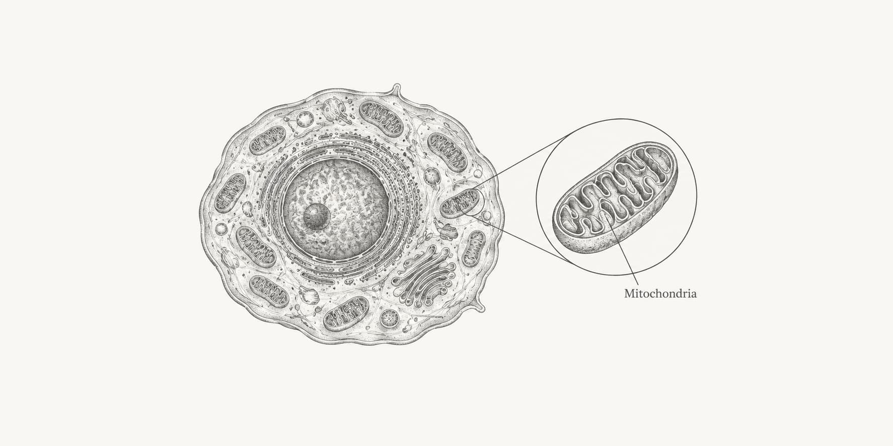
Many human cells contain hundreds of mitochondria, and cells with high energy demands can contain thousands. A liver cell has about 1,000 to 2,000 mitochondria.
There is no single average for the human body. Counts range from zero in mature red blood cells to about 100,000 in a mature egg cell, and even “one mitochondrion” can be hard to define when the organelles join into a network.
How many mitochondria are in a cell?
Many human cells contain hundreds of mitochondria, while a liver cell contains about 1,000 to 2,000.[1] The often quoted range of 100 to 2,000 is a useful rule of thumb, not a measured average across every human cell type.
One biochemical study of five mammalian cell types estimated 83 to 677 “virtual mitochondria” per cell. It found an eightfold spread in mitochondrial DNA content between cell types, showing why one average obscures more than it explains.[2]
A useful range has to name the cell:
| Human cell type | Approximate mitochondria per cell |
|---|---|
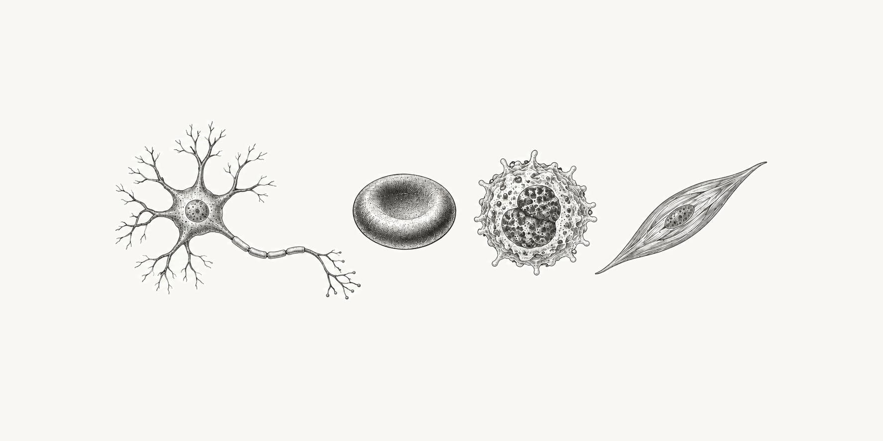
| Mature red blood cell | 0 |
| Resting platelet | 5 to 8 |
| Sperm cell | 50 to 75 |
| Liver cell | 1,000 to 2,000 |
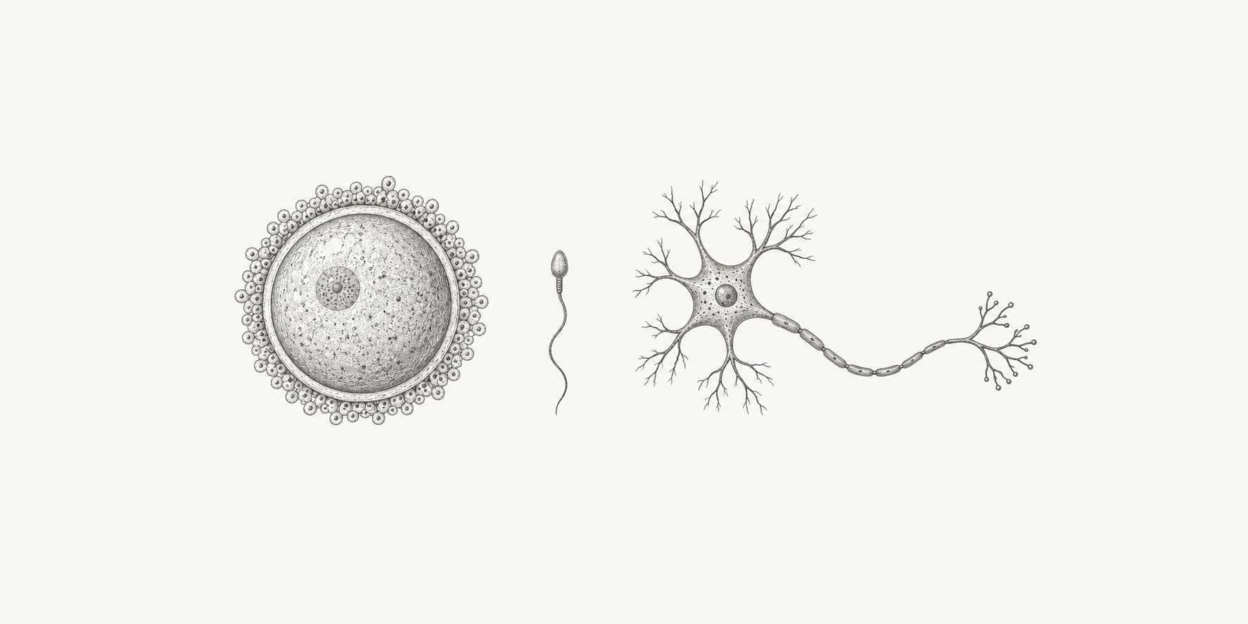
| Mature egg cell | Up to about 100,000 |
The table combines counts and estimates from microscopy, biochemical measurements, and reviews of the underlying studies.[1][3][4][5][6] They are representative orders of magnitude rather than universal constants. Age, health, activity, cell cycle, and the counting method can all change the result.
The chart uses the midpoint of each reported range for platelets, sperm cells, and liver cells. It uses 100,000 for the mature egg-cell estimate.[1][3][5][6] Mature red blood cells are omitted because zero cannot be plotted on a logarithmic axis.
Which cell has the most mitochondria?
The mature mammalian egg cell, or oocyte, has the largest known mitochondrial count among mammalian cell types, reaching about 100,000 organelles.[3]
That large supply supports egg maturation, fertilization, and the first stages of embryo development. The embryo initially divides the mitochondria inherited from the egg among its daughter cells rather than building a full new supply after every division.
Mature oocytes also contain hundreds of thousands of mitochondrial DNA molecules, but estimates vary widely.[3] This is one reason some sources give egg-cell figures above 100,000: they are reporting copies of mitochondrial DNA, not necessarily separate mitochondria.
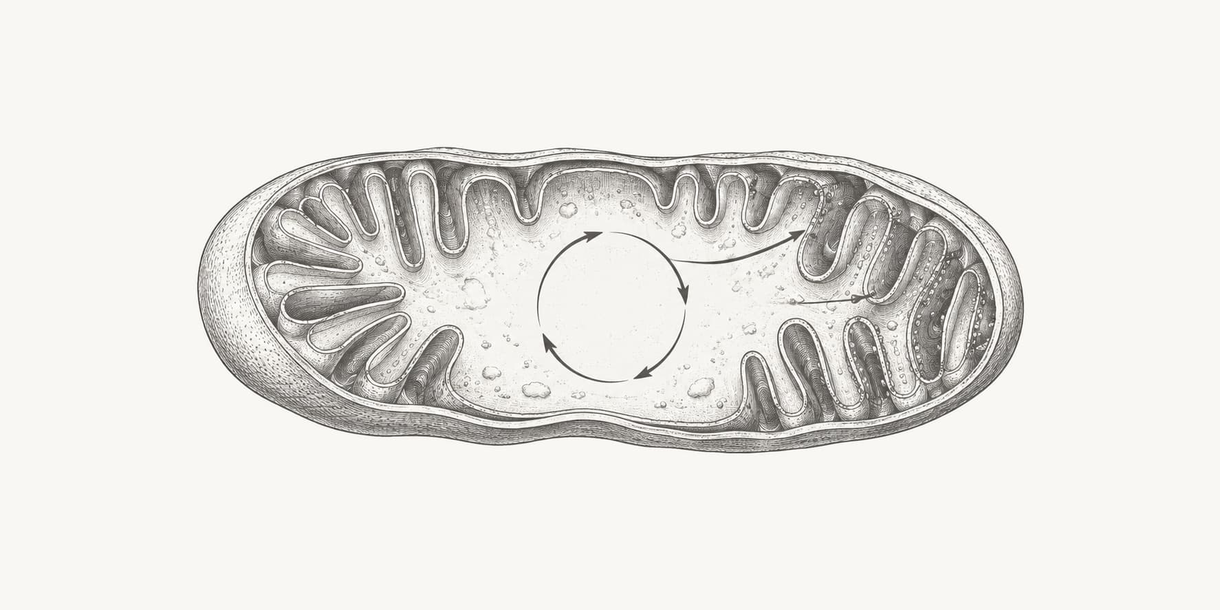
High-energy somatic cells are better compared by mitochondrial volume than by organelle count. In liver cells, mitochondria occupy about one-fifth of the cell. Heart muscle cells pack mitochondria between the contractile fibers where ATP is consumed, and their inner membranes contain more cristae than liver-cell mitochondria.[1]
Which human cells have no mitochondria?
Mature human red blood cells contain no mitochondria. Their precursors remove mitochondria and other organelles as they mature, leaving more room for hemoglobin and preventing the cells from consuming the oxygen they carry.[4]
Red blood cells dominate the number of cells in the human body, so a whole-body mitochondrial total cannot be found by multiplying one cell-level average by the body's cell count.
Platelets retain mitochondria despite having no nucleus. A resting platelet has about 5 to 8 mitochondria.[5] During platelet activation, those mitochondria can divide, so the number also depends on when the cell is measured.
Human sperm sit near the low end with about 50 to 75 mitochondria. They are arranged end to end in a sheath around the midpiece of the tail.[6]
How many copies of mitochondrial DNA are in a cell?
The mtDNA copy number per cell commonly ranges from hundreds to several thousand, while mature human egg cells can contain hundreds of thousands of mitochondrial genomes.
A study of five tissues from 50 people reported mean mtDNA copy numbers of 1,259 per cell in cerebellar cortex, 4,198 in frontal cortex and skeletal muscle, 4,617 in the caudate nucleus, and 9,235 in heart muscle.[9] These are tissue-study means. Individual cells can differ, and each tissue contains more than one cell type.
The chart reports the five means from that study.[9] A separate PCR-based analysis estimated 6,970 ± 920 mtDNA copies per diploid nuclear genome in heart muscle and 3,650 ± 620 in skeletal muscle.[10] The studies used different samples and assays, so their figures are not directly interchangeable.
Mitochondrial DNA copy number and mitochondria count measure different things. A mitochondrion can contain several mtDNA molecules, and connected mitochondria can share one network. In a biochemical study of five mammalian cell types, mtDNA ranged from 220 to 1,720 molecules per cell, while the estimated number of “virtual mitochondria” ranged from 83 to 677.[2]
Mature human oocytes sit far outside the somatic-cell range. One qPCR study of 87 metaphase II oocytes found an average of 795,000 ± 243,000 mtDNA copies, with substantial variation between oocytes from the same patient.[11]
MtDNA copy number qPCR estimates the ratio between a mitochondrial target and a nuclear reference sequence. The muscle study measured a segment of mitochondrial cytochrome b against the single-copy nuclear β-globin gene, then reported mitochondrial genomes per diploid nuclear genome.[10]
Why is the number of mitochondria hard to count?
Mitochondria constantly divide, fuse, move, and change shape, so a cell can shift between many separate organelles and a few connected networks without a comparable change in total mitochondrial mass.[7]
This makes the boundary of one mitochondrion less stable than a textbook drawing suggests. In some cells, interconnected tubules can behave as one continuous network. A microscope and its image-analysis settings may count that network as one object, several branches, or many fragments.
Mitochondrial DNA copy number is easier to measure than the number of separate organelles. Dividing genome copies by an assumed number per mitochondrion still produces an estimate rather than a direct count.
The distinction also matters in sequence analysis. Human mitochondrial genes use a different genetic code from most nuclear genes, so translating mitochondrial DNA requires the vertebrate mitochondrial code. The mitochondrial genome itself is only 16,569 base pairs long, compared with billions of base pairs in the human nuclear genome.[8]
For comparisons across tissues or conditions, mitochondrial volume, membrane area, DNA copy number, and respiratory capacity are often more reproducible than a count of separate objects. A mitochondrial number is most useful when the cell type, measurement method, and biological state are stated beside it.