How many genes are in mitochondrial DNA?
Human mitochondrial DNA has 37 canonical genes. Reports of additional small proteins explain why that count does not describe its entire coding potential.
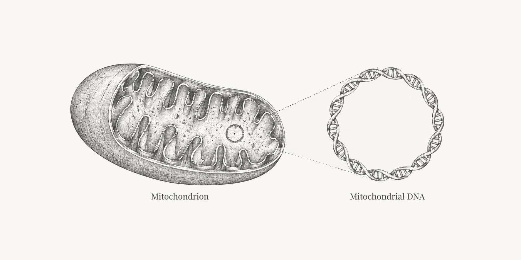
Human mitochondrial DNA contains 37 canonical genes: 13 protein-coding genes, 22 transfer RNA genes, and 2 ribosomal RNA genes. These are the standard annotated genes in a circular reference sequence spanning 16,569 base pairs.
The count describes distinct genes, rather than the thousands of mitochondrial genome copies that a cell may contain. It also needs a qualification: researchers have reported additional small proteins encoded within regions already assigned to other mitochondrial genes. We examined the evidence behind selected reports to explain what they do, and do not, change about the familiar count of 37.
How many genes are in mitochondrial DNA?
The standard human mitochondrial gene count is 37: 13 genes are annotated as protein-coding, while 24 encode RNA molecules used in protein synthesis.[1][2] Here, canonical refers to this established annotation, rather than every sequence proposed to encode a protein.
The 13 proteins form parts of the oxidative phosphorylation system, which couples energy released from nutrients to ATP production. Transfer RNAs (tRNAs) carry amino acids during protein synthesis, while ribosomal RNAs (rRNAs) form part of the mitochondrial ribosome that assembles proteins.[1]
| Protein complex | Mitochondrial genes | Gene count |
|---|---|---|
| Complex I: NADH dehydrogenase | MT-ND1, MT-ND2, MT-ND3, MT-ND4, MT-ND4L, MT-ND5, MT-ND6 | 7 |
| Complex III: cytochrome bc1 | MT-CYB | 1 |
| Complex IV: cytochrome c oxidase | MT-CO1, MT-CO2, MT-CO3 | 3 |
| Complex V: ATP synthase | MT-ATP6, MT-ATP8 | 2 |
The gene products are recorded in the human mitochondrial reference annotation; the table uses their standard human gene symbols.[2]
The phrase mitochondrial genes can also refer to a much larger group of genes whose proteins function in mitochondria, regardless of where the genes are located. MitoCarta3.0, for example, catalogues 1,136 human genes encoding proteins with strong evidence of mitochondrial localization. Most are nuclear genes, and their proteins must be imported into mitochondria. That inventory describes the mitochondrial protein complement, whereas 37 describes the genes physically encoded in mtDNA.[3]
Does mitochondrial DNA encode more than 13 proteins?
Published experiments support additional small proteins beyond the 13 canonical protein products. The discovery of humanin, followed by MOTS-c and other candidates, showed why a reference gene count and a genome's full coding potential are different questions.[4][5] A short open reading frame is a stretch of sequence that could encode a peptide. Finding one computationally does not establish that cells translate it.
We audited five discovery papers covering ten selected sequences: humanin, MOTS-c, SHLP1–6, SHMOOSE, and MTALTND4. We compared the reported lengths and genomic locations with evidence for naturally produced proteins and experiments addressing their mitochondrial origin. Our review is a structured synthesis of these papers, not a complete catalogue or a new experimental validation.
| Peptide | Length | Overlapping region | Evidence in the selected report |
|---|---|---|---|
| Humanin | 24 | MT-RNR2, encoding 16S rRNA | Activity of an expressed sequence and synthetic peptide; the original experiments do not settle the source of endogenous humanin. |
| MOTS-c | 16 | MT-RNR1, encoding 12S rRNA | Antibody detection and loss of signal after mitochondrial DNA or RNA depletion. |
| SHLP1–4 and SHLP6 | 20–38 | MT-RNR2 | Antibody detection and loss of signal in cells depleted of mtDNA. |
| SHLP5 | 24 | MT-RNR2 | Candidate sequence and transcript evidence; the study could not obtain a specific antibody. |
| SHMOOSE | 58 | Serine/leucine tRNA region and MT-ND5 | Antibody detection, two unique mass-spectrometry peptides, and an mtDNA-depletion control. |
| MTALTND4 | 99 | MT-ND4, in an alternative reading frame | Antibody detection, mass spectrometry, and mtDNA-depletion and translation-inhibition controls. |
Sources: Hashimoto et al. for humanin; Lee et al. for MOTS-c; Cobb et al. for SHLPs; Miller et al. for SHMOOSE; and Kienzle et al. for MTALTND4.[4][5][6][7][8] The downloadable evidence table records each sequence separately, with source locations and qualifications. Our audit scope and source notes explain the selection and counting rules.
The evidence is uneven even within a named peptide family. In the original SHLP report, five candidates were detected with antibodies; SHLP5 was not tested successfully with a specific antibody. The same paper amplified both nuclear and mitochondrial transcripts for SHLP2 and SHLP3, so transcript detection alone could not establish their origin.[6] Similar sequences in nuclear DNA are therefore a practical complication when assigning a peptide to mtDNA.
Translation also matters. The reported MOTS-c sequence requires the standard genetic code, despite its location in mitochondrial DNA; the discovery paper proposed translation in the cytoplasm.[5] SHMOOSE's translation compartment was left unresolved, while the reported MTALTND4 length depends on the study's stop-codon assumptions.[7][8] These qualifications support retaining 37 canonical genes, while acknowledging evidence for additional overlapping protein-coding sequences. Simply adding every reported peptide to 37 would mix annotation categories with candidates supported by different kinds of evidence.
How many genes are in nuclear DNA?
Human nuclear DNA contains about 19,400 protein-coding genes. Using GENCODE Release 50, we calculate 19,429 nuclear protein-coding genes by subtracting the 13 mitochondrial genes from the published total of 19,442.[2][9][10]
This subtraction is necessary because GENCODE includes the mitochondrial chromosome among its reference chromosomes. The result is a count for that annotation release, not a measurement of gene copies in one person's cells. It also compares protein-coding genes in both compartments, rather than comparing all 37 mitochondrial genes with only the protein-coding subset of nuclear genes.
Including non-coding RNA genes, pseudogenes, immune receptor segments, and other annotated categories produces a larger total. We calculate 78,696 nuclear gene entries from GENCODE's 78,733 total entries minus the 37 mitochondrial genes.[2][9][10] This broader count should not be interpreted as the number of protein-coding genes. Annotation scope and release date explain much of the variation among published estimates of how many genes humans have.
How many base pairs are in mitochondrial DNA?
The human mitochondrial reference sequence, RefSeq NC_012920.1, spans 16,569 base pairs, or about 16.6 kilobases.[2] This is the length of one reference genome, not the combined length of every mtDNA molecule in a cell.
By comparison, one haploid set of chromosomes in the human nuclear genome contains about 3.1 billion base pairs.[11] Using that rounded nuclear length, we calculate that one nuclear genome is roughly 187,000 times longer than one mitochondrial genome. The comparison uses one chromosome set, rather than the two sets present in a typical diploid body cell.
Mitochondrial genes are packed closely together, and some overlap. MT-ATP8 and MT-ATP6, for example, occupy partly overlapping stretches of DNA but encode different proteins.[2] The canonical mitochondrial protein-coding sequences use the vertebrate mitochondrial genetic code. In DNA translation, choosing this code matters: TGA, or UGA in RNA, specifies tryptophan in this code, whereas it normally signals a stop in the standard code.[12]
How many copies of mitochondrial DNA are in a cell?
Mitochondrial DNA copy number varies substantially among cells and tissues. A commonly cited range for mammalian cells is roughly 1,000 to 10,000 copies per cell, with values outside that range, including more than 100,000 in oocytes. This range is a broad description, not a measured average for every human cell type.[13]
A much broader comparison is now available. Rath and colleagues' 2024 study measured mtDNA in 52 human tissues from 952 donors in the Genotype-Tissue Expression project. They reported approximately 50-fold variation in tissue medians, alongside more than 200-fold variation among individuals within a tissue.[14] These are the authors' published findings; we have not recalculated their dataset.
The denominator is important: the 2024 measurements are mtDNA copies per diploid nuclear genome, estimated from the ratio of mitochondrial to nuclear DNA. That is a normalization to nuclear DNA content, not a direct count in each cell. Cell mixtures and differences in nuclear DNA content can affect how it relates to copies per cell.[14]
Earlier measurements provide useful context, but should remain separate. Frahm and colleagues examined five tissues from 50 people aged from eight weeks to 93 years. Their 2005 study reported mean estimates ranging from 1,259 copies in cerebellar cortex to 9,235 in heart muscle.[15] The following figure retains that study's means; it is not a subset of the newer dataset.
| Tissue | Mean mtDNA copies per cell |
|---|---|
| Heart muscle | 9,235 |
| Caudate nucleus | 4,617 |
| Frontal lobe cortex | 4,198 |
| Skeletal muscle | 4,198 |
| Cerebellar cortex | 1,259 |
Source: Frahm et al. (2005). These are estimates from tissue DNA, not counts made in each individual cell. They should not be treated as fixed values for all people or every cell within a tissue. The authors found no significant age-related change in absolute copy number in these five tissues.[15]
Genome copies can also differ in sequence within the same cell or tissue, a state called heteroplasmy. Fisher and colleagues' 2026 analysis of 47 tissue types from 947 donors examined this genetic variation across the body.[16] Gene count, copy number, and sequence variation therefore describe different properties: the genes a genome encodes, how many genome molecules are present, and how their sequences differ.
Copy number also differs from the number of mitochondria, because one mitochondrion can contain multiple genome copies.[1] Having more mtDNA copies does not, by itself, add new types of mitochondrial genes.