Primer dimers: how they form, how to predict them and how to stop them
Reading the 3′ end, the ΔG and the melt curve

Matic Broz, PhD18 min
Take two primers for human β-actin. Both are 20 or 21 nucleotides long, both are 55 to 57% GC, and their melting temperatures are 60.0 and 61.1 °C, about a degree apart. Every line of the usual primer checklist passes. But the last eight bases of the forward primer, TGTGGATC, are the reverse complement of the last eight bases of the reverse primer. When the two meet in the tube, each 3′ end lies on the other primer with a few bases of template to copy.
A polymerase that finds that duplex fills it in, and the result is a 33 bp piece of DNA that carries a binding site for each primer. From then on it amplifies like any other product, only faster, because it is short. That is a primer dimer. The oligo analyzer finds this one in seconds, and the rest of this page explains what it is looking for and what to do when it finds it.
What a primer dimer is
A primer dimer is a PCR artifact made when primers anneal to each other instead of to the template and the polymerase extends them.[1] A self-dimer forms between two copies of the same primer; a cross-dimer, also called a heterodimer, forms between the forward and the reverse primer. In a multiplex reaction every primer can pair with every other, so a set of ten primers has 55 possible dimers, counting each primer with itself.
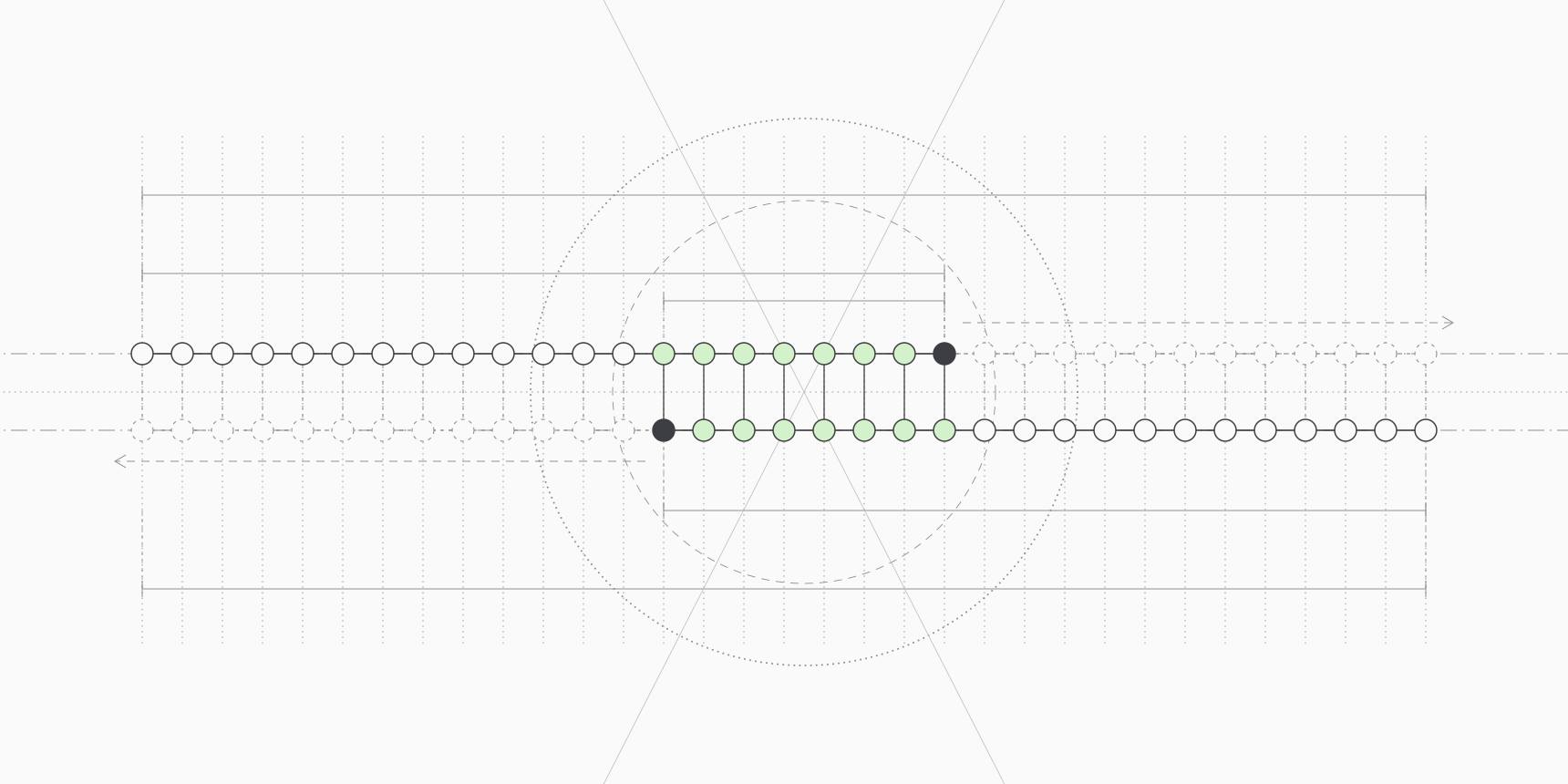
Not every primer-to-primer duplex can be extended. A polymerase adds bases only to a paired 3′ end, and only where the partner strand continues past it to serve as a template. The β-actin pair above is the dangerous geometry, with the two 3′ ends overlapping:
forward  5'-CGCAAGTACTCCGTGTGGATC-3'
                         ||||||||
reverse               3'-ACACCTAGTCGTTCGTCCTC-5'
Each 3′ end is paired and each has a 5′ tail of the other primer beyond it, so both strands can be extended to full length. The product is 21 + 20 − 8 = 33 bp. If the complementary stretch sat at the 5′ ends instead, the 3′ ends would hang free and nothing could be copied. Internal complementarity is in between: it ties up primer, but only a paired 3′ end gives the polymerase somewhere to start.
Why the 3′ end matters most
A dimer that cannot be extended is a temporary nuisance. It sequesters some primer while it lasts, then melts at the next denaturation step and leaves nothing behind. An extended dimer is permanent. Once the polymerase has copied across, the new strand contains the full sequence of one primer and the complement of the other, which makes it a perfect template for both primers in every later cycle.[1]
That is why dimer checks single out the 3′ end. Primer3 scores complementarity anywhere in two primers and, separately, complementarity anchored at a 3′ end, and PREMIER Biosoft's rules of thumb are stricter for 3′ dimers than for internal ones.[3][6]
The same logic explains why a GC clamp, one or two G or C bases at the 3′ end, is a trade-off. It helps the primer hold its target, but a GC-rich or self-complementary 3′ end, such as the GATC that ends the β-actin forward primer, also holds a partner primer more firmly.
How primer dimers show up
On an agarose gel, primer dimers run as a faint, often fuzzy band near the bottom of the lane, below about 100 bp.[1] They can be hard to separate from unused primers, and they are easy to miss next to a strong product. When they compete well, the specific band weakens or disappears.
In SYBR Green qPCR, the dye binds any double-stranded DNA, so dimers add fluorescence and distort the quantification.
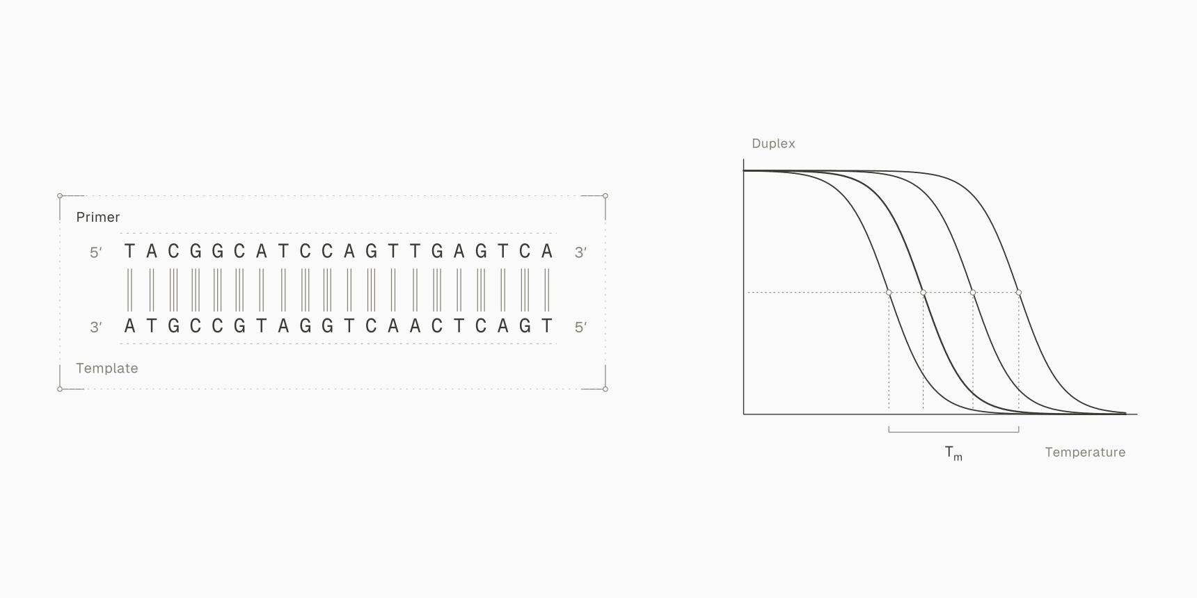
A melt curve at the end of the run separates them. Ririe and colleagues showed that the position and shape of a product's melting curve depend on its length, GC content and sequence, enough to separate products less than 2 °C apart.[7] A primer dimer of 30 to 50 bp melts below a longer specific product, so it appears as an extra peak at a lower temperature.
The no-template control is the cleanest test. With no template, the only DNA in the well is primer, so any amplification there comes from primers acting on each other, or from contamination. A late signal in the no-template control with a low-temperature melt peak is the classic primer-dimer signature.
Dimers matter most when the target is scarce: in Chou and colleagues' experiments, suppressing primer dimerization improved amplification most clearly below roughly 1,000 target copies.[9]
Prediction and outcome do not always agree. Brownie and colleagues tried to make primer dimers deliberately from primers with different kinds of complementarity, and could not get detectable dimers from a single primer on its own, even with 3′ complementarity.[8] A predicted dimer is a risk to check, not a forecast that the reaction will fail.
How dimer stability is scored
Dimer checkers score a primer-to-primer duplex the same way a Tm calculator scores a primer on its target: with nearest-neighbor thermodynamics. SantaLucia's unified parameters assign a free energy to each of the ten possible stacks of two adjacent base pairs, measured in 1 M NaCl, and sum them with initiation terms for the two ends:[2]
Here is how many times stack occurs, each initiation term applies to an end closed by that type of base pair, and the symmetry term, kcal/mol, applies only to self-complementary duplexes. More negative means a more stable duplex.
For the eight-base overlap of the β-actin pair, TGTGGATC paired with its complement, the seven stacks add up to kcal/mol. The ends are one T·A pair () and one C·G pair (), so kcal/mol in 1 M NaCl. A PCR buffer has far less salt. SantaLucia's correction is:[2]
where is half the number of phosphates in the duplex, 7 for an 8 bp duplex. Primer3's default buffer of 50 mM monovalent cations, 1.5 mM Mg²⁺ and 0.6 mM dNTPs counts as about 164 mM Na⁺ after von Ahsen's Mg²⁺ conversion, which gives kcal/mol.[13][3] Primer3's own alignment reports kcal/mol for the same dimer, slightly more stable, because it also counts the unpaired bases dangling next to each end of the helix.
Why the temperature matters
A free energy is only meaningful at a stated temperature, because and the entropy term grows as the tube heats up. The same β-actin dimer is kcal/mol at 25 °C, kcal/mol at 37 °C and kcal/mol at 60 °C. The oligo analyzer reports ΔG at 37 °C by default, and IDT does not state the temperature behind its values, so a ΔG copied from one tool cannot be compared with a threshold from another unless both used the same temperature.
The structure's melting temperature avoids that problem. For two different strands at equal concentration, SantaLucia's Tm is:[2]
with the total strand concentration and the gas constant. Tm does not depend on the analysis temperature, but it does depend on concentration. The β-actin dimer melts at 15.8 °C at Primer3's default 50 nM and at 22.4 °C at 500 nM, the starting primer concentration of the protocol behind that default.[3] That puts it close to room temperature while the reaction is being set up, which is exactly when an enzyme without hot start can extend it.
Rules of thumb for "too stable"
There is no single cut-off, and the published ones disagree. They are worth reading as screens from the tool that states them, at that tool's own conditions.
| Source | What is flagged | β-actin pair |
|---|---|---|
| IDT OligoAnalyzer guidance | Self- or heterodimer ΔG of −9 kcal/mol or more negative | Passes |
| PREMIER Biosoft design guidelines | 3′ end self- or cross-dimer more stable than about −5 kcal/mol; internal dimer more stable than about −6 kcal/mol | Flagged |
| Primer3 defaults | Any or 3′-anchored dimer with a structure Tm above 47 °C | Passes |
IDT treats a dimer of −9 kcal/mol or more negative as likely to cause trouble, and asks you to check the most stable of the self-dimer and heterodimer structures it lists.[5] PREMIER Biosoft is stricter at the 3′ end, tolerating about −5 kcal/mol for a 3′ dimer and −6 kcal/mol for an internal one, with −2 and −3 kcal/mol for 3′ and internal hairpins.[6] Primer3 sets its limits as melting temperatures instead, 10 °C below its minimum primer Tm.[3]
The β-actin pair lands between them. It is a reminder that a pass from one checker is not a clean bill of health, and that the geometry of the dimer, here a fully extendable 3′ overlap, matters as much as the number.
Primer3's self-complementarity and 3′ complementarity scores
Primer3 has two families of dimer settings. The older ones score a simple alignment, 1 for each complementary base, −1 for a mismatch and −2 for a gap, and the newer ones, used by default, run a thermodynamic alignment and report the melting temperature of the most stable structure.[3] The thermodynamic alignment considers mismatches, loops and dangling ends, not just perfect runs of pairs.[3][4]
| What it limits | Thermodynamic setting (default) | Alignment-score setting (default) |
|---|---|---|
| A primer binding a copy of itself anywhere | PRIMER_MAX_SELF_ANY_TH (47.0) | PRIMER_MAX_SELF_ANY (8.00) |
| A primer binding a copy of itself at its 3′ end | PRIMER_MAX_SELF_END_TH (47.0) | PRIMER_MAX_SELF_END (3.00) |
| Left primer binding right primer anywhere | PRIMER_PAIR_MAX_COMPL_ANY_TH (47.0) | PRIMER_PAIR_MAX_COMPL_ANY (8.00) |
| Left primer binding right primer at a 3′ end | PRIMER_PAIR_MAX_COMPL_END_TH (47.0) | PRIMER_PAIR_MAX_COMPL_END (3.00) |
| A primer folding on itself | PRIMER_MAX_HAIRPIN_TH (47.0) | None |
The manual describes the 3′ setting as the one that tries to predict primer-dimer formation, by anchoring the alignment at the 3′ end.[3] Primer3 also has PRIMER_MAX_END_STABILITY, a cap on how tightly the last five 3′ bases bind their target, in kcal/mol. Its default of 100 leaves it off, and it describes how readily a primer's 3′ end primes anywhere rather than how it pairs with another primer.[3]
In Primer3 on ProteinIQ, the same values appear as Left Self-Complementarity Tm (C), 3' Pair Complementarity Tm (C) and their neighbors, with the 47 °C limits under the design settings. The Check existing primers task scores primers you already have against your template.
Worked example: a β-actin primer pair
We took the human ACTB mRNA (RefSeq NM_001101.5), searched its coding sequence for primer pairs whose 3′ ends overlap, and checked the strongest one with Primer3 2.6.1 through primer3-py 2.2.0 at Primer3's default conditions.[14][4] We then asked Primer3 to design new pairs around the same stretch of the gene. The pairs below are a calculation, not a tested assay. The script and its full output are in public/data/guides/primer-dimers/.
| Property | Dimer-prone pair | Redesigned pair |
|---|---|---|
| Forward primer, 5′ to 3′ | CGCAAGTACTCCGTGTGGATC | CGCAAGTACTCCGTGTGGAT |
| Reverse primer, 5′ to 3′ | CTCCTGCTTGCTGATCCACA | CGGACTCGTCATACTCCTGC |
| Length, nt | 21 and 20 | 20 and 20 |
| GC content | 57.1% and 55.0% | 55.0% and 60.0% |
| Primer Tm | 61.1 and 60.0 °C | 60.1 and 60.0 °C |
| Product | 81 bp, bases 1087 to 1167 | 94 bp, bases 1087 to 1180 |
| 3′ ends paired with each other | 8 bases, extendable to a 33 bp dimer | None |
| Cross-dimer ΔG at 37 °C | −6.85 kcal/mol | −3.00 kcal/mol |
| Cross-dimer Tm, 50 nM and 500 nM | 15.8 and 22.4 °C | −31.9 and −22.2 °C |
| Primer3 3′ pair complementarity Tm | 15.8 °C | 0.1 °C |
The dimer-prone pair is unremarkable on every single-primer measure. Its self-dimers are weak, with structure Tm values of −9.9 and 1.1 °C, and the only notable single-primer structure is a small hairpin in the forward primer that melts at 45.3 °C, below Primer3's 47 °C limit and well below the 60 °C annealing step. Its problem exists only when the two primers are together.
The redesign keeps the forward primer's binding site, drops its last base and moves the reverse primer 13 bases downstream. The strongest cross-structure Primer3 finds is now a four-base internal pairing with both 3′ ends unpaired:
forward         5'-CGCAAGTACTCCGTGTGGAT-3'
                            ||||
reverse  3'-CGTCCTCATACTGCTCAGGC-5'
Nothing in that structure can be extended, and at −3.00 kcal/mol it passes all three rules in Table 1. The amplicon grows from 81 to 94 bp, still a comfortable size for qPCR.
The same Primer3 run also shows why a pair needs checking even when a program designed it. Its top-ranked pair was the redesign above, but its second and third choices went back to the original reverse primer site and kept 6 and 7 bases of 3′ overlap, at −5.09 and −5.66 kcal/mol. All of them passed the default 47 °C limits, because a short 3′ overlap at 50 nM melts far below 47 °C even when it is fully extendable.
Design rules that keep dimers away
Most dimer problems are prevented at the design stage, with a handful of rules that also keep primers specific:
- Length of about 18 to 24 nucleotides. Lorenz gives 15 to 30 as the workable range, PREMIER Biosoft 18 to 22 as optimal, and Primer3 defaults to 18 to 27 with 20 as the target.[1][6][3]
- GC content of 40 to 60%.[1][6] Primer3's default window, 20 to 80%, is wider, so tighten it for routine PCR.[3] The GC content calculator checks a list of primers at once.
- A modest GC clamp. Ending in a G or C helps priming, but more than three G or C bases among the last five makes a sticky 3′ end that primes from short matches.[1][6]
- Matched melting temperatures, within about 5 °C of each other, so that one annealing temperature suits both primers.[1][6] Primer3 does not enforce this by default; its PRIMER_PAIR_MAX_DIFF_TM is 100 °C.[3]
- No complementarity between 3′ ends, within a primer or across the set. Watch for palindromes such as
GATCorGAATTCat the 3′ end, which pair with a copy of themselves.
A reverse primer entered in the wrong orientation is a different and common mistake. Both primers are ordered 5′ to 3′, and the reverse complement tool converts a sequence copied from the top strand into the form you order.
For a multiplex panel, check every primer against every other. The number of pairs grows quickly, and a dimer between two primers from different assays is as damaging as one within an assay.
How to fix primer dimers in the lab
When a gel or melt curve already shows dimers, start with the reaction before ordering new primers.
Use a hot-start polymerase. Dimers often form while the reaction sits at room temperature, before the first denaturation step. Chou and colleagues withheld a reagent until the tube reached 60 to 80 °C and saw putative primer oligomers suppressed even where mispriming was negligible.[9]
Antibody-blocked enzymes do the same job without opening the tube: Kellogg and colleagues used a monoclonal antibody that holds Taq polymerase inactive until heat releases it.[10]
Raise the annealing temperature. Lorenz suggests starting about 5 °C below the primers' Tm.[1] A higher annealing temperature leaves less time and less stability for weak primer-to-primer duplexes in each cycle, though it does nothing for dimers made during setup. A gradient run is the quickest way to find the highest temperature that still gives a strong product.
Lower the primer concentration. When primers are in large excess over template, they are more likely to find each other than the target, so Lorenz recommends adjusting the primer-to-template ratio first.[1] The same logic explains why dimers are worst in low-template samples and no-template controls.
Try touchdown PCR. Don and colleagues introduced it to avoid spurious priming: the first cycles anneal above the primers' Tm and the temperature then steps down, so the correct product gets a head start.[11] Korbie and Mattick note that any Tm difference between correct and incorrect annealing gives the correct product a twofold advantage per cycle.[12] A typical program starts 10 °C above the calculated Tm and drops 1 °C every second cycle.[1]
Titrate Mg²⁺. Higher Mg²⁺ generally raises yield but lowers specificity and fidelity, and Lorenz names Mg²⁺ titration and annealing temperature as the two changes most likely to fix a reaction.[1] Mg²⁺ also stabilizes every duplex, including dimers, which is why calculators ask for it. Lorenz also lists DMSO as an additive that can help.[1]
Redesign as the last resort, and keep the good half. As the β-actin example shows, moving one primer a few bases is often enough. For large multiplex panels, Brownie and colleagues suppressed dimer accumulation with tailed primers whose short dimer products fold into pan-handles that block further priming.[8]
Primer dimer tools on ProteinIQ
The oligo analyzer checks primers you already have. It runs Primer3's thermodynamic functions on up to 96 oligos on paid plans, and with Analyze interactions on, scores every pair of up to 24 for heterodimers and the 3′ end stability in both directions.
The Structures tab draws each alignment with its ΔG in kcal/mol and its Tm, and the Analysis temperature (°C) setting, 37 °C by default, sets the temperature for every ΔG. How it compares with IDT's OligoAnalyzer and other calculators, and why their Tm values differ, is covered in the oligo analyzer comparison.
Primer3 designs new pairs from a template with the complementarity limits in Table 2 and reports their structure Tm values for each pair; its Check existing primers task scores a given pair against the template. RNAcofold predicts the joint structure of two RNA strands with RNA energy parameters, which suits RNA oligos and probes rather than DNA primers.
Primer dimers also cost time: a failed run means another hour or more on the cycler, which how long PCR takes breaks down step by step.
Frequently asked questions
What is the difference between a self-dimer and a cross-dimer?
A self-dimer forms between two copies of the same primer. A cross-dimer, or heterodimer, forms between two different primers, usually the forward and reverse primer of one pair. Both become a problem when their 3′ ends are paired and can be extended.
What size is a primer dimer on a gel?
Usually below 100 bp, near the bottom of the lane.[1] An extended dimer is roughly the two primer lengths added together, minus the overlap, so two 20-mers that overlap by 8 bases give a 32 bp product.
What ΔG value is too low for a primer dimer?
It depends on whose rule you use and at what temperature. IDT flags dimers of −9 kcal/mol or more negative, PREMIER Biosoft treats about −5 kcal/mol as the limit for a 3′ dimer, and Primer3 limits the structure's Tm to 47 °C by default.[5][6][3] Weigh a 3′ dimer more heavily than an internal one of the same ΔG.
Can primer dimers form in a no-template control?
Yes, and that is where they are easiest to see. With no template to bind, primers meet each other more often, and any signal with a low-temperature melt peak points to dimers rather than your product.
Do primer dimers matter for TaqMan probe assays?
Less than for SYBR Green. A hydrolysis probe only reports when it binds its target sequence, and a primer dimer does not contain that sequence, so the dimer adds no fluorescence. It still competes with the target for primers and polymerase, which matters most when the target is scarce.
Is a hairpin the same as a primer dimer?
No. A hairpin is a primer folding on itself; a dimer needs two molecules. Primer3 and the oligo analyzer report both, and a hairpin that melts below the annealing temperature, like the 45.3 °C hairpin in the β-actin forward primer, is usually harmless.