How to use AutoDock Vina online
Run small-molecule docking in a browser by preparing a receptor and ligand, defining a search box, choosing Vina settings, and inspecting ranked poses.
TL;DR
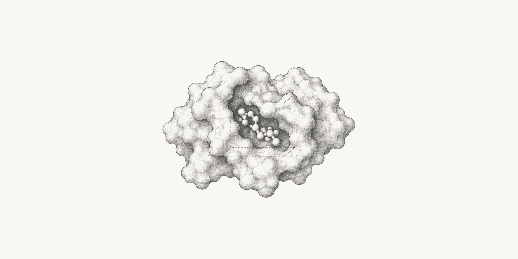
- AutoDock Vina online needs a receptor, a small-molecule ligand, and a search box around a plausible binding site. Start with Vina scoring, exhaustiveness 8, and nine poses.
- The search box is as important as the scoring function: make it large enough for the ligand to move, but no larger than the evidence requires.
- Vina scores are approximate rankings in kcal/mol, not experimental binding free energies or direct predictions of Kd and IC50.
- Validate a protocol by redocking a known ligand, repeating searches, inspecting pose families, and checking key interactions before screening new compounds.
AutoDock Vina predicts how a small molecule may fit inside a receptor binding site and ranks the poses it finds. You can run it online without installing Vina, Meeko, AutoGrid, or a command-line environment.
For a first run:
Open AutoDock Vina online
Open the ProteinIQ AutoDock Vina webserver.
Add the receptor and ligand
Upload the prepared receptor and the small molecule you want to dock.
Define the binding pocket
Set a search box that covers the intended site without making the search region unnecessarily large.
Choose the settings and submit
For a first test, use the Vina scoring function, exhaustiveness 8, and
nine output poses.
Inspect the docked poses
Compare the returned structures and interactions instead of relying only on the top score.
What is AutoDock Vina?
AutoDock Vina is an open-source molecular docking engine in the AutoDock suite. Given a receptor structure, a ligand, and a search region, Vina generates possible binding poses and assigns each pose an estimated affinity score.[1][2]
Vina was introduced by Trott and Olson in 2010 with a new scoring function, an efficient search method, and multithreading. Version 1.2 added Python bindings and specialized methods for simultaneous multi-ligand docking, hydrated docking, macrocycles, and AutoDock4-based zinc coordination. The current ProteinIQ implementation runs AutoDock Vina 1.2.7 and prepares common receptor and ligand formats for you.[3]
A normal result contains several ranked poses, a score in kcal/mol for each pose, and RMSD bounds relative to the best-ranked output pose. Vina treats the receptor as rigid by default while allowing the ligand to translate, rotate, and change its rotatable bonds. Selected receptor side chains can be made flexible, but Vina does not model full backbone motion or large induced-fit changes.[4]
Vina is designed for receptor-ligand docking. It is not a protein-protein docking program, and its documentation recommends software built for that problem instead.[5]
How does AutoDock Vina work?
Vina must search a large conformational space for plausible poses, then score those poses well enough to rank them. The search is stochastic, so separate runs can return different answers unless you fix the random seed and keep the inputs and settings unchanged.[1][5]
It searches inside a defined box
The search box tells Vina where the ligand is allowed to move. Vina explores ligand positions, orientations, and torsion angles inside that region. A smaller box is easier to search thoroughly, but a box that is too small can exclude the correct pose. A box that is too large spreads the same search effort across much more space. The official FAQ advises keeping the search region below about 30 × 30 × 30 Å when possible, or raising exhaustiveness for larger searches.[5]
It alternates random changes with local optimization
Each independent docking run starts from a random ligand conformation. Vina perturbs its position, orientation, or torsion angles, then refines the new pose with Broyden-Fletcher-Goldfarb-Shanno local optimization. A Metropolis acceptance test decides whether the search keeps the new pose, and promising minima are saved while the process repeats.[1]
Exhaustiveness controls how much independent search effort Vina applies. Raising it usually improves the chance of finding a good minimum in a difficult search space, but it does not correct a wrong receptor, ligand, scoring function, or search box.[4]
random pose
    ↓
change position, orientation, or torsion
    ↓
BFGS local optimization
    ↓
accept or reject the new pose
    ↓
save promising minima and repeat
    ↓
cluster and rank the final poses
It scores each pose
The standard Vina scoring function combines two attractive Gaussian terms, repulsion, hydrophobic interactions, a non-directional hydrogen-bond term, and a conformational-entropy penalty tied to ligand flexibility. It does not include explicit electrostatic or solvation terms. The score is reported in kcal/mol, with more negative values ranked ahead of less negative values under the same protocol. It is an approximation, not an experimental free energy.[1][3]
Vina, Vinardo, and AutoDock4 use different scoring functions, so their numerical scores should not be compared directly. AutoDock Vina 1.2 also includes specialized hydrated-docking and zinc workflows based on AutoDock4 features. Those workflows do not make the standard Vina score universal across target classes.[3][4]
It clusters and ranks the results
Vina remembers promising intermediate poses, refines them, merges similar solutions, and sorts the final set. The num_modes setting caps how many poses can be returned; energy_range can exclude poses that score too far above the best result. The RMSD lower and upper bounds compare each mode with the best output mode. They do not tell you how close a pose is to a crystallographic ligand unless you calculate that comparison separately.[2][5]
How to use AutoDock Vina online
The online workflow handles installation, file conversion, receptor and ligand preparation, execution, and result display. You still need to make the scientific choices that determine whether the run is meaningful.
1. Open AutoDock Vina
Go to AutoDock Vina online. You can use an experimental receptor structure, a prepared model, or a structure predicted with AlphaFold 2.
For the cleanest first run, start with one receptor, one ligand, and a known binding site. Add batch or flexible-receptor settings only after the basic protocol works.
2. Add the receptor
You can upload a receptor as PDB, ENT, CIF, mmCIF, or PDBQT, or fetch a structure directly from the RCSB Protein Data Bank by its PDB ID.
Before docking, check that you have the intended chain, biological state, and binding-site conformation. Experimental structures may contain alternative locations, missing atoms, nonstandard residues, crystallization additives, ions, waters, cofactors, and old ligands.
Remove what does not belong in your model, but do not automatically delete a metal, cofactor, or conserved water that helps form the binding site. When the structure has missing atoms or other repairable problems, PDBFixer can help repair it before receptor preparation. Complete structures can proceed directly to preparation.
If several receptor structures are available, prefer one with a relevant bound ligand and a pocket conformation close to the state you want to study. If the site is unknown, a pocket detector such as fpocket can generate candidates, but biological evidence should decide which pocket to test.
3. Add the ligand
The webserver accepts PDBQT, SDF, MOL, MOL2, SMILES, SMI, TXT, and CSV ligand inputs. You can also paste a SMILES string, fetch a compound from PubChem, or draw the molecule in the browser.
Prefer SDF or MOL2 over PDB for small molecules because those formats preserve bond orders and related chemical information. The official Vina tutorial warns against preparing ligands from PDB files for the same reason.[2]
Check the ligand's stereochemistry, protonation state, formal charge, tautomer, and 3D geometry. One misplaced proton can change hydrogen-bond donors, acceptors, and the predicted pose.
When you start from SMILES, the webserver generates a 3D conformer before docking, but you should still confirm that the chemical form matches the experiment you want to model.
4. Choose the run mode
| Mode | What it does | Best use |
|---|---|---|
Single ligand | Docks one ligand into one receptor | Pose prediction, protocol testing, and redocking |
Batch docking | Docks each ligand independently against the same receptor | Comparing a small set of compounds or running a small virtual screen |
Simultaneous co-docking | Places several ligands in the pocket during the same search | Fragment-growing hypotheses or systems where several ligands may bind together |
Batch docking and simultaneous docking answer different questions. A batch produces a separate result for each ligand. Simultaneous docking produces poses and a score for the combined multi-ligand system, so it should not be used as a shortcut for ranking independent compounds. These multi-ligand modes are part of the expanded AutoDock Vina 1.2 workflow.[3]
For hundreds or thousands of ligands, use a dedicated high-throughput docking workflow instead of uploading ten compounds at a time.
5. Define the binding pocket
A good scoring function cannot rescue a box that excludes the binding site. The webserver gives you four ways to define the pocket:
| Pocket method | Use it when | What happens |
|---|---|---|
Bound ligand | The receptor contains a co-crystallized ligand in the relevant site | The server builds a box around the selected ligand, adds padding, and removes that ligand before docking |
Selected residues | You know the key binding-site residues | The server builds a box around those residues and leaves them in the receptor |
Manual coordinates | You already know the box center and dimensions | The coordinates and sizes are passed to Vina in Å |
Whole protein | You have no defensible site hypothesis | The box covers the receptor, which greatly increases the search burden |
A bound ligand is usually the best starting point because it anchors the box to an observed site. Selected residues are useful when the receptor has no ligand but the active site is known from mutagenesis, homologs, or prior structures. Manual coordinates work well when you are reproducing a published protocol.[4]
Whole-protein docking is a last resort. When the pocket is unknown, run a detector such as fpocket first. Compare its predicted sites with biological evidence, then dock into the best-supported region.
The online tool uses 5 Å of padding by default for bound-ligand and selected-residue boxes. Preview the box and confirm that the ligand can fit with enough room to rotate, without covering unrelated cavities or most of the protein.
6. Choose the docking settings
Vina's defaults work for a focused pocket and an ordinary ligand. Larger boxes and more flexible systems need more search effort.
| Setting | First run | Change it when |
|---|---|---|
Scoring function | Vina | Try Vinardo as an alternative model; use AD4 when your protocol or a specialized hydrated-ligand or zinc workflow requires it |
Exhaustiveness | 8 | Raise it for a larger box, a flexible ligand, flexible side chains, or inconsistent repeated runs |
Number of poses | 9 | Raise it when you need more alternative binding modes |
Energy range | 3 kcal/mol | Raise it when plausible alternatives are being excluded |
Random seed | 0, which allows a new seed | Set a nonzero integer when you need an exactly repeatable run |
Flexible residues | None | Add a small number of known mobile side chains when rigid-receptor docking cannot represent the site |
Exhaustiveness increases search effort, not scoring accuracy. If the scoring function prefers the wrong pose, running the same setup longer will not repair the model. Avoid changing many settings at once. Keep a record of the receptor preparation, ligand state, box, scoring function, and seed so you know what caused a result to change.[4]
The online tool allows exhaustiveness values from 1 to 64 and up to 50 output poses. Batch docking supports up to 10 ligands, and simultaneous co-docking supports up to five ligands.
7. Run the docking job
Submit the job after checking the 3D box preview. Runtime depends mainly on the box volume, ligand size and flexibility, exhaustiveness, flexible receptor residues, and the number of ligands.
A small single-ligand run with a focused pocket is the best protocol check. If that run fails to place a known ligand sensibly, fix the preparation or box before spending more computation on a batch.
8. Inspect and download the results
The results page shows the ranked poses in a 3D viewer alongside their Vina scores and RMSD bounds. Start by looking at several top poses rather than accepting mode 1 automatically.
Check whether:
- the ligand sits inside the intended pocket;
- the pose has obvious clashes or strained geometry;
- known catalytic or recognition residues make plausible contacts;
- charged and polar groups face a chemically sensible environment;
- several top poses form the same pose family;
- the result changes sharply when you repeat the run.
Download the ranked PDBQT poses, prepared structures, and preparation records. Those files matter when you need to reproduce the calculation or explain why two runs differ.
AutoDock Vina examples
The following examples show how the same result interface supports a single-ligand run, a joint multi-ligand search, and an independent batch. The scores are useful only within the stated receptor preparation, box, scoring function, and random seed.
Example 1: c-Abl kinase docking with imatinib
This single-ligand example docks imatinib into the ATP-binding site of the c-Abl kinase domain, using the co-crystallized ligand in PDB 1IEP to define the search region. You can open the example run.
- Inputs: c-Abl kinase domain from PDB
1IEP(2.1 Å) and imatinib from PubChem CID5291 - Non-default setting:
Random seed=42to make the stochastic search reproducible
AutoDock Vina returned five poses in the configured energy window. The top-ranked pose has an Affinity of −12.512 kcal/mol, while the other visible poses range from −11.290 to −9.775 kcal/mol. The viewer supports inspection of the proposed placement in the kinase pocket, but the score is a docking ranking rather than an experimental binding free energy or proof that the pose is correct.
Example 2: methotrexate and NADPH co-docking with human DHFR
This example searches for joint placements of methotrexate and the NADPH cofactor in one human dihydrofolate reductase binding region. It demonstrates simultaneous co-docking, where both ligands move within the same Vina search rather than being docked as independent jobs. You can open the example run.
- Inputs: human DHFR from PDB
1U72(1.9 Å), methotrexate from PubChem CID126941, and NADPH from PubChem CID5884 - Non-default settings:
Ligand mode=Simultaneous co-docking;Binding pocket=Manual coordinateswith center(26.624, 14.574, 5.689) Åand size(26.204, 28.087, 32.768) Å;Random seed=42
The structure view places both ligands in the DHFR receptor context and lists all nine joint poses. The top two poses score −12.243 and −12.209 kcal/mol, so their score difference is small even though their returned RMSD bounds indicate different joint geometries.
The Data view reports the best pose at −12.243 kcal/mol and the second pose with an RMSD lower bound of 2.105 Å and RMSD upper bound of 3.381 Å relative to the best returned mode. These values compare Vina's joint pose solutions; they do not establish that both molecules bind simultaneously in an experimental system.
Example 3: batch docking a c-Abl inhibitor panel
This focused batch runs imatinib, nilotinib, and ponatinib independently against the same c-Abl receptor and search box. Holding the receptor preparation and Vina setup constant makes the returned scores useful for an initial within-run comparison. You can open the example run.
- Inputs: c-Abl kinase domain from PDB
1IEP(2.1 Å), with imatinib, nilotinib, and ponatinib as three independent ligands - Non-default settings:
Ligand mode=Batch docking;Binding pocket=Manual coordinateswith center(15.190, 53.902, 16.917) Åand size(18.664, 26.739, 23.526) Å;Random seed=42
The structure view combines the 16 successful poses returned across the three ligands and lets each result be inspected in the shared c-Abl receptor context.
The leading Affinity values are −13.615 kcal/mol for nilotinib, −12.512 kcal/mol for imatinib, and −12.438 kcal/mol for ponatinib. Nilotinib ranks first under this specific preparation, search box, scoring function, and seed, but that ordering is a computational prioritization result, not a measured affinity series. Check it with pose inspection, repeated searches, and experimental evidence.
How should you interpret AutoDock Vina scores?
An AutoDock Vina score is an approximate ranking score in kcal/mol. Within the same receptor, ligand preparation method, search box, and scoring function, a more negative score usually indicates a pose that Vina considers more favorable.[4]
What is a good AutoDock Vina score?
There is no universal “good Vina score.” A score of −8 kcal/mol may be useful for one target and unremarkable for another. Ligand size, pocket chemistry, receptor conformation, protonation, and the scoring model all affect the number.[5]
Do not convert a Vina score directly into a dissociation constant and report it as predicted experimental affinity. Docking scores are better suited to pose generation, rough prioritization, and comparisons made under a controlled protocol.[1][6]
The common result columns answer different questions:
| Output | What it tells you |
|---|---|
| Affinity | The score used to rank poses under the selected scoring function |
| RMSD lower bound | A lower estimate of the pose's distance from the best output mode |
| RMSD upper bound | A corresponding upper estimate of that distance |
| Mode | The pose's rank in the returned set |
RMSD values near zero mean that the pose resembles the best output pose. They are not evidence that either pose matches the experimentally observed binding mode.[2]
How do you validate an AutoDock Vina protocol?
Start with redocking when a co-crystal structure is available. Remove the experimental ligand, dock it back into the same receptor, and compare the predicted pose with the observed one. Look at both geometric agreement and recovery of the interactions that define the binding mode.[6][5]
Then test whether the protocol can separate known actives from suitable inactive or decoy compounds. The official Vina FAQ recommends evaluating accuracy on the target you care about before using docking to select compounds.[5]
A useful validation sequence is:
- Redock a native ligand into the observed pocket.
- Repeat the run with different seeds and, when needed, higher exhaustiveness.
- Test known ligands and matched decoys under the same settings.
- Try plausible protonation or tautomer states when the chemistry is uncertain.
- Check whether essential waters, metals, or cofactors improve or damage pose recovery.
- Inspect shortlisted poses with a geometry checker such as PoseBusters.
- Confirm important predictions with experimental data.
A stable docking protocol should recover chemically plausible pose families and give similar conclusions across reasonable preparation choices. A single attractive score is not validation.
Common AutoDock Vina mistakes
Making the search box too large
A larger box does not make the result safer. It makes the search harder. Cover the pocket and enough surrounding space for the ligand to move, then stop.[5]
Docking the wrong chemical form
A flat 2D ligand, wrong stereoisomer, unsuitable tautomer, or incorrect protonation state can redirect the whole run. Inspect the prepared ligand before interpreting the score.
Removing every non-protein atom
Some waters, ions, metals, and cofactors are part of the binding environment. Decide what to retain from structural and biochemical evidence rather than deleting all heteroatoms by habit.
Comparing scores from different protocols
Vina, Vinardo, and AD4 scores are not interchangeable. Scores from different receptor structures, protonation states, boxes, or preparation methods are not clean head-to-head comparisons.[4]
Treating mode 1 as the answer
Top-ranked poses can be wrong, and several poses may represent the same orientation. Inspect pose families, contacts, geometry, and consistency across repeated runs.
Using whole-protein docking at default effort
Blind docking may miss the relevant site or return a shallow surface pocket because the search space is too broad. Narrow the site with structural knowledge or pocket prediction whenever possible.
Using Vina for the wrong docking problem
Vina is built for receptor-ligand docking. It is not the right default for docking two proteins, predicting large backbone rearrangements, or replacing molecular dynamics and experimental binding measurements.
AutoDock Vina alternatives
AutoDock Vina works well for fast small-molecule docking, but other methods fit some jobs better.
Use AutoDock-GPU when you want GPU-accelerated AutoDock4-style docking or need to process a larger screen. Use GNINA when convolutional-neural-network scoring or rescoring is useful for pose selection. Use fpocket before docking when the binding site is unknown.
No docking program wins on every target. Compare methods on known ligands and choose the protocol that works for your receptor. The molecular docking tools page covers the available approaches and their use cases.
A practical AutoDock Vina checklist
Before you trust a result, check:
- Receptor: Are binding-site residues, cofactors, and relevant waters handled correctly?
- Ligand: Are stereochemistry, protonation, tautomer state, and salt fragments correct?
- Box: Is it centered on a plausible pocket and sized in Angstroms?
- Controls: Can you redock a native ligand or compare known actives and inactives?
- Settings: Is exhaustiveness reasonable for the box size and ligand flexibility?
- Interpretation: Did you inspect the pose, not just the score?
- Export: Did you save downloadable files for reproducibility?
AutoDock Vina tools on ProteinIQ
- AutoDock Vina: Dock small molecules into a prepared receptor, with single-ligand, batch, simultaneous co-docking, flexible-residue, and specialized AutoDock4 workflows.
- PDB Fixer: Repair common structure problems before receptor preparation.
- fpocket: Identify candidate pockets when no binding site is established.
- PoseBusters: Check the geometry and chemical validity of a selected docked pose after conversion to a supported molecular format.
AutoDock Vina FAQs
Can I use AutoDock Vina without installing it?
Yes. The AutoDock Vina webserver runs the job remotely and prepares common input formats. You do not need to install Vina, Meeko, AutoGrid, Python, or Linux tools locally.
What files do I need for AutoDock Vina?
You need a receptor structure, a ligand, and a defined search region. The online tool accepts PDB, CIF, mmCIF, ENT, or PDBQT receptors and common ligand formats including SDF, MOL2, PDBQT, and SMILES.
How do I choose the AutoDock Vina search box?
Center the box on the known or suspected binding site and keep it as small as scientifically reasonable. If you have a co-crystal ligand, use its center as the starting point. If the site is unknown, use pocket prediction or a broader exploratory workflow, then validate the result carefully.[5]
What does exhaustiveness do in AutoDock Vina?
Exhaustiveness controls search effort. Higher values run more independent search attempts and usually take longer. It can help when the ligand is flexible, the box is large, or repeated runs do not consistently recover the expected pose.[4]
Should I use Vina, Vinardo, or AutoDock4 scoring?
Use Vina as the default for a first run. Try Vinardo as an alternate Vina-family scoring option. Use AutoDock4 scoring when your protocol specifically needs AD4 maps or AD4 features such as certain zinc or hydrated docking workflows. Do not compare scores across scoring functions as if they were on the same scale.[3][4]
Can AutoDock Vina calculate IC50?
No. Vina reports predicted docking scores in kcal/mol. Those scores can help prioritize poses and compounds, but they do not directly calculate IC50. IC50 depends on assay conditions, mechanism, substrate concentration, and experimental system.