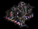
TL;DR
- AntiFold takes a fixed antibody variable-domain structure and predicts which amino acids and sequences are compatible with that backbone.
- Start with Number of sequences set to 0, inspect residue probabilities, then sample about 10 designs at temperature 0.2 in the smallest IMGT region that matches the design goal.
- A lower AntiFold score means stronger support from the model within that run, not proven affinity, stability, specificity, or developability.
- Use an IMGT-numbered structure for region-specific sampling, then refold and experimentally test shortlisted sequences.
AntiFold proposes antibody sequences that fit a submitted variable-domain backbone. Its main advantage is antibody-specific inverse folding; its main caveat is that structural compatibility is not the same as experimentally confirmed binding, stability, specificity, or developability.[2]
To use AntiFold online, add an antibody or nanobody structure to the AntiFold webserver, verify the chain roles, choose an IMGT design region, and either inspect residue probabilities or sample new sequences. Start with the probability-only default before generating a library.[1]
Open AntiFold online
Open the ProteinIQ AntiFold webserver.
Add the variable-domain structure
Upload a PDB, ENT, or mmCIF file, fetch a structure from RCSB, or use an official IMGT-numbered AntiFold fixture.
Assign the antibody chains
Choose paired antibody or nanobody mode and confirm the heavy, light, VHH, and optional antigen chain IDs.
Run probability analysis first
Keep Number of sequences at 0 and inspect the residue distributions before deciding where and how broadly to sample.
Sample a focused library
Select the smallest useful IMGT region, set a sequence count, and start at temperature 0.2 with a recorded seed.
Validate the candidates
Compare model support and mutation patterns, refold shortlisted sequences, screen liabilities, and test the intended function experimentally.
What is AntiFold?
AntiFold is an antibody-specific inverse folding model. Conventional structure prediction asks which structure a sequence may adopt. Inverse folding starts from a structure and asks which sequences are compatible with that backbone.
The model is fine-tuned from ESM-IF1, a structure-conditioned sequence model trained on millions of predicted protein structures. AntiFold specializes that model using solved antibody structures from SAbDab and predicted antibody structures from the Observed Antibody Space data used by the authors.[2][4][7]
The intended input is an antibody variable fragment with paired heavy and light variable domains, or a single-domain VHH nanobody. An antigen chain can be supplied as context. AntiFold keeps the submitted backbone fixed and predicts residue distributions or sampled antibody sequences; it does not generate a new backbone during the run.[2][3]
ProteinIQ currently runs AntiFold 0.3.1. The online implementation returns the source-native residue CSV, sampled FASTA when requested, optional NumPy embeddings, a run log, and an additive structured result for browsing and workflows.[1]
How does AntiFold work?
AntiFold follows four connected stages: structural conditioning, antibody-specific sequence modeling, residue-level scoring, and optional regional sampling.
The structure defines the design context
The model reads backbone coordinates from the selected antibody chains and, when provided, one antigen chain. Geometry tells the model which residues occupy buried, exposed, loop, interface, and framework environments. The sequence is then evaluated in that fixed structural context.
This is why input quality matters. Missing backbone atoms, incorrect chain assignments, unusual numbering, or a backbone that does not represent the intended binding state can change the distribution the model produces. AntiFold can work with solved or predicted structures, but a prediction should still be checked for plausible chain pairing and loop geometry.[2]
ESM-IF1 supplies a structure-conditioned sequence model
ESM-IF1 combines invariant geometric processing with a sequence-to-sequence transformer. It was trained to recover protein sequences from backbone coordinates and uses span masking so that partially missing coordinates can be tolerated in its general setting.[4]
AntiFold starts from this learned representation rather than training an antibody model from scratch. Fine-tuning adapts the residue preferences to antibody variable domains, where framework regions are relatively conserved and complementarity-determining regions (CDRs) have distinct structural and sequence behavior.[2]
Antibody-specific training improves CDR recovery
The AntiFold training strategy included solved and predicted antibody structures, CDR-weighted masking, regional span masking, and layer-wise learning-rate decay. In the paper's AbMPNN test set, AntiFold recovered 60% of native CDRH3 amino acids, compared with 56% for AbMPNN, 43% for ESM-IF1, and 35% for ProteinMPNN.[2]
Recovery is a benchmark of native-sequence reconstruction, not a guarantee that the native residue is always optimal or that a non-native design will bind. It is most useful for understanding how well a model has learned antibody-specific structure-sequence relationships.
Every residue receives a 20-amino-acid distribution
AntiFold writes one log probability for each standard amino acid at each residue. It also converts that distribution into positional perplexity, which behaves like the effective number of amino acids supported by the model. A concentrated distribution has lower perplexity; a broad distribution has higher perplexity.
Perplexity is comparative, not a universal traffic light. A high value can indicate several structurally plausible alternatives, but it does not show that every alternative preserves affinity, specificity, solubility, or expression.
Region masks and temperature control sampling
When sampling is enabled, AntiFold changes only positions in the selected IMGT region mask. IMGT numbering assigns comparable positions across immunoglobulin variable domains and defines framework and CDR boundaries, which makes a setting such as CDRH3 unambiguous across compatible structures.[5]
Temperature rescales the residue distribution before sampling:
- Lower temperatures concentrate choices around the highest-probability amino acids.
- The source default,
0.2, is a conservative starting point used in the AntiFold sequence-structure evaluation. - Higher temperatures broaden the sampled library and usually increase mutations, but also admit more weakly supported choices.[2][3]
The paper sampled 20 designs per antibody at temperature 0.2, refolded them with ABodyBuilder2, and reported a mean CDR backbone root-mean-square deviation of 0.95 Å from the experimental structures. That benchmark describes a test-set average, not the expected accuracy of every new design.[2]
Prepare a useful AntiFold input
Use the antibody variable domains
AntiFold is designed for variable heavy and variable light domains, or one VHH domain. Constant domains are outside its maintained design surface. A paired input needs at least two protein chains; nanobody mode is the correct choice for a single VHH chain.[1][3]
Accepted online files are .pdb, .ent, and .cif, up to 50 MiB. One structure is submitted per job. For several structures, use separate runs or a workflow rather than combining unrelated models into one file.[1]
Check IMGT numbering before regional sampling
IMGT numbering aligns structurally corresponding framework and CDR positions across antibody variable domains. AntiFold uses those position labels to decide which residues belong to settings such as CDRH3, light_cdrs, or FWH2.[5]
Use an IMGT-numbered structure from SAbDab when available, or renumber an antibody sequence or structure with ANARCI-compatible tooling before sampling. ANARCI supports IMGT, Kabat, Chothia, Enhanced Chothia, and AHo schemes, so confirm that the output scheme is specifically IMGT.[6][7]
ProteinIQ does not silently renumber the submitted coordinates. AntiFold reports a warning when the residue labels do not appear IMGT compatible. Treat that warning as a reason to verify the mask before trusting region-specific designs.
Assign chain roles explicitly when the file is complex
Blank heavy and light fields select the first two non-antigen chains in file order. That is convenient for a clean two-chain Fv, but explicit IDs are safer when the file contains an antigen, duplicate biological assemblies, constant domains, or extra proteins.
For a paired antibody-antigen complex:
- set
Heavy chain IDto the VH chain; - set
Light chain IDto the VL chain; - set
Antigen chain IDto one distinct antigen chain.
The official AntiFold documentation recommends one ideally small antigen chain. Adding an antigen changes structural context, but the AntiFold paper did not find a statistically significant affinity-prediction improvement from antigen context in its reported comparisons. Include it when the modeled interface is relevant, not because it guarantees better ranking.[2][3]
How to use AntiFold online
1. Add the structure
Open AntiFold online. Upload the structure or fetch it from RCSB. A raw RCSB structure is not necessarily IMGT numbered, so verify residue labels before regional sampling.
For an interface exercise with a known compatible input, use the official 6y1l_imgt.pdb fixture from the AntiFold repository. It contains paired variable domains and is already numbered for the source examples.[3]
2. Select paired antibody or nanobody mode
Choose Paired antibody (VH/VL) for an Fv or Fab variable-domain pair. Choose Nanobody / VHH for a single-domain antibody.
Confirm the chain IDs shown in the structure. Chain labels do not have to be literal H and L; labels such as A and B work when assigned to the correct roles. Light-chain region choices are unavailable in nanobody mode.
3. Start with probability analysis
Keep Number of sequences at 0 for the first run. This returns residue probabilities and perplexity without sampling a library. It answers three useful questions:
- Which positions have a sharply preferred residue?
- Which positions admit several alternatives?
- Does the model's preferred residue agree with the submitted sequence in the intended design region?
Probability analysis separates model inspection from library generation. It also enables Extract per-residue embeddings, which cannot be combined with sampling in AntiFold 0.3.1.[1]
4. Choose the smallest region that matches the experiment
The default CDRs only (CDR1, CDR2, CDR3) mask spans all six paired-antibody CDRs. A narrower mask usually makes the resulting library easier to interpret.
| Design goal | Useful starting mask | What remains fixed |
|---|---|---|
| Explore the heavy-chain third CDR | CDRH3 | Other CDRs and frameworks |
| Redesign all heavy-chain CDRs | heavy_cdrs | Light chain and all frameworks |
| Explore one loop on both chains | CDR1, CDR2, or CDR3 | Other named regions |
| Adjust framework residues | frameworks or one FWH or FWL region | All unselected regions |
| Broad variable-domain redesign | all | No selected antibody region is fixed |
Broad masks create a larger combinatorial space and can change more residues at once. Use them only when the validation plan can distinguish structural compatibility from unwanted effects on binding, humanness, or developability.
5. Choose count, temperature, and seed
For a first sampling run, use:
Number of sequences:10Sampling temperature:0.2Random seed:42
Ten sequences match the official source example and are enough to inspect whether the chosen mask and temperature produce the intended mutation density.[3]
If the designs are nearly identical, increase temperature in small steps before increasing both temperature and region breadth. If they mutate too aggressively, lower the temperature or narrow the region. Sequence count controls how many draws you inspect; it does not make an individual draw more accurate.
Keep the seed in the run record. On the reviewed ProteinIQ runtime, identical settings preserve the sampled FASTA sequences. Final GPU probability and embedding digits are not bitwise repeatable, so avoid tests or decisions that depend on their last decimal place.[1]
6. Submit and inspect all result views
Every successful run includes:
- a residue-probability CSV;
- a structured JSON result;
- the AntiFold run log.
Sampling adds a native FASTA file. Embedding extraction adds a native .npy array. The Results view aligns the original and sampled antibody sequences, the Residue probabilities view preserves the native table, and the Files view keeps every downloadable artifact.[1]
Reproducible example with the official 6Y1L fixture
The official repository uses 6y1l_imgt.pdb as its main paired-antibody example. It is suitable for checking the interface because its chain roles and numbering are known to AntiFold.[3]
Run two passes:
| Pass | Settings | Expected artifact types | Question answered |
|---|---|---|---|
| Probability map | paired mode, chains H and L, Number of sequences = 0, default CDR mask | CSV, JSON, log | Which residues and amino acids does the model support? |
| Focused sampling | same input, Number of sequences = 10, Sampling temperature = 0.2, Random seed = 42 | CSV, FASTA, JSON, log | What variants are drawn under the source example settings? |
This exercise intentionally does not quote numeric ProteinIQ results because there is no maintained public example job for the current webserver. Use the files generated by the run itself rather than copying values from a different runtime.
For antigen context, the source repository also includes 3hfm.pdb with heavy chain H, light chain L, and antigen chain Y. Use it to test chain assignment, not as proof that antigen-aware scoring will predict affinity.[3]
How to interpret AntiFold results
Residue log probabilities
The 20 amino-acid columns are log probabilities. Within one residue row, the value closest to zero has the greatest model support. top_res identifies that amino acid.
Compare alternatives at the same position before comparing raw values across different structures. Structural environment, chain context, numbering, and model uncertainty all change the distribution.
Perplexity
Positional perplexity summarizes the breadth of the 20-amino-acid distribution:
- low perplexity means the model concentrates support on a small set;
- high perplexity means support is spread across more alternatives.
Perplexity is not a mutation acceptance threshold. It does not identify which biochemical property limits a residue, and it cannot replace an assay-specific score. The 2026 antigen-aware benchmark found that inverse folding models can enrich some binders but do not consistently provide nuanced affinity prediction across realistic antibody datasets.[8]
Sequence score and global score
score is the mean negative log-likelihood over the selected design region. global_score is the mean negative log-likelihood over all selected antibody residues. Lower is better supported by AntiFold.
Use these values to rank closely related sequences from the same input, region, and settings. Do not read them as binding free energy, dissociation constant, expression yield, melting temperature, or probability of experimental success. The original paper reported useful but imperfect zero-shot correlations with binding measurements, and the later benchmark reinforces the need for independent validation.[2][8]
Sequence recovery and mutation count
seq_recovery is the fraction of antibody positions that match the submitted sequence. Mutation count reports how many positions changed.
Both describe distance from the input. Neither is a quality score. A high-recovery design may preserve the parent closely without improving the target property; a low-recovery design may be diverse but difficult to validate.
Per-residue embeddings
The optional .npy file contains AntiFold's source-native latent representation for each residue. It is useful for downstream clustering, visualization, or custom models, but the embedding dimensions do not have simple named biochemical meanings. Embedding extraction is available only when sequence sampling is off.[1][3]
How to check shortlisted sequences
Refold against the intended backbone
Predict the structure of each shortlisted sequence with an antibody-specific model such as ABodyBuilder3, then align it to the design backbone. Inspect the variable-domain orientation and each mutated CDR rather than relying only on a whole-Fv summary.
This mirrors the AntiFold paper's sequence-structure evaluation, which refolded sampled sequences and measured CDR backbone deviation. A good refold supports structural compatibility, but prediction agreement is still not experimental proof.[2]
Check numbering and sequence liabilities
Confirm that mutations map to the intended IMGT positions and that no chain boundary or insertion code was misread. Then check:
- unexpected cysteines or disulfide patterns;
- glycosylation and chemical-liability motifs;
- hydrophobic or charged surface patches;
- framework mutations near conserved packing residues;
- humanness and potential immunogenicity for therapeutic programs.
BioPhi can support antibody humanness and humanization analysis. These filters answer different questions from AntiFold and should not be collapsed into one composite score.
Reassess binding and specificity
If the design changes a CDR or interface residue, evaluate the antibody-antigen complex with an appropriate structural or experimental workflow. AntiFold support means the sequence is compatible with the supplied backbone distribution; it does not prove that the intended antigen interaction remains or improves.
Use biophysical and functional assays appropriate to the program, such as expression, monodispersity, thermal stability, affinity, competition, and specificity measurements. Test the parent and relevant controls alongside the designs.
Troubleshooting
| Symptom | Likely cause | What to do |
|---|---|---|
| Paired mode rejects the file | Fewer than two protein chains were detected | Use nanobody mode for a VHH or upload the paired variable domains. |
| A requested region appears empty | Residue labels do not match the IMGT mask | Renumber with IMGT labels and verify insertion codes before resubmitting. |
| Wrong chains are treated as VH and VL | Auto-detection followed file order | Set explicit heavy, light, and antigen chain IDs. |
| Embedding extraction is unavailable | Sequence sampling is enabled | Set Number of sequences to 0. |
| Designs are too similar | Temperature or region breadth is too low | Raise temperature gradually or broaden the mask, one change at a time. |
| Designs mutate too many positions | Temperature or region breadth is too high | Lower temperature or select a narrower IMGT region. |
| Scores look favorable but refolding changes the loop | The fixed-backbone assumption is weak for that sequence | Remove the candidate or validate with additional structure and experimental methods. |
| Repeated probability values differ in final digits | GPU inference is not bitwise repeatable | Compare scientifically meaningful precision and keep the run provenance. |
AntiFold alternatives
| Method | Best fit | Main difference from AntiFold |
|---|---|---|
| ProteinMPNN | General fixed-backbone protein sequence design | Handles broad protein targets and richer general constraints rather than antibody-specific IMGT masks.[9] |
| LigandMPNN | Sequence design around ligands, metals, nucleotides, or fixed side chains | Conditions explicitly on non-protein context and binding-site atoms. |
| DiffAb | Antibody CDR sequence and structure co-design | Can change CDR geometry as well as sequence instead of keeping the backbone fixed. |
| IgDesign | CDR sequence design from an antibody-antigen complex | Uses its own antibody-antigen design and sampling pipeline and requires complex-oriented chain roles. |
| ABodyBuilder3 | Antibody structure prediction from a proposed VH/VL sequence | Predicts the structure after sequence design rather than generating sequences from a structure. |
Choose AntiFold when the backbone is the design constraint and antibody-specific residue distributions are important. Choose a co-design method when loop geometry must change, or a general inverse folding method when the target is not an antibody variable domain.
Practical checklist
Before submitting:
- Confirm the file contains the intended VH/VL pair or VHH domain.
- Use IMGT numbering for region-specific sampling.
- Set explicit chain IDs for complexes or multi-chain files.
- Start with probability analysis and inspect the run warning log.
Before selecting sequences:
- Use the smallest design mask that matches the hypothesis.
- Compare scores only among closely matched runs and settings.
- Review mutation coordinates, perplexity, and residue probabilities together.
- Refold shortlisted sequences and inspect mutated loops.
- Screen humanness and developability separately.
- Confirm binding, specificity, stability, and expression experimentally.
Antibody design tools on ProteinIQ
- AntiFold for antibody-specific fixed-backbone inverse folding
- ABodyBuilder3 for VH/VL structure prediction
- DiffAb for CDR sequence and structure co-design
- IgDesign for antibody-antigen CDR sequence design
- BioPhi for humanness scoring and humanization
- ProteinMPNN for general fixed-backbone protein design
Frequently asked questions
Can AntiFold use a predicted antibody structure?
Yes. The AntiFold paper reported similar amino-acid recovery on solved structures and structures predicted with ABodyBuilder2, and it included an AlphaFold-predicted example. Check the predicted CDR geometry and chain orientation before treating the backbone as a design constraint.[2]
Does an AntiFold design have better binding affinity?
Not necessarily. AntiFold scores structural sequence support, not experimental affinity. The paper reported zero-shot correlations on several datasets, but both the original study and later benchmarking show that this signal is imperfect and dataset dependent.[2][8]
Does the structure have to be IMGT numbered?
IMGT numbering is required for dependable region-specific sampling because AntiFold maps CDR and framework masks from residue numbers. Probability analysis can still run on a parseable variable-domain structure, but a numbering warning means the region labels should not be trusted until corrected.[3][5]
Can AntiFold design only CDRH3?
Yes. Select Heavy-chain CDR3, which maps to CDRH3. The other antibody regions remain unchanged during sampling.[1]
How many sequences should a first run generate?
Ten is a practical first pass and matches the official source example. Increase the count after confirming that the region and temperature produce useful mutation patterns. More samples explore more draws but do not improve the reliability of any one score.[3]
What temperature should be used?
Start at 0.2, the source default and the temperature used in the paper's sequence-structure sampling benchmark. Lower it for more concentrated designs or raise it gradually for diversity.[2][3]
Should an antigen chain always be included?
No. Include one antigen chain when its interface geometry is part of the intended structural context. The original AntiFold study did not find a statistically significant affinity-prediction improvement from antigen context in its reported comparison, so antigen inclusion should not be treated as an automatic quality boost.[2]
Can AntiFold design a nanobody?
Yes. Choose Nanobody / VHH mode and assign the VHH chain, or leave it blank when the file contains one unambiguous protein chain. Light-chain region masks are not available in this mode.[1][3]
Are AntiFold results deterministic?
The random seed controls sequence sampling, and sampled FASTA designs are stable on the reviewed ProteinIQ runtime. GPU residue probabilities and embeddings can vary in their final digits between equivalent runs, so they are not bitwise deterministic.[1]
Sources▼
- Use AntiFold Online ProteinIQ · August 25, 2026. https://proteiniq.io/app/antifold
- AntiFold: improved structure-based antibody design using inverse folding Bioinformatics Advances · 2025. https://doi.org/10.1093/bioadv/vbae202
- AntiFold official repository GitHub (oxpig/AntiFold) · August 25, 2026. https://github.com/oxpig/AntiFold
- Learning inverse folding from millions of predicted structures Proceedings of Machine Learning Research · 2022. https://proceedings.mlr.press/v162/hsu22a.html
- IMGT unique numbering for all IG and TR V-REGION of all species IMGT · August 25, 2026. https://www.imgt.org/IMGTScientificChart/Numbering/IMGTnumbering.html
- ANARCI: antigen receptor numbering and receptor classification Bioinformatics · 2016. https://doi.org/10.1093/bioinformatics/btv552
- SAbDab: the structural antibody database Nucleic Acids Research · 2014. https://doi.org/10.1093/nar/gkt1043
- Benchmarking antigen-aware inverse folding methods for antibody design Bioinformatics Advances · 2026. https://doi.org/10.1093/bioadv/vbag081
- Robust deep learning based protein sequence design using ProteinMPNN Science · 2022. https://doi.org/10.1126/science.add2187

Founder and computational chemist, ProteinIQ
Dr. Matic Broz is the founder of ProteinIQ and a computational chemist. He completed a PhD focused on protein structure, molecular dynamics, and neural networks, and writes about structural biology and scientific software.