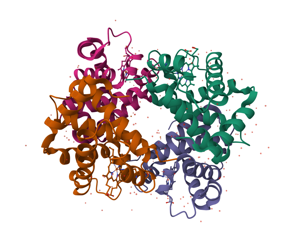
Physics-informed local sharpening for cryo-EM density maps. Learn more
Input
Unsharpened, unfiltered cryo-EM half-map.
Matching unsharpened, unfiltered cryo-EM half-map.
What is LocScale?
LocScale performs local density scaling for cryo-EM maps. It sharpens map regions according to local signal and reference information rather than applying one global B-factor across the whole volume.
ProteinIQ runs LocScale on uploaded MRC/MAP files and returns the sharpened map plus the processing log and generated map files. The first release focuses on the standard LocScale command for local sharpening with half-map or full-map inputs. Feature-enhanced maps, coordinate-model refinement, hybrid model completion, MPI runs, and PDF reports are tracked for future support.
Inputs
| Input | Description |
|---|---|
| Half Map 1 | First unsharpened, unfiltered half-map in MRC or MAP format. |
| Half Map 2 | Second matching unsharpened, unfiltered half-map. |
| Cryo-EM Map | Full unsharpened map for datasets without half-maps. |
| Mask | Optional MRC/MAP mask. LocScale estimates an FDR mask when this is omitted. |
| Reference Map | Optional MRC/MAP model map used directly as the LocScale reference. |
Half-map input is recommended when available because LocScale can use the pair for FSC-based resolution estimation.
ProteinIQ accepts MRC/MAP uploads up to 500 MB per file for this service. This is a ProteinIQ upload limit, not a LocScale file-format limit.
Settings
The default settings match LocScale defaults for local sharpening: one CPU process, FDR threshold 0.01, averaging filter size 3, EMmerNet batch size 8, cube size 32, and no input filtering. Optional resolution, window-size, FDR-window, FDR pre-filter, mask-threshold, and reference-map-resolution fields are passed to the corresponding LocScale command-line options only when set.
Outputs
LocScale returns a primary sharpened MRC map. ProteinIQ also returns the LocScale log and generated processing files such as FDR masks or local-scaling diagnostic maps when LocScale writes them.
Open the returned MRC files in ChimeraX, Coot, or another cryo-EM map viewer for inspection.
LocScale examples
TRPV1 half-maps with FSC filtering
This example demonstrates the recommended paired-half-map workflow on a compact region of the Rattus norvegicus TRPV1 channel dataset EMD-5778. It uses matching unfiltered half-map subvolumes, a data-derived mask, and the corresponding deposited reconstruction as a direct reference map.
- Inputs: Two 64 × 64 × 64 half-map subvolumes, matching mask, and reference map at 1.2156 Å per voxel
- Non-default settings:
CPU processes=4,Resolution cutoff (A)=3.4,Window size (pixels)=24, andApply FSC filter= enabled
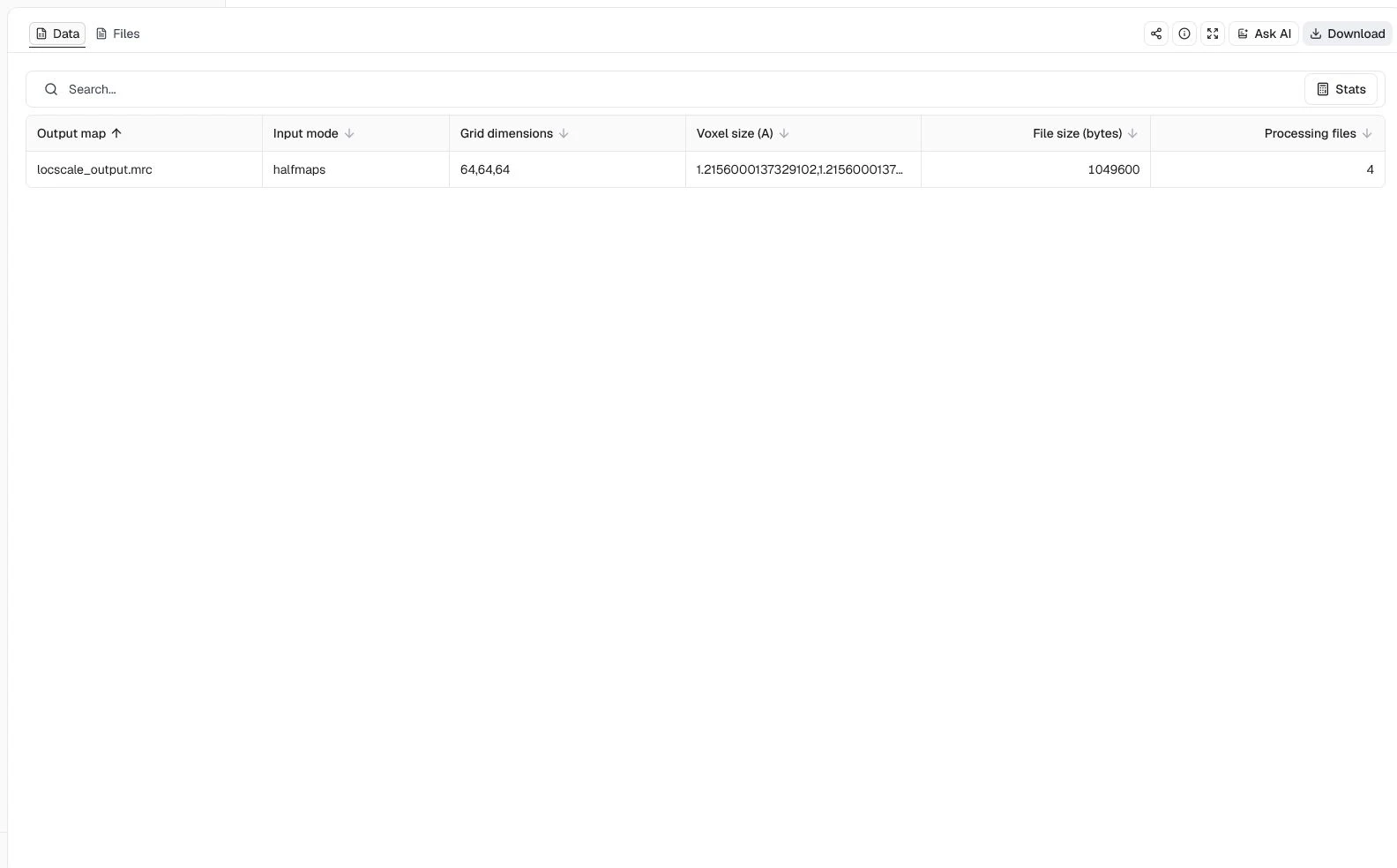
The completed job returns locscale_output.mrc on the original 64 × 64 × 64 grid, together with the reconstructed unfiltered map, bfactor_map.mrc, qfit_map.mrc, and the processing log. The table confirms that LocScale used halfmaps mode and preserved the input voxel spacing. This result shows that the paired-map and FSC-filtering workflow completed; it does not by itself establish improved interpretability. Compare the output with the input half-maps in a cryo-EM viewer before using it for model building.
β-galactosidase full-map reference scaling
This EMD-2984 example shows full-map operation on a high-resolution region of Escherichia coli β-galactosidase. The input is an average of matching deposited half-map subvolumes, accompanied by a data-derived mask and the corresponding deposited reconstruction as the direct scaling reference.
- Inputs: One 48 × 48 × 48 full-map subvolume, matching mask, and reference map at 0.637 Å per voxel
- Non-default settings:
Map input=Full map,CPU processes=4,Resolution cutoff (A)=2.2, andWindow size (pixels)=16
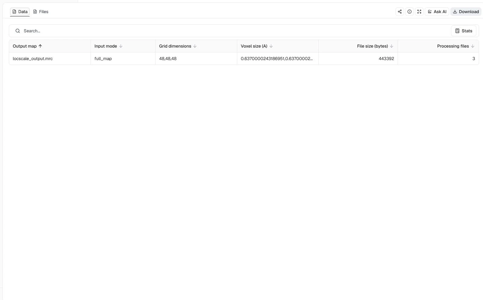
ProteinIQ returns a 48 × 48 × 48 sharpened MRC map at the same 0.637 Å voxel spacing, plus bfactor_map.mrc, qfit_map.mrc, and the LocScale log. The compact subvolume makes the reference-map workflow quick to inspect, but it is not a substitute for processing and evaluating the complete EMD-2984 reconstruction.
Masked and filtered γ-secretase full map
This example uses a compact region from the EMD-3061 human γ-secretase half-map pair to demonstrate full-map processing with both an explicit mask and input filtering. The supplied reference is the matching region from the deposited reconstruction.
- Inputs: One 48 × 48 × 48 full-map subvolume, matching mask, and reference map at 1.4 Å per voxel
- Non-default settings:
Map input=Full map,CPU processes=4,Resolution cutoff (A)=3.4,Window size (pixels)=24, andFilter input maps= enabled
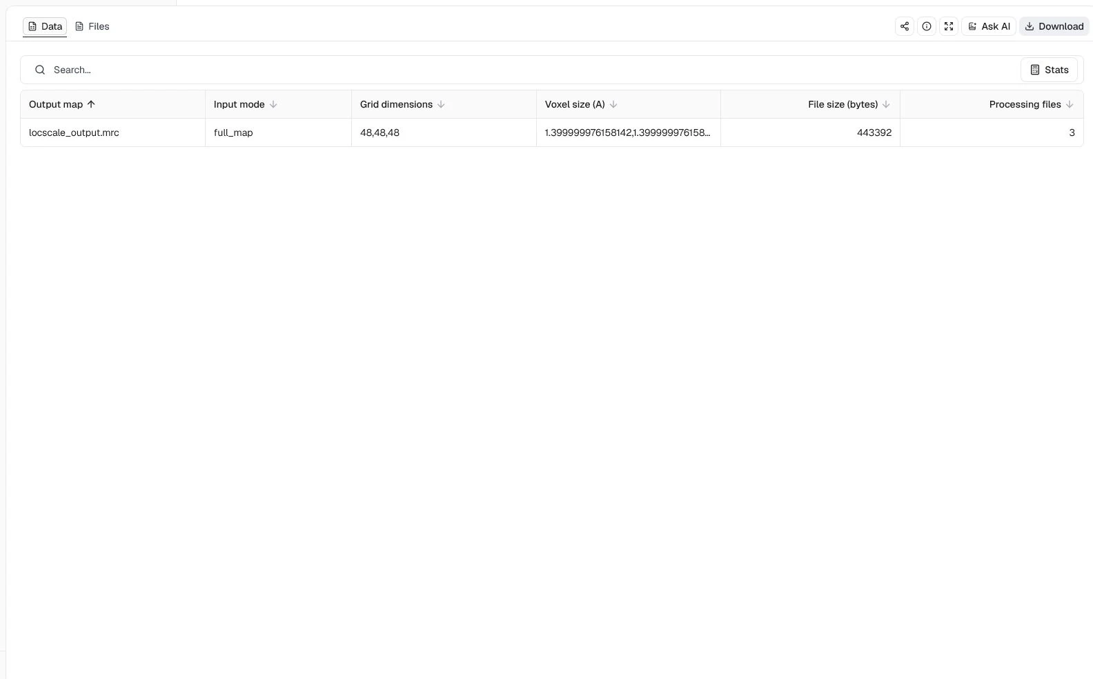
The output table records the expected 48 × 48 × 48 grid, 1.4 Å voxel spacing, and three processing files. This confirms that LocScale completed the masked, filtered full-map workflow and returned its sharpened map and diagnostics. Whether filtering improves local density for a particular structural interpretation still requires direct comparison with the unfiltered input and independent validation against the half-maps.
Limitations
- Inputs should be unsharpened and unfiltered. Pre-sharpened maps can produce misleading artifacts.
- Coordinate-model and hybrid LocScale modes require additional REFMAC/CCP4 runtime support and are not enabled in this release.
- Feature-enhanced maps and pVDDT confidence maps are not enabled in this release.
- Point-group symmetry options are held at the LocScale default of
C1until symmetry-specific runtime checks are added.
Related tools
AllMetal3D
Predict metal and water binding sites in protein structures using 3D convolutional neural networks (AllMetal3D + Water3D).
MolProbity
Validate protein structure quality with all-atom contact analysis, Ramachandran plots, rotamer assessment, and geometry checks.
PDBsum
Generate a downloadable PDBsum structural summary report archive for a single protein structure.
pyRMSD
Calculate pairwise RMSD matrices for PDB structure ensembles with pyRMSD, including the condensed matrix and source statistics files.
SuperWater
Predict protein hydration sites from a structure using a diffusion model with ESM features and a confidence-filtering head.
Aggrescan3D
Static-mode Aggrescan3D analysis for per-residue aggregation propensity from a single protein structure.
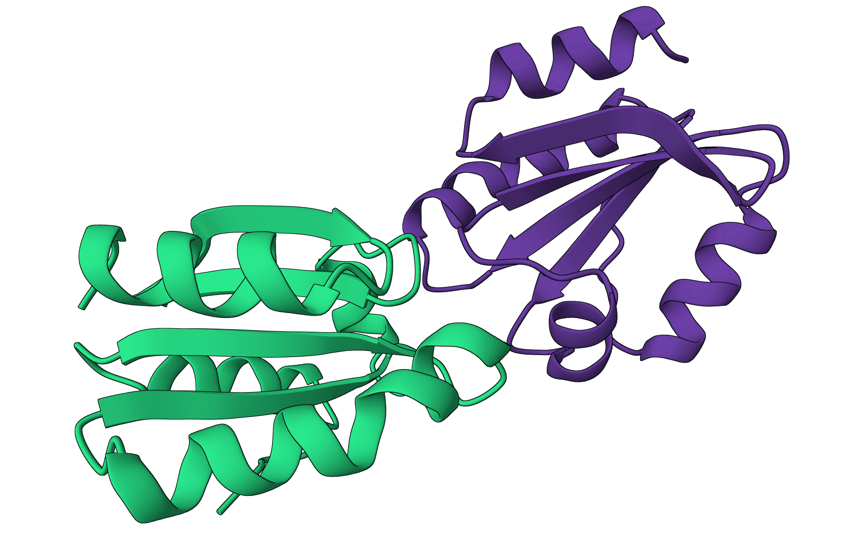
DockQ
Assess docking model quality by comparing predicted complexes against native references. DockQ v2.1.3 supports protein, nucleic-acid, and supported small-molecule interfaces with native metrics.
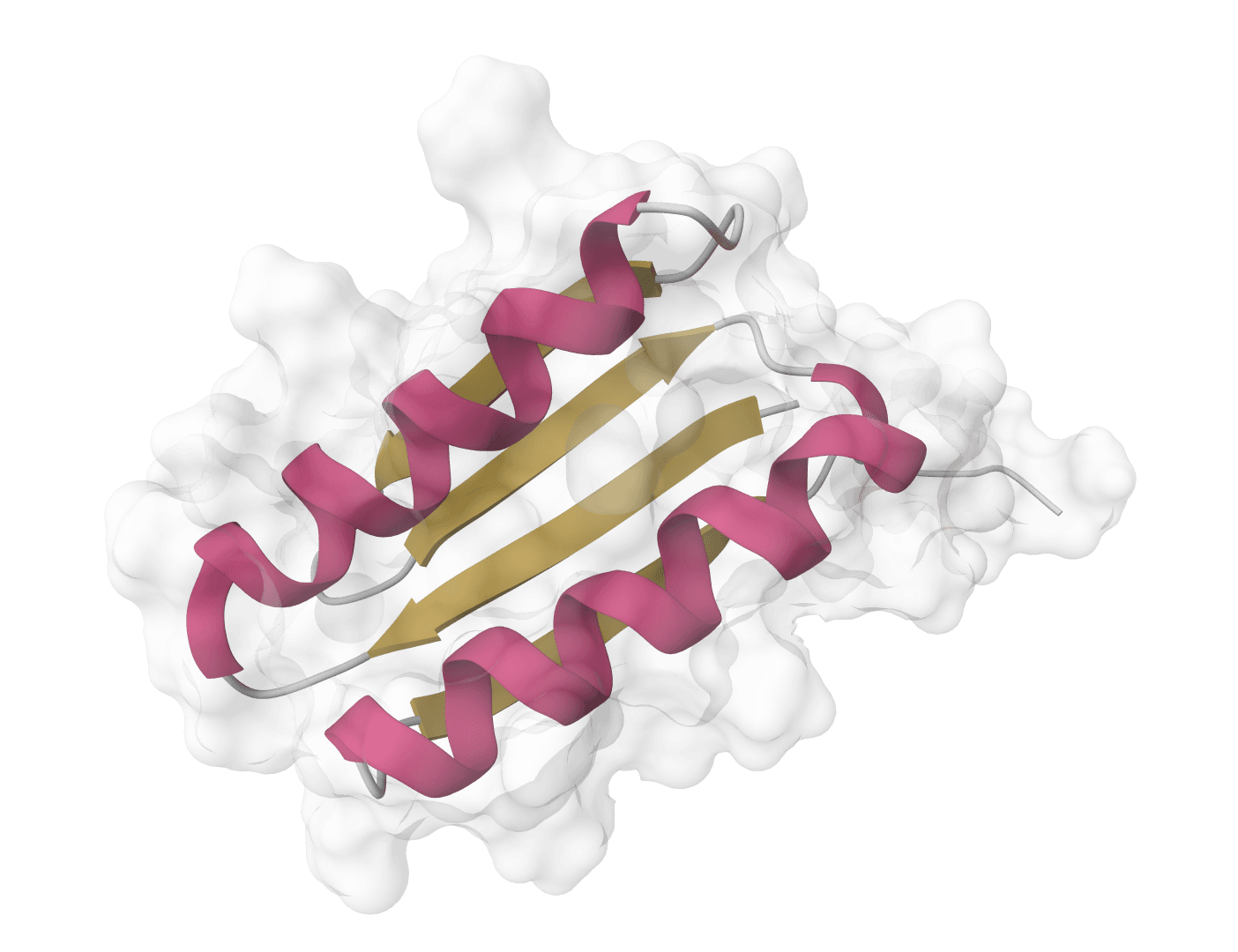
DSSP
Assign protein secondary structure with the source DSSP program and return annotated mmCIF, legacy DSSP, residue geometry, hydrogen-bond statistics, and native files.
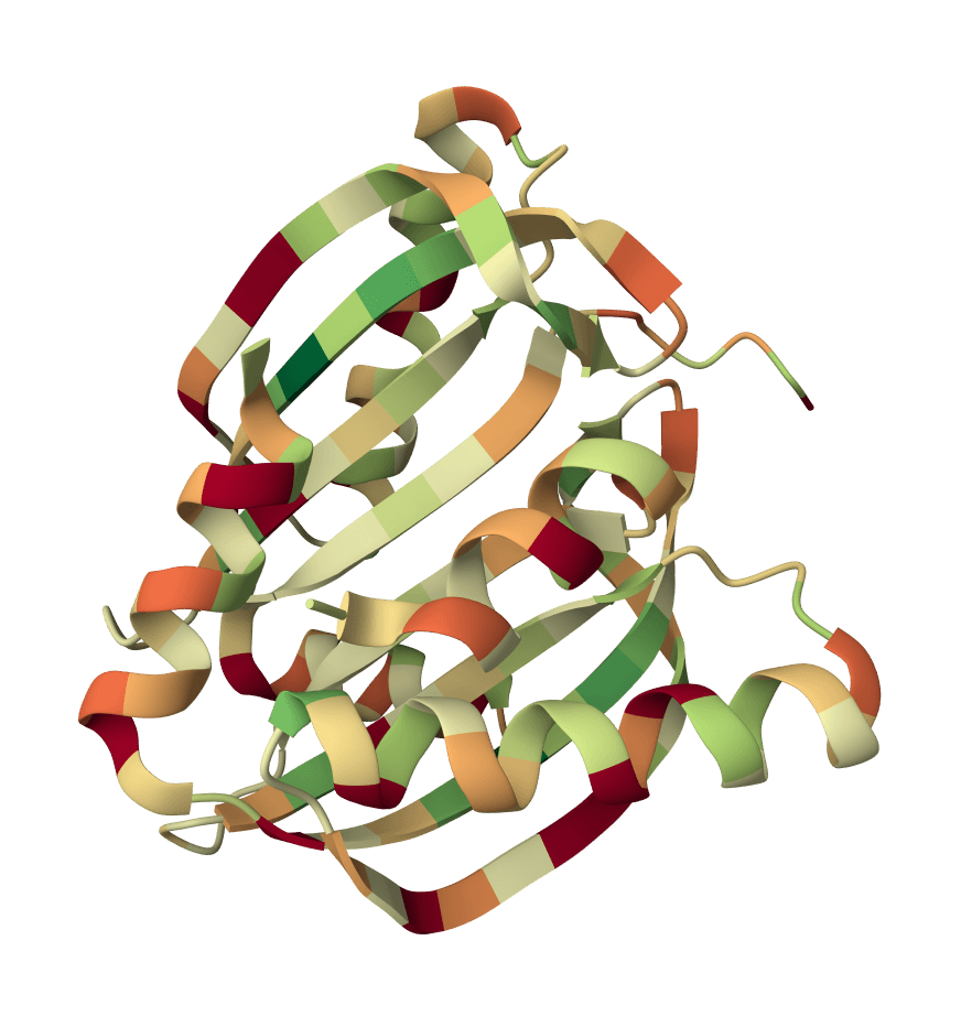
IPSAE
Scoring function for interprotein interactions in AlphaFold2, AlphaFold3 and Boltz predictions. Calculates ipSAE, ipTM, pDockQ, pDockQ2, and LIS scores to assess protein-protein interface quality.
PLIP
Analyze noncovalent interactions in protein-ligand complex structures with PLIP, including hydrogen bonds, hydrophobic contacts, pi interactions, salt bridges, water bridges, halogen bonds, and metal complexes.