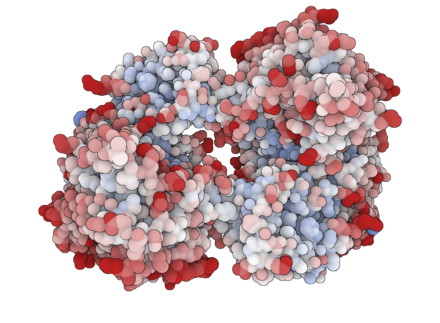
A hydrophobicity plot visualizes the local hydrophobicity along a protein sequence using a sliding window average. This smooths out residue-by-residue variations and reveals structural patterns that correlate with protein function and topology.
This tool provides 24 different hydrophobicity scales, each optimized for specific applications. Some scales derive from partition coefficients between water and organic solvents, others from accessible surface area measurements, and some from empirical correlations with membrane proteins or antigenic sites.
For the original Kyte-Doolittle hydropathy analysis, use our dedicated Hydropathy Plot tool. For a single-value summary of overall protein hydrophobicity, see GRAVY.
The algorithm assigns each amino acid a hydrophobicity index based on the selected scale. A sliding window then moves along the sequence, calculating the average hydrophobicity at each position.
For a window of size centered at position , the local hydrophobicity is calculated as:
where is the hydrophobicity value of the amino acid at position .
This averaging smooths out single-residue fluctuations. Smaller windows (5-7 residues) preserve local variations but produce noisier plots. Larger windows (15-21 residues) emphasize extended domains like transmembrane helices.
Most scales assign positive values to hydrophobic residues and negative values to hydrophilic residues. However, some scales like Hopp-Woods use the opposite convention, assigning positive values to hydrophilic residues to highlight potential epitopes.
Useful for predicting peptide behavior in reversed-phase chromatography:
Sliding window: The number of residues averaged at each position. Use 9 for surface region analysis and epitope prediction. Use 19 for transmembrane helix prediction, as this matches typical helix length spanning a lipid bilayer.
Hydrophobicity scale: Select based on your analysis goal. We recommend Eisenberg for general analysis, Hopp-Woods for epitope prediction, and Janin or Rao-Argos for transmembrane prediction.
The interactive chart displays hydrophobicity values along the sequence length. Hover over any point to see the exact position, residue, and calculated value.
Positive values indicate hydrophobic regions likely to be buried in the protein core or embedded in membranes. Extended peaks above +1.6 with window size 19 suggest potential transmembrane helices.
Negative values indicate hydrophilic regions typically found on protein surfaces or in aqueous environments.
Hopp-Woods and Welling use inverted conventions where positive values indicate hydrophilic residues. For these scales, peaks indicate surface-exposed, potentially antigenic regions suitable for antibody binding.
Download results as CSV for quantitative analysis or PNG for publication figures. The CSV includes position, residue identity, and hydrophobicity value for each data point.
| Application | Recommended scales |
|---|---|
| Transmembrane prediction | Eisenberg, Janin, Guy, Rao-Argos |
| Epitope prediction | Hopp-Woods, Welling, Parker |
| General analysis | Eisenberg, Fauchere-Pliska |
| Peptide chromatography | Wilson, Meek, Cowan-Whittaker |
Hydrophobicity plots assume that local sequence determines local properties. They cannot account for three-dimensional structure effects, long-range interactions, or post-translational modifications.
For dedicated transmembrane topology prediction, specialized tools like TMHMM or Phobius typically achieve higher accuracy by incorporating evolutionary profiles and topology grammar rules.
Plot net charge vs pH for protein sequences. Visualize how protein charge changes across pH 0-14 and identify the isoelectric point (pI) where the net charge crosses zero.
Generate Kyte-Doolittle hydropathy plots to visualize hydrophobic and hydrophilic regions along protein sequences. Identify transmembrane domains and surface-exposed regions.
Generate amino acid property profiles using 42 different scales spanning hydrophobicity, secondary structure propensity, flexibility, polarity, surface accessibility, antigenicity, and more.
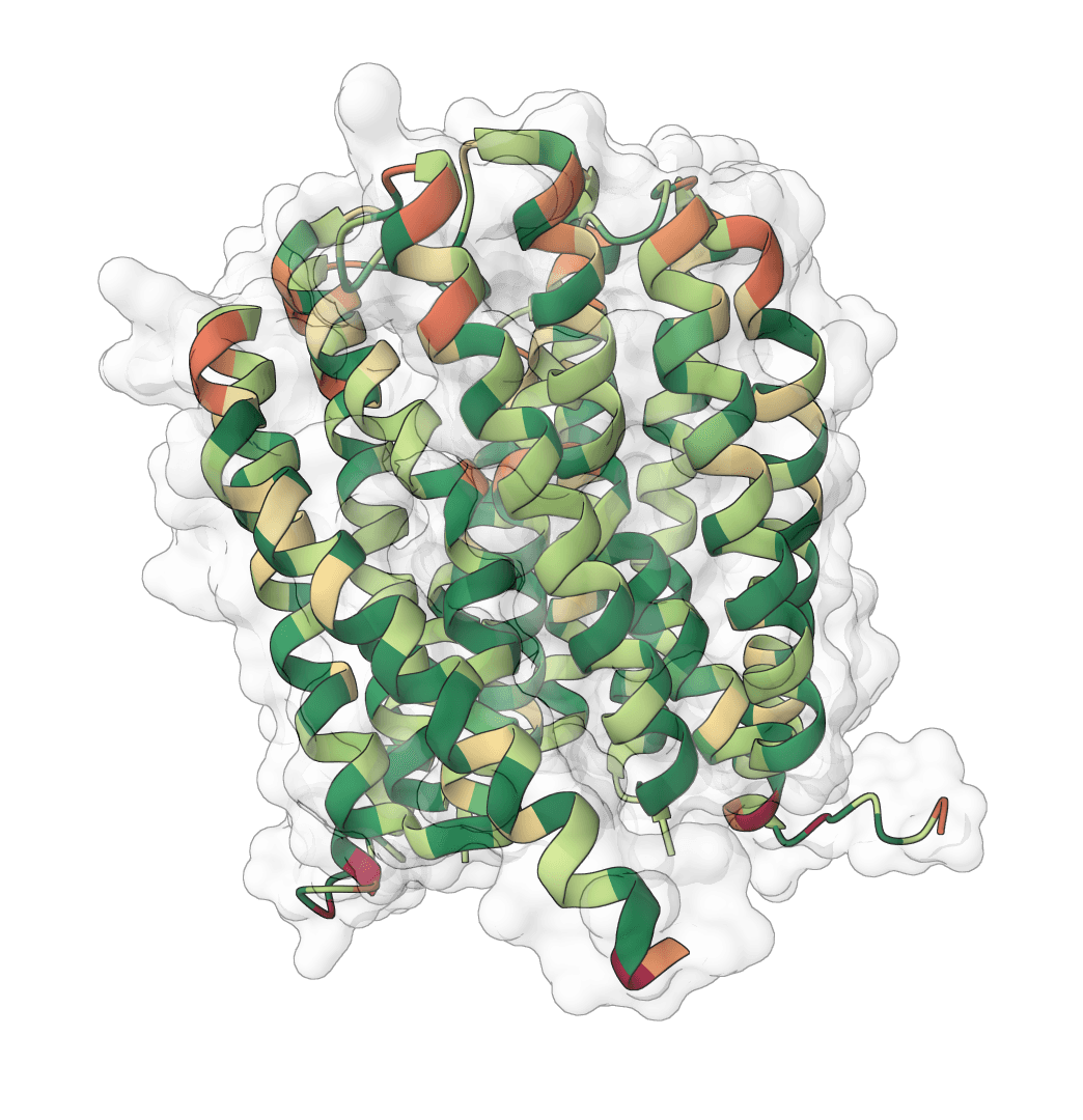
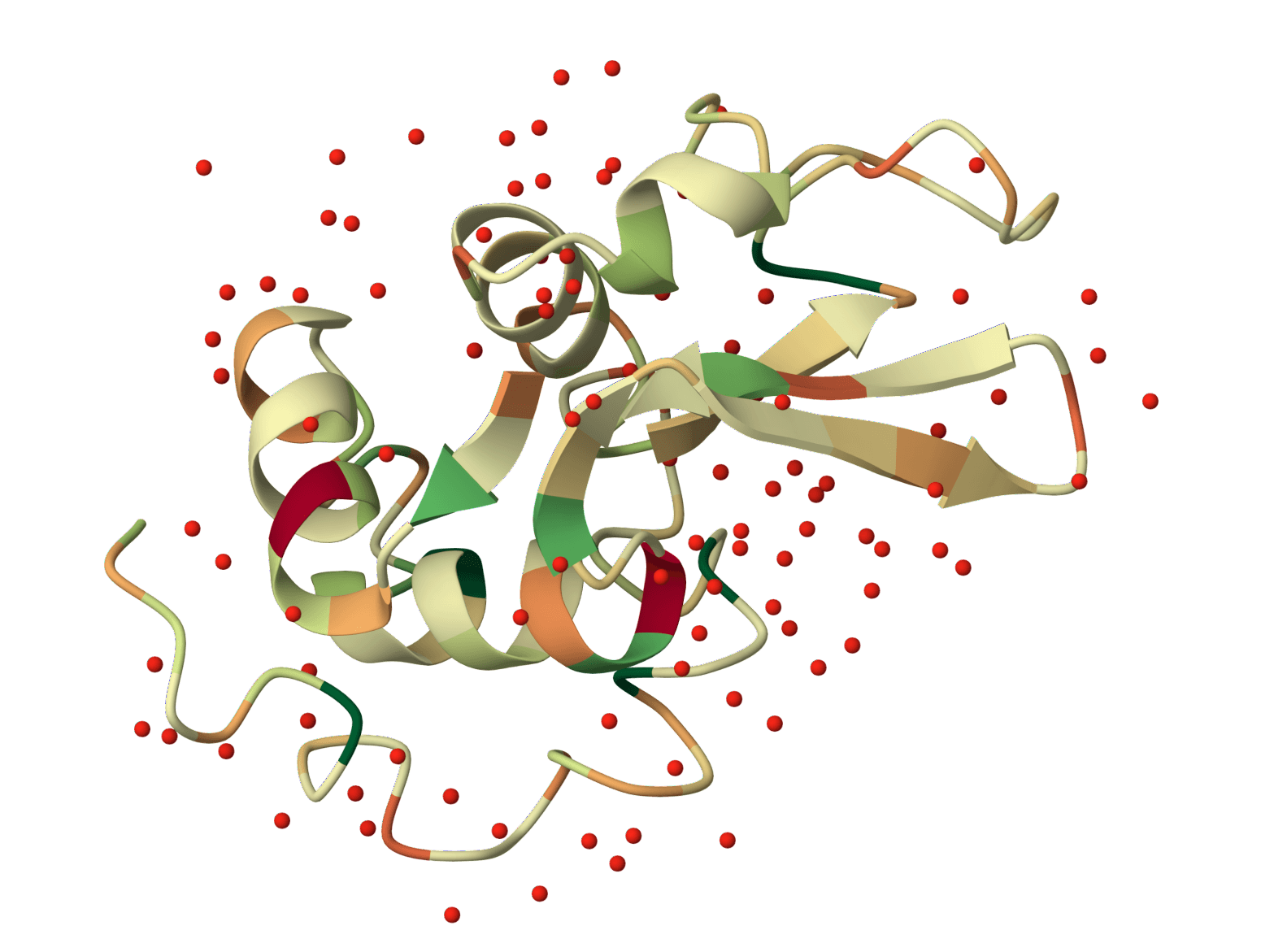
Faithful static-mode Aggrescan3D tool for per-residue aggregation propensity analysis from a single protein structure.
Match experimental peptide masses against theoretical digest fragments of a protein sequence. Identify peptides from mass spectrometry data by peptide mass fingerprinting.
Predict protease and chemical cleavage sites across a protein sequence for up to 39 enzymes simultaneously. Identify where each enzyme cuts, the cleavage residue, and context window around each site.
Cleave a protein sequence with a chosen protease and compute the masses of the resulting peptides. Supports multiple enzymes, missed cleavages, chemical modifications, and different ion types for mass spectrometry experiment planning.
Predict pKa values of ionizable groups in proteins and protein-ligand complexes from 3D structure. PROPKA calculates environment-driven pKa shifts for standard ionizable residues, terminal groups, and supported ligand atom types.
Calculate protein parameters, including molecular weight, theoretical pI, extinction coefficients, aromaticity, secondary structure fractions, atomic composition, estimated half-life, and several indices, including instability, aliphatic index, and GRAVY.
Isoelectric Point Calculator 2.0 - Predict protein/peptide isoelectric point (pI) using 18+ validated pKa scales, SVR models, and deep learning. Supports proteins, peptides, and comprehensive analysis.
A hydrophobicity plot visualizes the local hydrophobicity along a protein sequence using a sliding window average. This smooths out residue-by-residue variations and reveals structural patterns that correlate with protein function and topology.
This tool provides 24 different hydrophobicity scales, each optimized for specific applications. Some scales derive from partition coefficients between water and organic solvents, others from accessible surface area measurements, and some from empirical correlations with membrane proteins or antigenic sites.
For the original Kyte-Doolittle hydropathy analysis, use our dedicated Hydropathy Plot tool. For a single-value summary of overall protein hydrophobicity, see GRAVY.
The algorithm assigns each amino acid a hydrophobicity index based on the selected scale. A sliding window then moves along the sequence, calculating the average hydrophobicity at each position.
For a window of size centered at position , the local hydrophobicity is calculated as:
where is the hydrophobicity value of the amino acid at position .
This averaging smooths out single-residue fluctuations. Smaller windows (5-7 residues) preserve local variations but produce noisier plots. Larger windows (15-21 residues) emphasize extended domains like transmembrane helices.
Most scales assign positive values to hydrophobic residues and negative values to hydrophilic residues. However, some scales like Hopp-Woods use the opposite convention, assigning positive values to hydrophilic residues to highlight potential epitopes.
Useful for predicting peptide behavior in reversed-phase chromatography:
Sliding window: The number of residues averaged at each position. Use 9 for surface region analysis and epitope prediction. Use 19 for transmembrane helix prediction, as this matches typical helix length spanning a lipid bilayer.
Hydrophobicity scale: Select based on your analysis goal. We recommend Eisenberg for general analysis, Hopp-Woods for epitope prediction, and Janin or Rao-Argos for transmembrane prediction.
The interactive chart displays hydrophobicity values along the sequence length. Hover over any point to see the exact position, residue, and calculated value.
Positive values indicate hydrophobic regions likely to be buried in the protein core or embedded in membranes. Extended peaks above +1.6 with window size 19 suggest potential transmembrane helices.
Negative values indicate hydrophilic regions typically found on protein surfaces or in aqueous environments.
Hopp-Woods and Welling use inverted conventions where positive values indicate hydrophilic residues. For these scales, peaks indicate surface-exposed, potentially antigenic regions suitable for antibody binding.
Download results as CSV for quantitative analysis or PNG for publication figures. The CSV includes position, residue identity, and hydrophobicity value for each data point.
| Application | Recommended scales |
|---|---|
| Transmembrane prediction | Eisenberg, Janin, Guy, Rao-Argos |
| Epitope prediction | Hopp-Woods, Welling, Parker |
| General analysis | Eisenberg, Fauchere-Pliska |
| Peptide chromatography | Wilson, Meek, Cowan-Whittaker |
Hydrophobicity plots assume that local sequence determines local properties. They cannot account for three-dimensional structure effects, long-range interactions, or post-translational modifications.
For dedicated transmembrane topology prediction, specialized tools like TMHMM or Phobius typically achieve higher accuracy by incorporating evolutionary profiles and topology grammar rules.
Plot net charge vs pH for protein sequences. Visualize how protein charge changes across pH 0-14 and identify the isoelectric point (pI) where the net charge crosses zero.
Generate Kyte-Doolittle hydropathy plots to visualize hydrophobic and hydrophilic regions along protein sequences. Identify transmembrane domains and surface-exposed regions.
Generate amino acid property profiles using 42 different scales spanning hydrophobicity, secondary structure propensity, flexibility, polarity, surface accessibility, antigenicity, and more.
Faithful static-mode Aggrescan3D tool for per-residue aggregation propensity analysis from a single protein structure.
Match experimental peptide masses against theoretical digest fragments of a protein sequence. Identify peptides from mass spectrometry data by peptide mass fingerprinting.
Predict protease and chemical cleavage sites across a protein sequence for up to 39 enzymes simultaneously. Identify where each enzyme cuts, the cleavage residue, and context window around each site.
Cleave a protein sequence with a chosen protease and compute the masses of the resulting peptides. Supports multiple enzymes, missed cleavages, chemical modifications, and different ion types for mass spectrometry experiment planning.
Predict pKa values of ionizable groups in proteins and protein-ligand complexes from 3D structure. PROPKA calculates environment-driven pKa shifts for standard ionizable residues, terminal groups, and supported ligand atom types.
Calculate protein parameters, including molecular weight, theoretical pI, extinction coefficients, aromaticity, secondary structure fractions, atomic composition, estimated half-life, and several indices, including instability, aliphatic index, and GRAVY.
Isoelectric Point Calculator 2.0 - Predict protein/peptide isoelectric point (pI) using 18+ validated pKa scales, SVR models, and deep learning. Supports proteins, peptides, and comprehensive analysis.