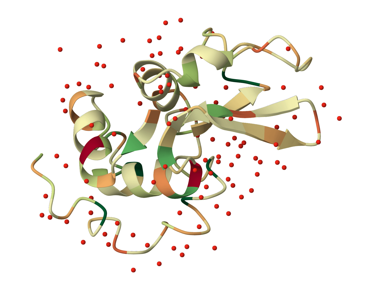
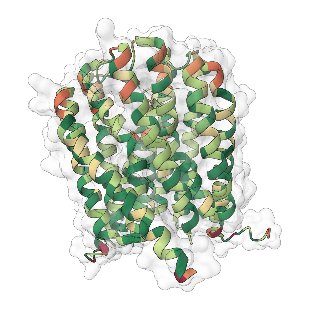
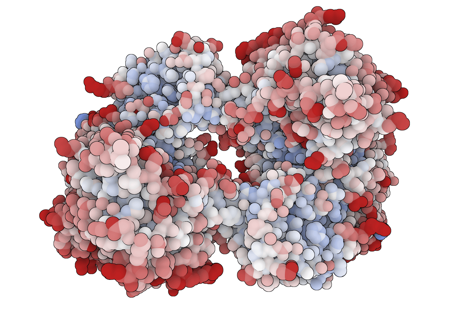
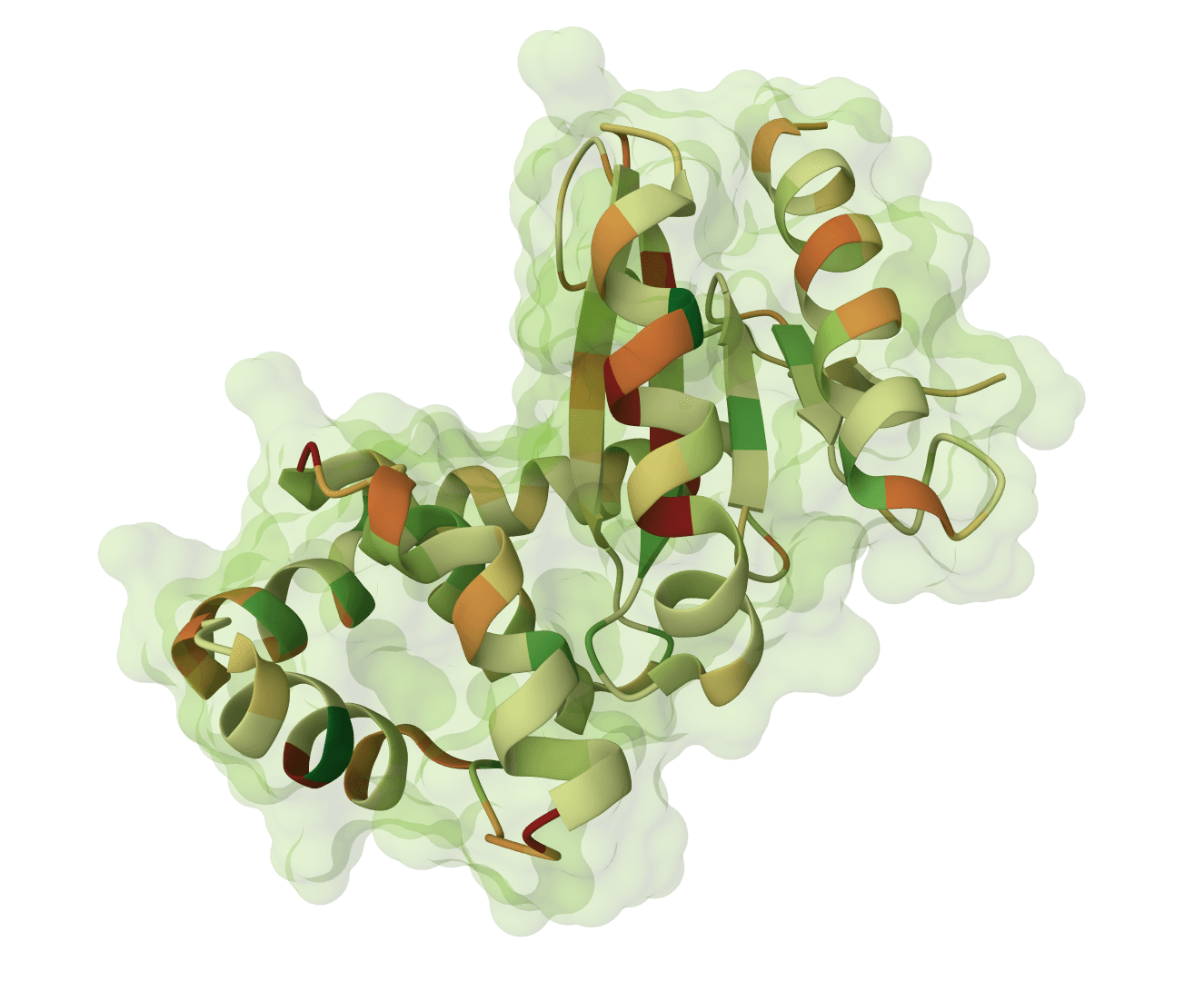
Hydropathy describes how amino acids interact with water. Hydrophobic ("water-fearing") residues avoid aqueous environments and tend to bury themselves in protein cores or lipid membranes. Hydrophilic ("water-loving") residues prefer contact with water and are typically found on protein surfaces.
This property is one of the most important determinants of protein structure. The tendency of hydrophobic residues to cluster away from water drives protein folding, while hydrophilic residues on the surface enable solubility and interactions with other molecules.
A hydropathy plot is a graphical representation of the hydrophobic and hydrophilic character of a protein sequence. It uses a sliding window approach to calculate average hydropathy values at each position, revealing patterns that correlate with protein structure and function.
The method was introduced by Jack Kyte and Russell Doolittle in 1982 and has become one of the most widely used tools in protein sequence analysis. Their original paper has been cited over 40,000 times, making it one of the most influential publications in computational biology.
Hydropathy plots are commonly used to identify transmembrane domains, predict surface-exposed regions, and locate potential antigenic sites for antibody binding.
The algorithm assigns each amino acid a hydropathy index based on its physicochemical properties. Hydrophobic residues like isoleucine, valine, and leucine receive positive scores, while hydrophilic residues like arginine, lysine, and aspartate receive negative scores.
A sliding window moves along the protein sequence, calculating the average hydropathy value for all residues within the window at each position. This averaging smooths out single-residue fluctuations and highlights broader structural patterns.
Smaller window sizes (5-7 residues) preserve local variations but produce noisier plots. Larger windows (15-21 residues) smooth the signal and emphasize extended domains like transmembrane helices.
Regions above zero (positive values) indicate hydrophobic stretches. Extended peaks above +1.6 with a window size of 19 suggest potential transmembrane helices, as these regions must be hydrophobic enough to span the lipid bilayer.
Regions below zero (negative values) indicate hydrophilic stretches. These are typically surface-exposed loops or regions that interact with the aqueous environment.
Three hydropathy scales are available, each optimized for different applications.
The Kyte-Doolittle scale is the original and most widely used hydropathy scale. It was derived from experimental measurements of amino acid transfer free energies between water and vapor phases.
This scale ranges from +4.5 (isoleucine, most hydrophobic) to -4.5 (arginine, most hydrophilic). We recommend this scale for general hydropathy analysis and transmembrane domain prediction.
The Hopp-Woods scale was designed specifically to predict antigenic epitopes and surface-exposed regions. Unlike Kyte-Doolittle, it assigns positive values to hydrophilic residues.
Use this scale when searching for potential antibody binding sites or identifying surface loops that may be immunogenic. The scale ranges from approximately -3.4 to +3.0.
The Cornette scale was optimized for detecting amphipathic helices, which have distinct hydrophobic and hydrophilic faces. These structures are common in membrane-associated proteins and antimicrobial peptides.
This scale accounts for the periodicity of alpha-helices (approximately 3.6-3.7 residues per turn) and is useful for identifying membrane-interacting regions that don't fully span the bilayer.
Sliding window: The number of residues averaged at each position. We recommend 9 for surface region analysis and 19 for transmembrane helix prediction.
Hydropathy scale: The amino acid hydropathy values to use. Kyte-Doolittle is best for general analysis and transmembrane prediction, Hopp-Woods for epitope prediction, and Cornette for amphipathic helix detection.
The interactive chart displays hydropathy values along the sequence length. Hover over any point to see the exact position, residue, and hydropathy value.
Transmembrane domains: Look for extended peaks above +1.6 spanning approximately 20 residues. These likely represent membrane-spanning helices.
Surface loops: Regions consistently below zero, especially sharp negative peaks, often correspond to surface-exposed loops that may be flexible or involved in protein-protein interactions.
Amphipathic regions: Oscillating patterns with moderate positive and negative values can indicate amphipathic helices that associate with, but don't fully span, membranes.
Results can be downloaded as CSV for further analysis or as PNG for publication figures. The CSV export includes position, residue identity, and hydropathy value for each data point.
| Scale | Best for | Positive values indicate |
|---|---|---|
| Kyte-Doolittle | Transmembrane domains | Hydrophobic (buried) |
| Hopp-Woods | Epitope prediction | Hydrophilic (exposed) |
| Cornette | Amphipathic helices | Optimized for helix periodicity |
Hydropathy plots provide useful first-pass predictions but have important limitations. They cannot account for tertiary structure effects, post-translational modifications, or membrane protein topology rules.
For transmembrane prediction specifically, dedicated tools like TMHMM or Phobius that incorporate evolutionary information typically achieve higher accuracy than hydropathy plots alone.
Plot net charge vs pH for protein sequences. Visualize how protein charge changes across pH 0-14 and identify the isoelectric point (pI) where the net charge crosses zero.
Generate hydrophobicity plots using 24 different amino acid scales. Visualize hydrophobic and hydrophilic regions for protein analysis, epitope prediction, and membrane protein studies.
Generate amino acid property profiles using 42 different scales spanning hydrophobicity, secondary structure propensity, flexibility, polarity, surface accessibility, antigenicity, and more.
Faithful static-mode Aggrescan3D tool for per-residue aggregation propensity analysis from a single protein structure.
Match experimental peptide masses against theoretical digest fragments of a protein sequence. Identify peptides from mass spectrometry data by peptide mass fingerprinting.
Predict protease and chemical cleavage sites across a protein sequence for up to 39 enzymes simultaneously. Identify where each enzyme cuts, the cleavage residue, and context window around each site.
Cleave a protein sequence with a chosen protease and compute the masses of the resulting peptides. Supports multiple enzymes, missed cleavages, chemical modifications, and different ion types for mass spectrometry experiment planning.
Predict pKa values of ionizable groups in proteins and protein-ligand complexes from 3D structure. PROPKA calculates environment-driven pKa shifts for standard ionizable residues, terminal groups, and supported ligand atom types.

Calculate protein parameters, including molecular weight, theoretical pI, extinction coefficients, aromaticity, secondary structure fractions, atomic composition, estimated half-life, and several indices, including instability, aliphatic index, and GRAVY.
Isoelectric Point Calculator 2.0 - Predict protein/peptide isoelectric point (pI) using 18+ validated pKa scales, SVR models, and deep learning. Supports proteins, peptides, and comprehensive analysis.
Hydropathy describes how amino acids interact with water. Hydrophobic ("water-fearing") residues avoid aqueous environments and tend to bury themselves in protein cores or lipid membranes. Hydrophilic ("water-loving") residues prefer contact with water and are typically found on protein surfaces.
This property is one of the most important determinants of protein structure. The tendency of hydrophobic residues to cluster away from water drives protein folding, while hydrophilic residues on the surface enable solubility and interactions with other molecules.
A hydropathy plot is a graphical representation of the hydrophobic and hydrophilic character of a protein sequence. It uses a sliding window approach to calculate average hydropathy values at each position, revealing patterns that correlate with protein structure and function.
The method was introduced by Jack Kyte and Russell Doolittle in 1982 and has become one of the most widely used tools in protein sequence analysis. Their original paper has been cited over 40,000 times, making it one of the most influential publications in computational biology.
Hydropathy plots are commonly used to identify transmembrane domains, predict surface-exposed regions, and locate potential antigenic sites for antibody binding.
The algorithm assigns each amino acid a hydropathy index based on its physicochemical properties. Hydrophobic residues like isoleucine, valine, and leucine receive positive scores, while hydrophilic residues like arginine, lysine, and aspartate receive negative scores.
A sliding window moves along the protein sequence, calculating the average hydropathy value for all residues within the window at each position. This averaging smooths out single-residue fluctuations and highlights broader structural patterns.
Smaller window sizes (5-7 residues) preserve local variations but produce noisier plots. Larger windows (15-21 residues) smooth the signal and emphasize extended domains like transmembrane helices.
Regions above zero (positive values) indicate hydrophobic stretches. Extended peaks above +1.6 with a window size of 19 suggest potential transmembrane helices, as these regions must be hydrophobic enough to span the lipid bilayer.
Regions below zero (negative values) indicate hydrophilic stretches. These are typically surface-exposed loops or regions that interact with the aqueous environment.
Three hydropathy scales are available, each optimized for different applications.
The Kyte-Doolittle scale is the original and most widely used hydropathy scale. It was derived from experimental measurements of amino acid transfer free energies between water and vapor phases.
This scale ranges from +4.5 (isoleucine, most hydrophobic) to -4.5 (arginine, most hydrophilic). We recommend this scale for general hydropathy analysis and transmembrane domain prediction.
The Hopp-Woods scale was designed specifically to predict antigenic epitopes and surface-exposed regions. Unlike Kyte-Doolittle, it assigns positive values to hydrophilic residues.
Use this scale when searching for potential antibody binding sites or identifying surface loops that may be immunogenic. The scale ranges from approximately -3.4 to +3.0.
The Cornette scale was optimized for detecting amphipathic helices, which have distinct hydrophobic and hydrophilic faces. These structures are common in membrane-associated proteins and antimicrobial peptides.
This scale accounts for the periodicity of alpha-helices (approximately 3.6-3.7 residues per turn) and is useful for identifying membrane-interacting regions that don't fully span the bilayer.
Sliding window: The number of residues averaged at each position. We recommend 9 for surface region analysis and 19 for transmembrane helix prediction.
Hydropathy scale: The amino acid hydropathy values to use. Kyte-Doolittle is best for general analysis and transmembrane prediction, Hopp-Woods for epitope prediction, and Cornette for amphipathic helix detection.
The interactive chart displays hydropathy values along the sequence length. Hover over any point to see the exact position, residue, and hydropathy value.
Transmembrane domains: Look for extended peaks above +1.6 spanning approximately 20 residues. These likely represent membrane-spanning helices.
Surface loops: Regions consistently below zero, especially sharp negative peaks, often correspond to surface-exposed loops that may be flexible or involved in protein-protein interactions.
Amphipathic regions: Oscillating patterns with moderate positive and negative values can indicate amphipathic helices that associate with, but don't fully span, membranes.
Results can be downloaded as CSV for further analysis or as PNG for publication figures. The CSV export includes position, residue identity, and hydropathy value for each data point.
| Scale | Best for | Positive values indicate |
|---|---|---|
| Kyte-Doolittle | Transmembrane domains | Hydrophobic (buried) |
| Hopp-Woods | Epitope prediction | Hydrophilic (exposed) |
| Cornette | Amphipathic helices | Optimized for helix periodicity |
Hydropathy plots provide useful first-pass predictions but have important limitations. They cannot account for tertiary structure effects, post-translational modifications, or membrane protein topology rules.
For transmembrane prediction specifically, dedicated tools like TMHMM or Phobius that incorporate evolutionary information typically achieve higher accuracy than hydropathy plots alone.
Plot net charge vs pH for protein sequences. Visualize how protein charge changes across pH 0-14 and identify the isoelectric point (pI) where the net charge crosses zero.
Generate hydrophobicity plots using 24 different amino acid scales. Visualize hydrophobic and hydrophilic regions for protein analysis, epitope prediction, and membrane protein studies.
Generate amino acid property profiles using 42 different scales spanning hydrophobicity, secondary structure propensity, flexibility, polarity, surface accessibility, antigenicity, and more.
Faithful static-mode Aggrescan3D tool for per-residue aggregation propensity analysis from a single protein structure.
Match experimental peptide masses against theoretical digest fragments of a protein sequence. Identify peptides from mass spectrometry data by peptide mass fingerprinting.
Predict protease and chemical cleavage sites across a protein sequence for up to 39 enzymes simultaneously. Identify where each enzyme cuts, the cleavage residue, and context window around each site.
Cleave a protein sequence with a chosen protease and compute the masses of the resulting peptides. Supports multiple enzymes, missed cleavages, chemical modifications, and different ion types for mass spectrometry experiment planning.
Predict pKa values of ionizable groups in proteins and protein-ligand complexes from 3D structure. PROPKA calculates environment-driven pKa shifts for standard ionizable residues, terminal groups, and supported ligand atom types.
Calculate protein parameters, including molecular weight, theoretical pI, extinction coefficients, aromaticity, secondary structure fractions, atomic composition, estimated half-life, and several indices, including instability, aliphatic index, and GRAVY.
Isoelectric Point Calculator 2.0 - Predict protein/peptide isoelectric point (pI) using 18+ validated pKa scales, SVR models, and deep learning. Supports proteins, peptides, and comprehensive analysis.