What is EquiDock?
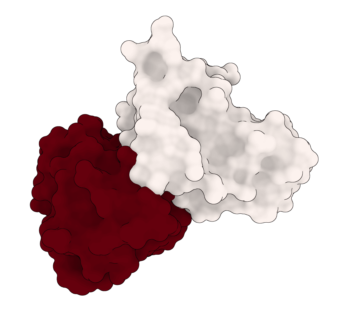
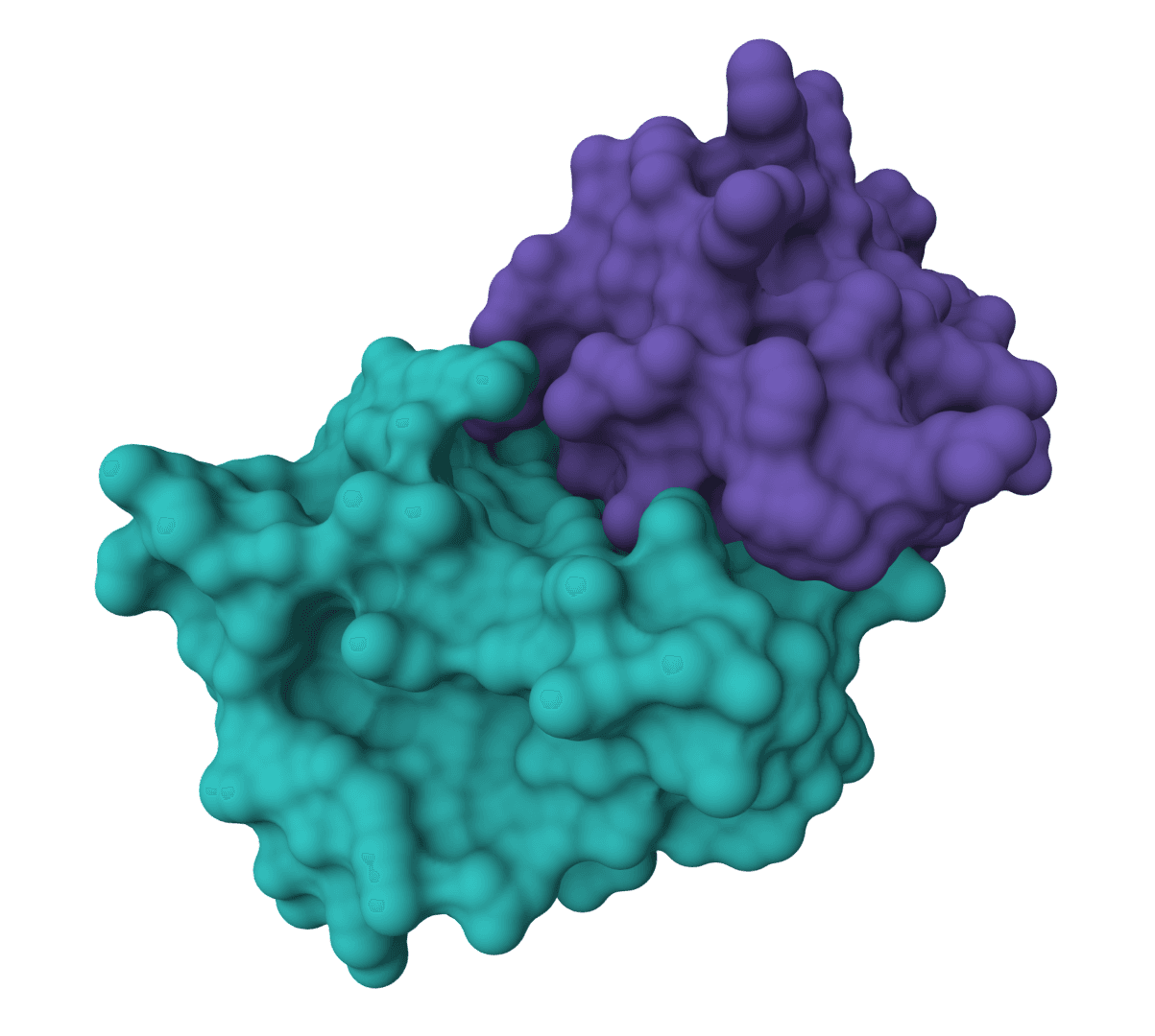
EquiDock predicts how two proteins bind together. Given the 3D structures of two unbound proteins, it outputs a docked complex showing their predicted binding pose.
The method uses SE(3)-equivariant graph neural networks, which means the predicted complex is always identical regardless of how the input structures are oriented. Rotate the inputs however you like, and the output remains consistent. This mathematical property makes predictions reliable and reproducible.
Developed at MIT and ETH Zurich, EquiDock was published as a spotlight paper at ICLR 2022. It runs 80-500x faster than traditional docking software by avoiding heavy sampling and refinement steps. The tradeoff is that it assumes rigid binding: neither protein changes shape upon contact.
How does EquiDock work?
EquiDock combines three techniques to predict binding poses:
Keypoint prediction: An attention mechanism identifies representative points on each protein that likely participate in binding. These keypoints approximate where the binding interface will form.
Optimal transport alignment: The model matches keypoints between the two proteins using optimal transport, finding correspondences between binding regions.
Differentiable Kabsch: Given matched keypoints, the algorithm computes the optimal rotation and translation to superimpose one protein onto the other, producing the final docked complex.
The neural network architecture extends Graph Matching Networks with E(3)-equivariant coordinate updates. Each protein is represented as a graph where nodes are residues and edges connect nearby residues. The network exchanges information both within each protein graph and between the two graphs to learn the binding transformation.
Confidence scoring
EquiDock computes confidence scores from keypoint alignment quality. Lower keypoint RMSD between predicted binding regions indicates higher confidence that the proteins dock well at that configuration.
How to use EquiDock online
ProteinIQ provides GPU-accelerated EquiDock predictions directly in the browser.
Inputs
| Input | Description |
|---|---|
Receptor | PDB file or RCSB PDB ID. Typically the larger protein. Max 1000 residues. |
Ligand | PDB file or RCSB PDB ID. Typically the smaller protein. Max 1000 residues. |
Both proteins should be unbound structures (not already in a complex). The tool expects standard ATOM records with backbone atoms (N, CA, C).
Settings
| Setting | Description |
|---|---|
Model variant | DIPS (default): trained on ~42,000 complexes for general use. DB5: optimized for the Docking Benchmark 5 dataset. |
Number of poses | How many binding configurations to generate: 1 (fastest), 3, or 5 (most thorough). Additional poses use random initial orientations. |
Results
EquiDock returns predicted complex structures and quality metrics:
| Output | Description |
|---|---|
| Complex PDB files | Receptor + ligand in predicted binding poses, ranked by confidence |
| Confidence score | Keypoint alignment quality (0-1, higher = better) |
| Contact map | Binary matrix showing residue-residue contacts at the interface |
| Interface residues | Lists of receptor and ligand residues within 8 Angstroms of each other |
Interpreting confidence
| Confidence | Interpretation |
|---|---|
| > 0.5 | Good alignment, likely reasonable pose |
| 0.3 - 0.5 | Moderate, review the interface manually |
| < 0.3 | Poor alignment, pose may be unreliable |
Additional metrics help assess pose quality:
- Min CA distance: Closest approach between receptor and ligand backbone atoms. Values under 3 Angstroms suggest clashes.
- Interface contacts: Number of residue pairs within 10 Angstroms. More contacts generally indicate a larger binding interface.
- Clash count: Residue pairs closer than 3 Angstroms. Should be zero or very low.
Limitations
EquiDock performs rigid docking only. Both proteins retain their input conformations. For proteins that undergo significant conformational changes upon binding, consider flexible docking methods.
The method predicts a single binding mode per pose. It does not model:
- Conformational flexibility or induced fit
- Allosteric effects
- Multiple simultaneous binding sites
Predictions work best for proteins with clear, localized binding interfaces. Extended or diffuse interfaces may produce lower confidence scores.
Related tools
AF2Dock
AF2Dock adapts AlphaFold2-style co-folding for structure-based protein-protein docking. It docks receptor and ligand protein structures with flow-matching refinement and ranks sampled complexes by iPTM.
DFMDock
DFMDock (Denoising Force Matching Dock) is a diffusion model that unifies sampling and ranking for protein-protein docking within a single framework. It predicts docked poses for protein-protein complexes from unbound structures using denoising score matching with optional clash force guidance.
GeoDock
GeoDock predicts flexible protein-protein docking complexes from two separate protein structures using a multi-track iterative transformer and the DIPS 0.3 checkpoint from the Gray Lab release.
ColabDock
ColabDock is a protein-protein docking framework that uses AlphaFold2 to predict complex structures guided by experimental restraints from cross-linking mass spectrometry, NMR, or other sources.
HADDOCK3
HADDOCK (High Ambiguity Driven protein-protein DOCKing) is an integrative modeling platform for biomolecular complexes. It uses experimental data and bioinformatic predictions to guide the docking process, generating accurate protein-protein complex structures.
LightDock
LightDock is a protein-protein, protein-peptide, and protein-DNA docking framework using Glowworm Swarm Optimization (GSO). It predicts macromolecular binding modes and interfaces for biological complexes.
ParaSurf
ParaSurf is a state-of-the-art surface-based deep learning model for predicting interactions between antibodies and antigens. It identifies paratope binding sites on antibody structures with high accuracy across multiple benchmark datasets.
SurfDock
SurfDock is a surface-informed diffusion generative model for protein-ligand docking, published in Nature Methods 2024. It leverages protein surface geometry to guide a diffusion process for reliable and accurate protein-ligand complex prediction.
DiffDock-L
DiffDock-L is a state-of-the-art molecular docking tool that uses diffusion models to predict how small molecule ligands bind to protein targets. It generates multiple binding poses with confidence scores.
DynamicBind
DynamicBind is an AI-powered protein-ligand binding prediction tool that recovers ligand-induced conformational changes from unbound protein structures. It predicts both ligand binding poses and protein conformational changes.